. 2014 Mar;42(5):3409-18.
doi: 10.1093/nar/gkt1279. Epub 2013 Dec 11.

Structure of the mammalian ribosomal pre-termination complex associated with eRF1.eRF3.GDPNP

Affiliations

Structure of the mammalian ribosomal pre-termination complex associated with eRF1.eRF3.GDPNP

Amédée des Georges et al. Nucleic Acids Res. 2014 Mar.

Abstract

Eukaryotic translation termination results from the complex functional interplay between two release factors, eRF1 and eRF3, in which GTP hydrolysis by eRF3 couples codon recognition with peptidyl-tRNA hydrolysis by eRF1. Here, we present a cryo-electron microscopy structure of pre-termination complexes associated with eRF1•eRF3•GDPNP at 9.7 -Å resolution, which corresponds to the initial pre-GTP hydrolysis stage of factor attachment and stop codon recognition. It reveals the ribosomal positions of eRFs and provides insights into the mechanisms of stop codon recognition and triggering of eRF3's GTPase activity.


Figures

Figure 1.
Figure 1.
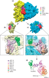
Unsupervised 3D classification. The 195 432 particles were first refined with RELION (27) to give a 9.1-Å ribosome reconstruction having low occupancy for the factors and tRNAs (top). The particles were then classified with RELION (27) starting with 10 seeds. The 10 resulting maps of the ribosome (bottom) are shown from the P stalk side (top vignette), with a close-up on the eRF1-eRF3 binding site (middle vignette), and from the 40S solvent side (bottom vignette). Bottom table: strength of the electron density for each domain of eRF1, eRF3 and tRNAs, as well as position of the L1 stalk.
Figure 2.
Figure 2.
Overview of the complex. (a) Overview of the map showing the 40S (yellow), 60S (blue), eRF1 (Magenta), eRF3 (red), P-site tRNA (green) and mRNA (coral). (b) Atomic model fitted into its density viewed from the 60S side with the 60S density removed. (c) Atomic model fitted into its density viewed from the 40S side with the 40S density removed. (d) Atomic model in the same orientation as in c, showing eRF1, eRF3, the P-site tRNA and mRNA path.
Figure 3.
Figure 3.
Close-up of the eRF1 N-domain and mRNA showing the TASNIKS amino-acids (blue), GTS loop (red) and YxCxxxF motif (green), and their positions relative to the approximate location of the stop codon bases represented as slabs. Codon positions are indicated in orange: first position, yellow: second position and blue: third position.
Figure 4.
Figure 4.
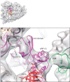
Close-up of eRF1-C and the mini-domain, showing the interaction of eRF1-C with H44 of the P stalk, and the interaction of the mini-domain with the 40S beak. rpL12 and rpLP0 not shown.
Figure 5.
Figure 5.
(a) Close-up of eRF3 showing the eRF1-eRF3 model fitted in the electron density together with the human 80S ribosome model (PDBID: 3J3A/B/D/F) [18S rRNA (purple), small subunit ribosomal proteins (beige), 28S rRNA (green), large subunit ribosomal proteins (light blue)]. (b) The 70S ribosome/EF-Tu/tRNA/GDPNP from (39) compared with the 80S ribosome-eRF1-eRF3 in (a).
Figure 6.
Figure 6.
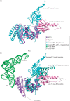
(a) Superimposition onto domain C of eRF1 in the crystal structure (7) (PDBID: 1DT9) and bound to the pre-TC ribosome. The mini-domain is not displayed for clarity. (b) Comparison between the positions of eRF1-M in the eRF1-eRF3-ribosome structure, the eRF1-eRF32–3 (15) (PDBID: 3E1Y) and the eRF1 (7) (PDBID: 1DT9) crystal structures with their domain C superimposed. Position of the P-site tRNA in the context of the pre-termination complex is shown to compare the distance necessary to be travelled for the GGQ loop to reach the peptidyl transfer center (PTC) with the amplitude of movement between the different conformations of eRF1 observed. Only the N domain of eRF1 in the pre-TC cryo-EM structure is shown for clarity.

Similar articles

Cited by

References

    1. Jackson RJ, Hellen CU, Pestova TV. Termination and post-termination events in eukaryotic translation. Adv. Protein Chem. Struct. Biol. 2012;86:45–93. - PubMed
    1. Alkalaeva EZ, Pisarev AV, Frolova LY, Kisselev LL, Pestova TV. In vitro reconstitution of eukaryotic translation reveals cooperativity between release factors eRF1 and eRF3. Cell. 2006;125:1125–1136. - PubMed
    1. Salas-Marco J, Bedwell DM. GTP hydrolysis by eRF3 facilitates stop codon decoding during eukaryotic translation termination. Mol. Cell. Biol. 2004;24:7769–7778. - PMC - PubMed
    1. Pisareva VP, Pisarev AV, Hellen CU, Rodnina MV, Pestova TV. Kinetic analysis of interaction of eukaryotic release factor 3 with guanine nucleotides. J. Biol. Chem. 2006;281:40224–40235. - PubMed
    1. Mitkevich VA, Kononenko AV, Petrushanko IY, Yanvarev DV, Makarov AA, Kisselev LL. Termination of translation in eukaryotes is mediated by the quaternary eRF1*eRF3*GTP*Mg2+ complex. The biological roles of eRF3 and prokaryotic RF3 are profoundly distinct. Nucleic Acids Res. 2008;34:3947–3954. - PMC - PubMed

Publication types

MeSH terms

Associated data