. 2014 Mar;111(5):1065-76.
doi: 10.1152/jn.00611.2013. Epub 2013 Dec 11.

Reorganization of hippocampal functional connectivity with transition to chronic back pain

Affiliations

Reorganization of hippocampal functional connectivity with transition to chronic back pain

Amelia A Mutso et al. J Neurophysiol. 2014 Mar.

Abstract

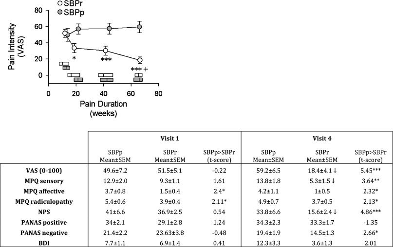
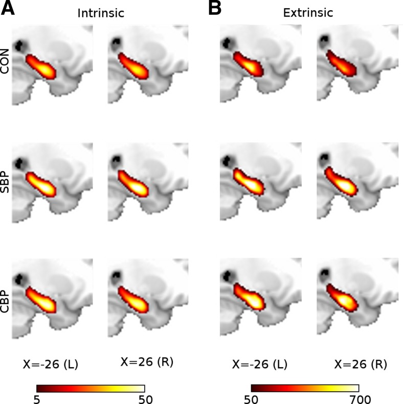
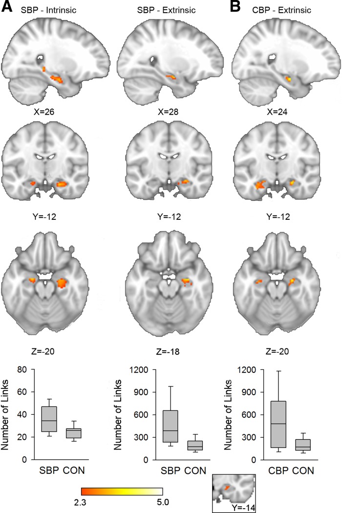
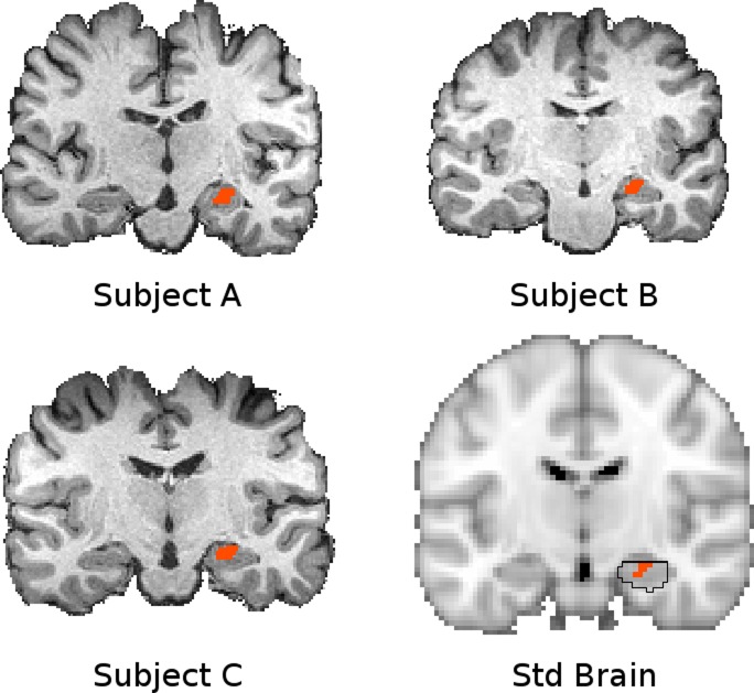
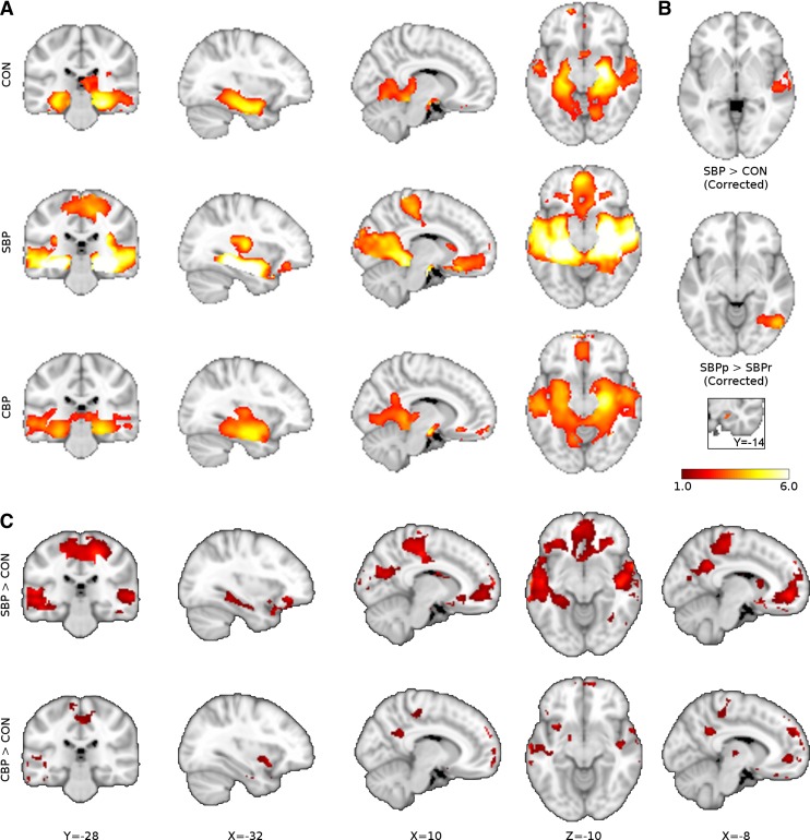
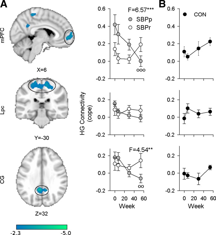
The hippocampus has been shown to undergo significant changes in rodent models of neuropathic pain; however, the role of the hippocampus in human chronic pain and its contribution to pain chronification have remained unexplored. Here we examine hippocampal processing during a simple visual attention task. We used functional MRI to identify intrinsic and extrinsic hippocampal functional connectivity (synchronous neural activity), comparing subacute back pain (SBP, back pain 1-4 mo) and chronic back pain (CBP, back pain >10 yr) patients to control (CON) subjects. Both groups showed more extensive hippocampal connectivity than CON subjects. We then examined the evolution of hippocampal connectivity longitudinally in SBP patients who recovered (SBPr, back pain decreased >20% in 1 yr) and those with persistent pain (SBPp). We found that SBPp and SBPr subjects have distinct changes in hippocampal-cortical connectivity over 1 yr; specifically, SBPp subjects showed large decreases in hippocampal connectivity with medial prefrontal cortex (HG-mPFC). Furthermore, in SBP patients the strength of HG-mPFC reflected variations in back pain over the year. These relationships were replicated when examined in a different task performed by SBP patients (rating fluctuations of back pain), indicating that functional connectivity of the hippocampus changes robustly in subacute pain and the nature of these changes depends on whether or not patients recover from SBP. The observed reorganization of processing within the hippocampus and between the hippocampus and the cortex seems to contribute to the transition from subacute to chronic pain and may also underlie learning and emotional abnormalities associated with chronic pain.

Keywords: back pain; functional connectivity; hippocampus; medial prefrontal cortex; transition to chronic pain.


Figures

Fig. 1.
Fig. 1.
Pain characteristics in subacute back pain (SBP) patients, as a function of grouping and time. Top: graph shows that SBP patients whose pain resolves (SBPr, n = 16) report decreases in pain on a visual analog scale (VAS) over the course of the study and ultimately differ significantly from SBP patients whose pain persists (SBPp, n = 14). Horizontal bars represent median and interquartile range of pain durations for each group. rm-ANOVA showed a significant group × time effect (P < 10−5). Post hoc (Tukey) tests: +P <0.05, within-group comparison to visit 1; *P < 0.05, ***P < 0.001, comparison between groups at corresponding times. Bottom: table shows pain and mood parameter differences for SBPp and SBPr. Significant decreases between visit 1 (at entry into the study) and visit 4 (1 yr after entry into study) (paired t-test, P < 0.01) are displayed as ↓. MPQ, McGill Pain Questionnaire; NPS, Neuropathic pain scale; BDI, Beck's Depression Inventory. PANAS, Positive Affect Negative Affect Scale. *P < 0.05, **P < 0.01, ***P < 0.001, unpaired t-test.
Fig. 2.
Fig. 2.
Mean intrinsic and extrinsic hippocampal connectivity in control (CON, n = 15), SBP (n = 32), and chronic back pain (CBP, n = 17) at visit 1 during a standard visual task. A: intrinsic hippocampal functional connectivity was computed for each voxel by identifying the number of other hippocampal voxels to which it was functionally connected. Extent of connectivity averaged across each group is illustrated with a heat map overlaid on a standard brain. B: extrinsic connectivity was computed by counting the number of nonhippocampal voxels to which hippocampal voxels were connected. Two voxels were considered functionally connected if their time series showed a correlation of r > 0.3. Hippocampal voxels colored yellow/white are functionally connected to more target region voxels than voxels in red/orange. Color bars indicate link count.
Fig. 3.
Fig. 3.
SBP and CBP patients show greater hippocampal connectivity than CON subjects. A: SBP patients (n = 32) show greater intrinsic (left) and extrinsic (right) hippocampal connectivity than CON subjects (n = 15) bilaterally and in the right hippocampus, respectively [bootstrap test, z > 2.3, threshold-free cluster enhancement (TFCE) cluster P < 0.05, corrected for age and sex]. Mean value of significant voxels was computed in each subject post hoc, and the connectivity across each group is shown as a graph below corresponding slices (post hoc Mann-Whitney U-test: intrinsic connectivity P < 0.001, extrinsic connectivity P = 0.007). B: CBP patients (n = 17) show greater extrinsic hippocampal connectivity than CON subjects bilaterally (bootstrap test, z > 2.3, TFCE P < 0.05 for multiple comparisons, data controlled for age and sex; post hoc U-test P = 0.007). Inset illustrates the conjunction of the 3 parametric maps shown in A and B. Bar graphs indicate median, quartile, and range of number of links.
Fig. 4.
Fig. 4.
The hippocampal seed shown in standard space (bottom right, orange, Y = −14) and native space for 3 subjects. The region of interest (ROI) was defined in standard space according to a region that showed greater connectivity extent in pain patients compared with CON subjects. This region is circumscribed by the hippocampus defined in the Harvard-Oxford subcortical atlas (bottom right, black outline), which identifies the likelihood of any particular voxel belonging to a particular subcortical structure (black outline indicates P > 0.5 likelihood for hippocampus). This ROI was transformed into native space for seed-based analysis in each subject. The fidelity of this transformation is indicated with T1 volumes from 3 representative subjects with the corresponding native space seed overlaid. Connectivity maps were transformed back into standard space subsequently for higher-level analysis.
Fig. 5.
Fig. 5.
Hippocampal-cortical connectivity differences between pain patients and CON subjects. A: whole brain functional connectivity of hippocampal seed (inset) in CON (n = 15), SBP (n = 32), and CBP (n = 17) at visit 1 during a standard visual task (mixed-effects analysis controlling for age and sex, voxel z > 2.3, cluster P < 0.05). B: hippocampal functional connectivity is distinct at visit 1 between groups. Middle temporal lobe connectivity to parahippocampus is greater in SBP than CON subjects, while SBPr (n = 17) show greater fusiform gyrus connectivity to parahippocampus than SBPp (n = 15; voxel z > 2.3, cluster P < 0.05). C: contrasts of CBP and SBP with CON performed at a low threshold (z > 1.0) without cluster correction reveal that the increases in cortical connectivity to the hippocampal seed are weak but widespread. Color bar illustrates z statistic of group mean hippocampal connectivity and group contrasts.
Fig. 6.
Fig. 6.
Longitudinally, SBPp and SBPr subjects show different changes in connectivity over 1 yr. A: right hippocampal gyrus (HG) in SBPp subjects (n = 14) shows significant changes in connectivity to medial prefrontal cortex (mPFC), paracentral lobule (Lpc), and cingulate gyrus (CG) over 1 yr (mixed effects, paired t-test visit 1 vs. visit 4, z > 2.3, cluster P < 0.05). Post hoc analysis controlling for age and sex showed that time evolution differs between SBPp and SBPr (n = 16) for mPFC and CG [rm-ANCOVA, group × time interactions: mPFC, F(3,75) = 6.57, P < 0.00053; CG, F(3,75) = 4.54, P = 0.0056]. Graphs are aligned to corresponding ROI. B: healthy CON subjects (n = 15) show no significant changes in HG to mPFC, Lpc, and GF over 1 yr [rm-ANOVA, F(3,42) = 2.24, P = 0.10 and F(3,42) = 1.1, P = 0.36, respectively]. **P < 0.01, ***P < 0.001, group × time interactions after controlling for sex and age. Mean ± SE shown for each visit.
Fig. 7.
Fig. 7.
HG connectivity reflects past and future pain in SBP (n = 29). A: HG-mPFC at visit 1 (v1) is significantly anticorrelated with decrease in pain by visit 4 (v4) 1 yr later (r2 = 0.21, P = 0.013, significant after correcting for multiple comparisons), while HG-CG at visit 4 correlates significantly with the amount that pain has decreased since visit 1 (r2 = 0.18, P = 0.02, uncorrected, not significant after correcting for multiple comparisons). B: decrease in connectivity shows a stronger anticorrelation to decrease in pain for both mPFC and CG. The more HG-mPFC and HG-CG connectivity drops over the course of 1 yr, the more persistent the pain at visit 4 (mPFC r2 = 0.35, P = 0.0007, CG r2 = 0.42, P = 0.00014), supporting the notion of an early hippocampal contribution to pain chronification. *P < 0.05, ***P < 0.001.
Fig. 8.
Fig. 8.
Decrease in HG-mPFC connectivity during a spontaneous pain-rating (SP) task (n = 29) shows the same relationship with pain identified in the standard visual (SV) task. A: decreases in HG-mPFC connectivity from visit 1 to visit 4 are significantly correlated between both the SV and SP tasks in SBP patients (r2 = 0.35, P = 0.001). B: in the SP task in SBP patients this connectivity decrease also shows a significant anticorrelation with decrease in pain (r2 = 0.134, P = 0.05). *P < 0.05, ***P < 0.001.

Similar articles

Cited by

References

    1. Abrahams S, Morris RG, Polkey CE, Jarosz JM, Cox TC, Graves M, Pickering A. Hippocampal involvement in spatial and working memory: a structural MRI analysis of patients with unilateral mesial temporal lobe sclerosis. Brain Cogn 41: 39–65, 1999 - PubMed
    1. Achard S, Salvador R, Whitcher B, Suckling J, Bullmore E. A resilient, low-frequency, small-world human brain functional network with highly connected association cortical hubs. J Neurosci 26: 63–72, 2006 - PMC - PubMed
    1. Alvarez RP, Biggs A, Chen G, Pine DS, Grillon C. Contextual fear conditioning in humans: cortical-hippocampal and amygdala contributions. J Neurosci 28: 6211–6219, 2008 - PMC - PubMed
    1. Apkarian AV. Pain perception in relation to emotional learning. Curr Opin Neurobiol 18: 464–468, 2008 - PMC - PubMed
    1. Apkarian AV, Baliki MN, Geha PY. Towards a theory of chronic pain. Prog Neurobiol 87: 81–97, 2009 - PMC - PubMed

Publication types