. 2014 Feb 13;123(7):1021-31.
doi: 10.1182/blood-2013-04-490847. Epub 2013 Dec 12.

SAMHD1 is mutated recurrently in chronic lymphocytic leukemia and is involved in response to DNA damage

Affiliations

SAMHD1 is mutated recurrently in chronic lymphocytic leukemia and is involved in response to DNA damage

Ruth Clifford et al. Blood. .

Abstract

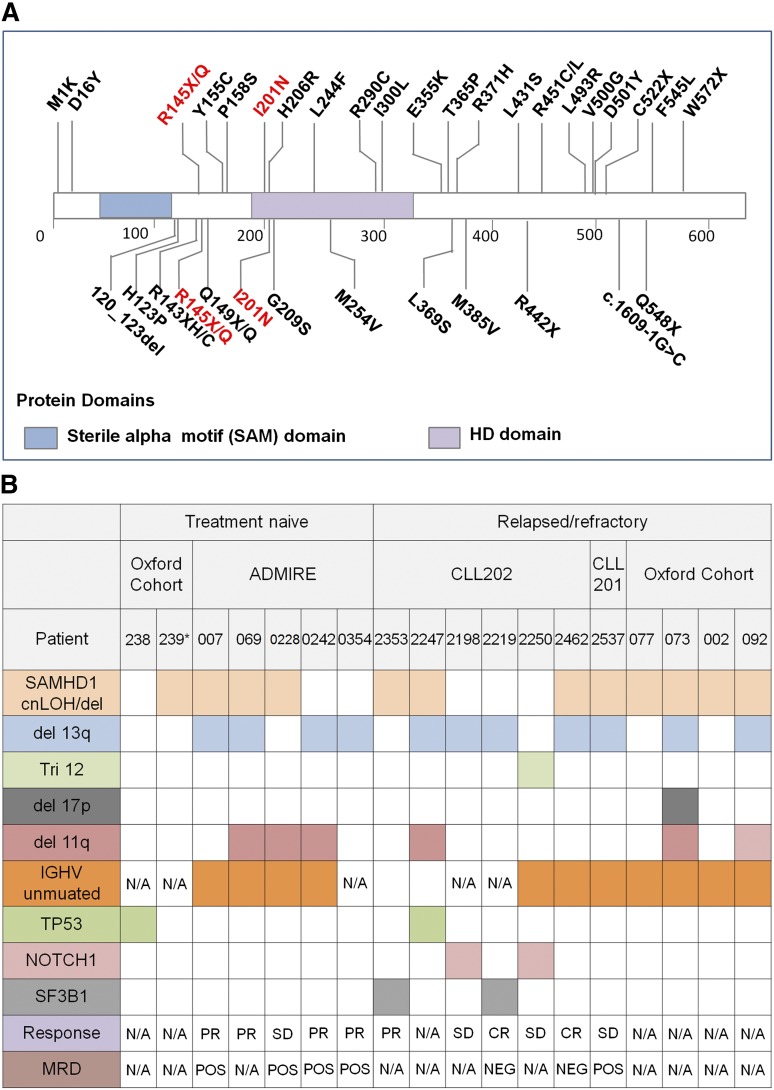
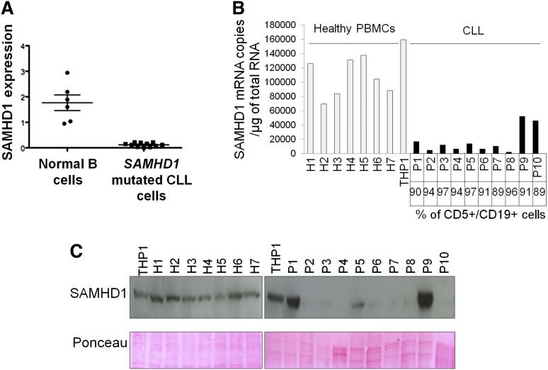
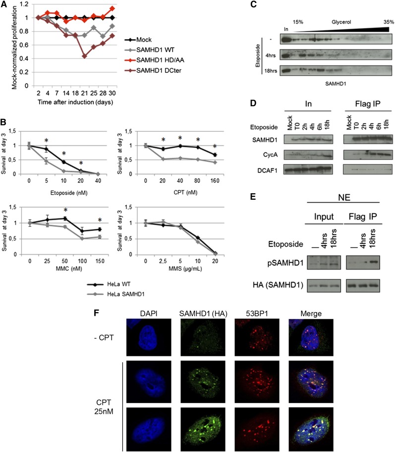
SAMHD1 is a deoxynucleoside triphosphate triphosphohydrolase and a nuclease that restricts HIV-1 in noncycling cells. Germ-line mutations in SAMHD1 have been described in patients with Aicardi-Goutières syndrome (AGS), a congenital autoimmune disease. In a previous longitudinal whole genome sequencing study of chronic lymphocytic leukemia (CLL), we revealed a SAMHD1 mutation as a potential founding event. Here, we describe an AGS patient carrying a pathogenic germ-line SAMHD1 mutation who developed CLL at 24 years of age. Using clinical trial samples, we show that acquired SAMHD1 mutations are associated with high variant allele frequency and reduced SAMHD1 expression and occur in 11% of relapsed/refractory CLL patients. We provide evidence that SAMHD1 regulates cell proliferation and survival and engages in specific protein interactions in response to DNA damage. We propose that SAMHD1 may have a function in DNA repair and that the presence of SAMHD1 mutations in CLL promotes leukemia development.


Figures

Figure 1
Figure 1
Analysis of an AGS patient. (A) Flow cytometry plot from whole peripheral blood of an AGS patient showing (left) forward-sideward scatter and (right) CD19 PE/CD5 APC scatter gated on lymphoid cells. (B) Whole genome array of chromosome 20 from leukemia cells of an AGS patient, visualized in Nexus (BioDiscovery). The log2 ratio indicates copy number changes, and the B-allele frequency plot indicates loss of heterozygosity.
Figure 2
Figure 2
Representative examples of somatic SAMHD1 mutations. (A) Electropherogram of SAMHD1 mutations using paired germ-line and tumor DNA. (Upper) Representative results from 2 patients are depicted. (Lower) Whole genome arrays showing (left) copy neutral loss of heterozygosity (cnLOH) or (right) a heterozygous deletion over chromosome 20 involving the SAMHD1 locus. (B) Multiple sequence alignment of SAMHD1 around the mutated residue (arrow) of each patient. Degree of conservation is reflected by background color code (dark blue, highly conserved to white, not conserved). (C) Whole genome array showing a small homozygous deletion at the 5′ end of the SAMHD1 locus in patient CLL 095.
Figure 3
Figure 3
Schematic representation of SAMHD1 mutations and relationship to other genetic lesions. (A) Schematic representation of SAMHD1 protein. (Upper) Mutations found in current study. (Lower) Known mutations in AGS patients. Mutations common to both diseases are shown in red. (B) Associations between SAMHD1 mutations and other genetic aberrations and clinical outcome. cnLOH, copy neutral loss of heterozygosity; CLL239*, AGS patient; Response, clinical response according to the iwCLL guidelines; PR, partial response; CR, complete response; SD, stable disease; MRD, minimal residual disease; N/A, not available.
Figure 4
Figure 4
Expression profiles of SAMHD1 in primary CLL cells. (A) cDNA pools from B cells (CD19+, CD5) sorted from peripheral blood of healthy donors and SAMHD1-mutated CLL cells (CD19+, CD5+) were subjected to quantitative PCR analysis using ABL as a control gene. (B) Measurement of SAMHD1 mRNA by quantitative RT-PCR. The percentage of leukemia cells (CD5+/CD19+) is indicated. Statistical comparison of SAMHD1 mRNA levels between each CLL sample and the mean of the healthy PBMCs was performed. The black bars indicate P < .01. (C) Measurement of SAMHD1 protein levels by western blot in 10 CLL samples and 7 healthy PBMCs. Ponceau dye staining was used to control protein load.
Figure 5
Figure 5
SAMHD1 effects on cell proliferation and cell viability in response to DNA damage agents. (A) HeLa cells stably expressing an inducible empty vector (Mock) or a vector encoding for SAMHD1 wild-type (WT) SAMHD1 mutated in the HD domain (HD/AA) or truncated at amino acid 575 (ΔCter) were induced with doxycycline. Results are expressed as the proliferation ratio of SAMHD1-transduced cells divided by mock-transduced cell. A representative experiment out of 3 is shown. (B) HeLa and HeLa cells stably expressing SAMHD1 were treated with increasing concentrations of DNA damaging agents, and the percentage of living cells was measured 3 days after treatment. Each graph shows the mean of 3 independent experiments. *A t-test P < .05. (C) HeLa cells stably expressing SAMHD1 were mock treated or treated with 5 nM of etoposide for 4 or 18 hours. Nuclear extracts were prepared and separated on a glycerol gradient. The localization of SAMHD1 along the gradient was assessed by western blot. (D) HeLa (Mock) and HeLa cells stably expressing Flag-HA–tagged SAMHD1 were treated with 5 nM etoposide for the indicated time. Flag IPs were performed and SAMHD1 interactions with CycA and DCAF1 were assessed by western blot. (E) Flag IPs were performed using nuclear extract of HeLa cells stably expressing Flag-HA–tagged SAMHD1 after treatment with 5 nM etoposide. SAMHD1 and SAMHD1 phosphorylated on threonine 592 (pSAMHD1) levels were determined by western blot. (F) SAMHD1 colocalizes with 53BP1 at the site of DSBs. HeLa cells expressing SAMHD1-HA were mock-treated or treated with 25 nM CPT. SAMHD1 and 53BP1 intranuclear localizations were determined by immunofluorescence using specific antibodies and confocal microscopy.

Comment in

  • SAMHD1: a new gene for CLL.
    Rossi D. Rossi D. Blood. 2014 Feb 13;123(7):951-2. doi: 10.1182/blood-2013-12-545384. Blood. 2014. PMID: 24526775 No abstract available.

Similar articles

Cited by

References

    1. Döhner H, Stilgenbauer S, Benner A, et al. Genomic aberrations and survival in chronic lymphocytic leukemia. N Engl J Med. 2000;343(26):1910–1916. - PubMed
    1. Quesada V, Conde L, Villamor N, et al. Exome sequencing identifies recurrent mutations of the splicing factor SF3B1 gene in chronic lymphocytic leukemia. Nat Genet. 2012;44(1):47–52. - PubMed
    1. Wang L, Lawrence MS, Wan Y, et al. SF3B1 and other novel cancer genes in chronic lymphocytic leukemia. N Engl J Med. 2011;365(26):2497–2506. - PMC - PubMed
    1. Puente XS, Pinyol M, Quesada V, et al. Whole-genome sequencing identifies recurrent mutations in chronic lymphocytic leukaemia. Nature. 2011;475(7354):101–105. - PMC - PubMed
    1. Landau DA, Carter SL, Stojanov P, et al. Evolution and impact of subclonal mutations in chronic lymphocytic leukemia. Cell. 2013;152(4):714–726. - PMC - PubMed

Publication types

MeSH terms

Substances

Supplementary concepts