. 2014;13(4):622-31.
doi: 10.4161/cc.27484. Epub 2013 Dec 12.

JNK signaling is needed to tolerate chromosomal instability

Affiliations

JNK signaling is needed to tolerate chromosomal instability

Heidi W-S Wong et al. Cell Cycle. 2014.

Abstract

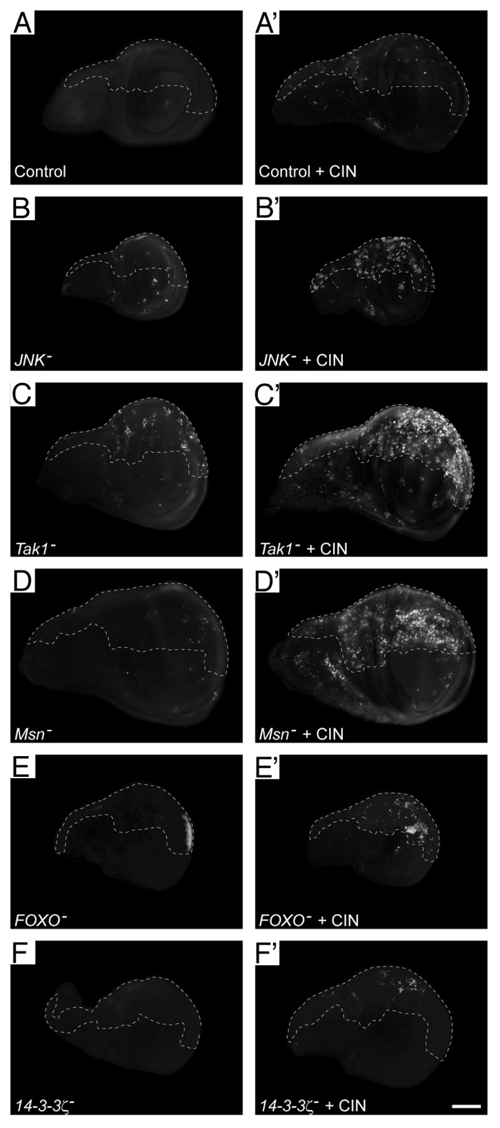
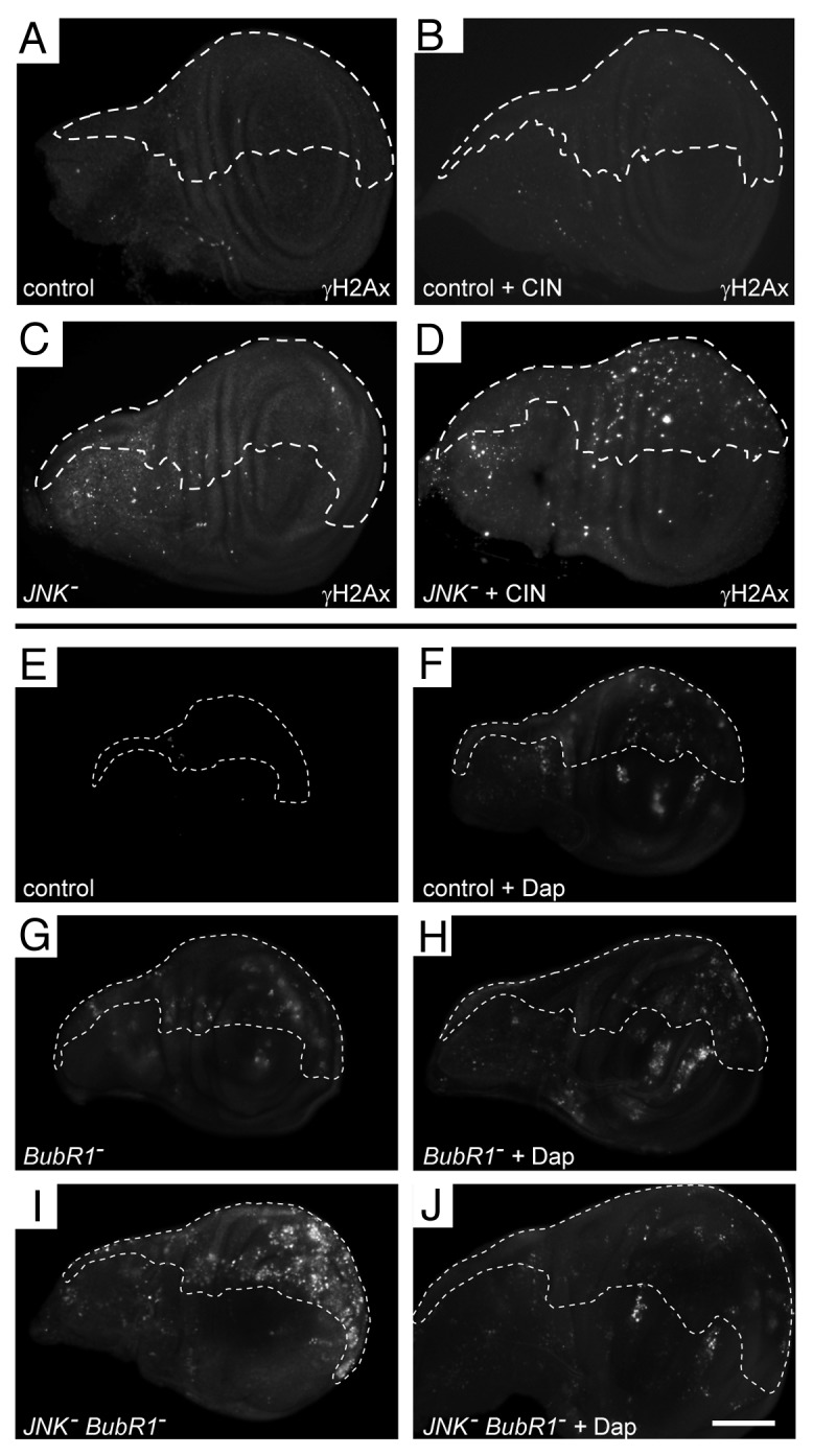
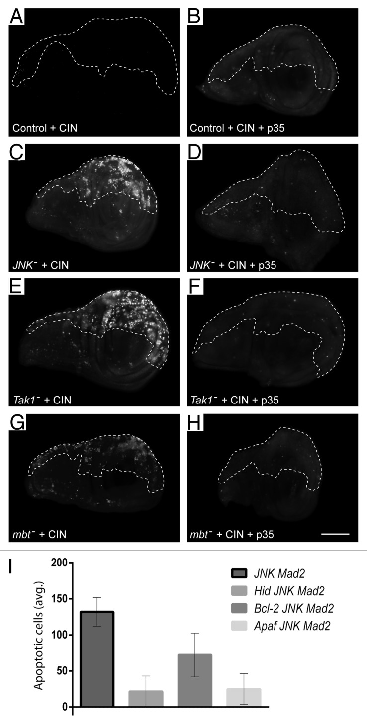
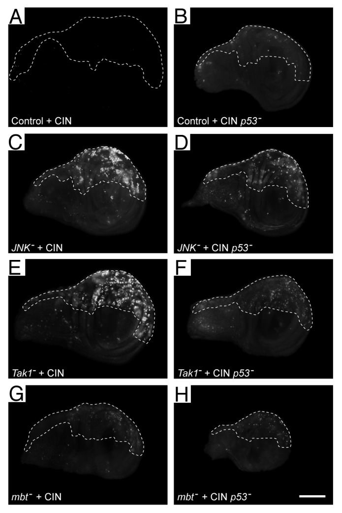
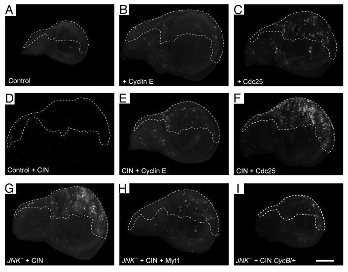
Chromosomal instability (CIN), as a common feature of tumors, represents a potential therapeutic target if ways can be found to specifically cause apoptosis in unstably dividing cells. We have previously shown that if signaling through the JNK pathway is reduced, apoptosis is triggered in models of chromosomal instability induced by loss of the spindle checkpoint. Here we identify components upstream and downstream of JNK that are able to mediate this effect, and test the involvement of p53 and DNA damage in causing apoptosis when JNK signaling is reduced in CIN cells. We show that cell cycle progression timing has a strong effect on the apoptosis seen when JNK signaling is reduced in genetically unstable cells: a shortened G 2 phase enhances the apoptosis, while lengthening G 2 rescues the JNK-deficient CIN cell death phenotype. Our findings suggest that chromosomal instability represents a significant stress to dividing cells, and that without JNK signaling, cells undergo apoptosis because they lack a timely and effective response to DNA damage.

Keywords: Drosophila; JNK; Mad2; apoptosis; chromosomal instability.


Figures

None
Figure 1. Knockdown of the JNK signaling pathway leads to cell death in CIN cells. Wing discs were stained with Acridine Orange to indicate cell death. In every disc, the unmarked region (anterior compartment) is the control tissue, in which no RNAi constructs are expressed, while the dashed line shows the posterior compartment in which JNK pathway members were knocked down by RNAi expression with or without CIN induced by mad2-RNAi expression. In control discs (A), induction of CIN caused little cell death (A’). For members of the JNK signaling pathway, the left column (B–F) shows discs in which JNK, Tak1, Msn, FOXO, or 14-3-3ζ have been reduced in the posterior compartment (dashed), with little cell death in each case. The right column (B’–F’) shows discs in which CIN has been induced (mad2-RNAi) in the posterior compartment along with knockdown of the indicated JNK pathway member. For each of these JNK pathway members, RNAi knockdown in CIN cells causes cell death that is not seen in the normally dividing control cells. The scale bar shows 100 μm
None
Figure 4. DNA damage is induced when JNK is reduced in CIN cells, and the cell death induced by knockdown of the JNK pathway in CIN cells is cell cycle-dependent. (A–D) Wing discs were stained for double-stranded DNA breaks (anti-P-H2AvD/γH2Ax). In every disc, the unmarked region does not express RNAi constructs, while the dashed line shows the posterior compartment cells that are affected by CIN (mad2-RNAi) and/or JNK knockdown. Control discs (A and B) show little DNA damage, even when CIN was induced (B). Knockdown of JNK also shows little DNA damage (C), but this is increased when JNK is reduced in CIN cells (D). (E–J) Wing discs were stained with Acridine Orange to show cell death. In each disc, the unmarked region does not express RNAi constructs, while the dashed line shows the area affected by overexpression of Dacapo and/or reduction of the indicated gene by RNAi. Control discs (E and F) show little cell death, although cell cycle arrest by overexpression of Dacapo (F) does cause some cell death, including death outside the overexpressing region (also seen in H and J). Induction of CIN by reducing BubR1 (G) causes some cell death, which is not altered by Dacapo overexpression (H). Knocking down JNK in CIN cells (I) gives considerable cell death, which is greatly reduced by overexpression of Dacapo (J). The scale bar shows 100 μm.
None
Figure 2. The cell death observed when the JNK pathway is knocked down in CIN cells can be greatly reduced by expressing the caspase inhibitor p35 or blocking the canonical apoptosis pathway. Wing discs were stained with Acridine Orange to show cell death. In every disc, the unmarked region does not express RNAi constructs, while the dashed line shows the posterior compartment affected by CIN (mad2-RNAi) and/or p35 overexpression. In control discs (A and B) CIN produced little cell death. When JNK was reduced in CIN cells (C), high levels of cell death were observed. This cell death was blocked by the caspase inhibitor p35 (D). The same inhibition of cell death by p35 was seen in CIN cells knocked down for the upstream JNK regulators Tak1 (E and F) and Mbt (G and H). The scale bar shows 100 μm. (I) The effect of knocking down the canonical apoptosis mediators Hid, Bcl-2, or Apaf on the amount of cell death seen in JNK-reduced CIN cells. The graph indicates the normalized average number of apoptotic cells per affected wing half for each genotype. Error bars indicate the 95% CI.
None
Figure 3. Some of the cell death observed when the JNK pathway is reduced in CIN cells is independent of p53. Wing discs were stained with Acridine Orange to show cell death. In every disc, the unmarked region does not express RNAi constructs, while the dashed line shows the area affected by CIN (induced by the expression of mad2-RNAi) and/or kncockdown of p53. Control wings (A and B) show little cell death when CIN is induced (A) or when p53 is knocked down in CIN cells (B). (C, E, and G) Imaginal discs in which members of the JNK signaling pathway, (JNK, Tak1, or Mbt) have been knocked down in CIN cells, giving rise to high levels of cell death. (D, F, and H) Imaginal discs showing that cell death is reduced but not eliminated by knockdown of p53 in CIN cells that are also knocked down for the indicated JNK pathway member. The scale bar shows 100 μm.
None
Figure 5. Cell death is increased in CIN cells when G2 is shortened and decreased when G2 is lengthened. Wing discs were stained with Acridine Orange to show cell death. In every disc, the unmarked region does not express RNAi constructs, while the dashed line shows the posterior compartment that is affected by CIN (mad2-RNAi) and/or gene overexpression. Control wings (A and B) show little cell death, even when CIN is induced (B). Cells with a shorter G1 phase from overexpression of Cyclin E (C and D) also show little cell death even when CIN is induced (D). Cells with a shorter G2 phase from overexpression of Cdc25 (stg-RNAi) (E and F) show some cell death, which is strongly enhanced by the induction of CIN (F). Cell death induced by JNK knockdown in CIN cells (G, H, and I) is reduced by lengthening G2 either by Myt1 overexpression (H) or loss of one copy of Cyclin B (I). The scale bar shows 100 μm.

Similar articles

Cited by

References

    1. Bakhoum SF, Compton DA. Chromosomal instability and cancer: a complex relationship with therapeutic potential. J Clin Invest. 2012;122:1138–43. doi: 10.1172/JCI59954. - DOI - PMC - PubMed
    1. Negrini S, Gorgoulis VG, Halazonetis TD. Genomic instability--an evolving hallmark of cancer. Nat Rev Mol Cell Biol. 2010;11:220–8. doi: 10.1038/nrm2858. - DOI - PubMed
    1. Gerlinger M, Swanton C. How Darwinian models inform therapeutic failure initiated by clonal heterogeneity in cancer medicine. Br J Cancer. 2010;103:1139–43. doi: 10.1038/sj.bjc.6605912. - DOI - PMC - PubMed
    1. Kolodner RD, Cleveland DW, Putnam CD. Cancer. Aneuploidy drives a mutator phenotype in cancer. Science. 2011;333:942–3. doi: 10.1126/science.1211154. - DOI - PMC - PubMed
    1. Lee AJ, Endesfelder D, Rowan AJ, Walther A, Birkbak NJ, Futreal PA, Downward J, Szallasi Z, Tomlinson IP, Howell M, et al. Chromosomal instability confers intrinsic multidrug resistance. Cancer Res. 2011;71:1858–70. doi: 10.1158/0008-5472.CAN-10-3604. - DOI - PMC - PubMed

Publication types

LinkOut - more resources