. 2014 Mar;42(5):3104-18.
doi: 10.1093/nar/gkt1292. Epub 2013 Dec 13.

Genetic analysis of the Replication Protein A large subunit family in Arabidopsis reveals unique and overlapping roles in DNA repair, meiosis and DNA replication

Affiliations

Genetic analysis of the Replication Protein A large subunit family in Arabidopsis reveals unique and overlapping roles in DNA repair, meiosis and DNA replication

Behailu B Aklilu et al. Nucleic Acids Res. 2014 Mar.

Abstract

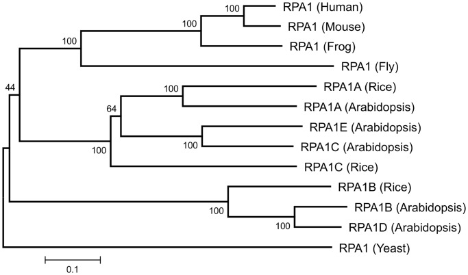
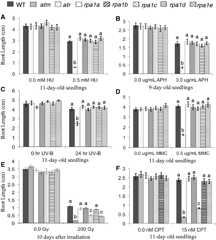
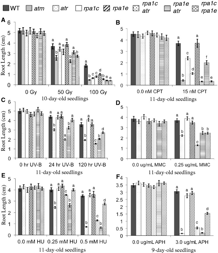
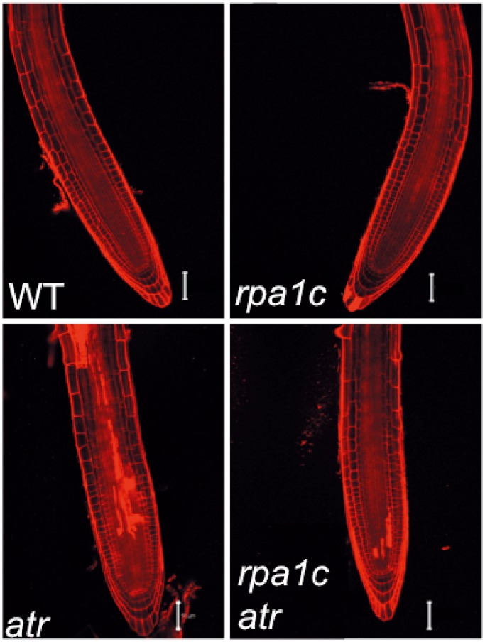
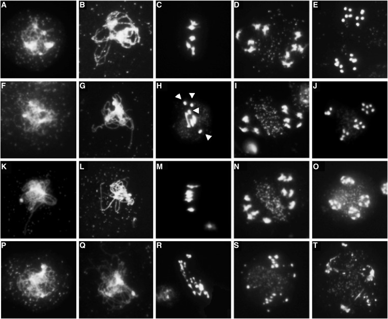
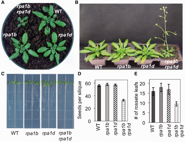
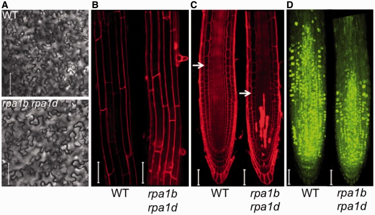
Replication Protein A (RPA) is a heterotrimeric protein complex that binds single-stranded DNA. In plants, multiple genes encode the three RPA subunits (RPA1, RPA2 and RPA3), including five RPA1-like genes in Arabidopsis. Phylogenetic analysis suggests two distinct groups composed of RPA1A, RPA1C, RPA1E (ACE group) and RPA1B, RPA1D (BD group). ACE-group members are transcriptionally induced by ionizing radiation, while BD-group members show higher basal transcription and are not induced by ionizing radiation. Analysis of rpa1 T-DNA insertion mutants demonstrates that although each mutant line is likely null, all mutant lines are viable and display normal vegetative growth. The rpa1c and rpa1e single mutants however display hypersensitivity to ionizing radiation, and combination of rpa1c and rpa1e results in additive hypersensitivity to a variety of DNA damaging agents. Combination of the partially sterile rpa1a with rpa1c results in complete sterility, incomplete synapsis and meiotic chromosome fragmentation, suggesting an early role for RPA1C in promoting homologous recombination. Combination of either rpa1c and/or rpa1e with atr revealed additive hypersensitivity phenotypes consistent with each functioning in unique repair pathways. In contrast, rpa1b rpa1d double mutant plants display slow growth and developmental defects under non-damaging conditions. We show these defects in the rpa1b rpa1d mutant are likely the result of defective DNA replication leading to reduction in cell division.


Figures

Figure 1.
Figure 1.
Neighbor-joining distance tree of RPA1-like protein sequences. All known sequences for rice and Arabidopsis are shown. Numbers above each branch indicate bootstrap values (percentage) of 1000 replicates. Scale bar represents the expected number of amino acid substitutions per site.
Figure 2.
Figure 2.
Hypersensitivity analysis of rpa1 single mutants. Root-length measurements of (A) plants grown in the absence or presence of 0.5 mM hydroxyurea (HU); (B) plants grown in the absence or presence of 3.0 μg/mL APH (USBiological, Swampscott, MA, USA); (C) plants grown for 5 days, treated with 0.4 J/s/m2 UV-B for 24 h or left untreated and grown for an additional 5 days; (D) plants grown in the absence or presence of 0.5 μg/mL MMC; (E) seeds gamma-irradiated (0 or 200 Gy) and grown for 10 days; (F) plants grown in the absence or presence of 15 nM CPT. Data are mean ± SE (n > 30). To analyze statistical difference with in each treatment group F-test (ANOVA) and LSD were carried out at P ≤ 0.05. Bars with different letters indicate significant differences.
Figure 3.
Figure 3.
Hypersensitivity analysis of rpa1c, rpa1e and atr mutant combinations. Root-length measurements of (A) 5-day-old seedlings were gamma-irradiated (0, 50 or 100 Gy) and grown for 5 days; (B) plants grown in the absence or presence of 15 nM CPT; (C) plants grown for 5 days, treated with 0.4 J/s/m2 UV-B for 24 h, 120 h or left untreated, and grown for an additional 5 days; (D) plants grown in the absence or presence of 0.25μg/mL MMC; (E) plants grown in the absence or presence of 0.25 mM, 0.5 mM hydroxyurea (HU); (F) plants grown in the absence or presence of 3.0 μg/mL APH (A.G. Scientific Inc., San Diego, CA, USA). Data are mean ± SE (n > 30). To analyze statistical difference with in each treatment group F-test (ANOVA) and LSD were carried out at P ≤ 0.05. Bars with different letters indicate significant differences.
Figure 4.
Figure 4.
PI viability staining of HU-treated root tips. WT, rpa1c, atr and rpa1c atr mutant lines were grown for 4 days, treated with 0.25 mM HU for 24 h, then transferred to media without HU for 24 h. Roots were stained with 5 mg/mL PI immediately prior to laser-scanning confocal microscopy. PI-filled cells represent dead cells. Bars = 50 µm.
Figure 5.
Figure 5.
Meiotic stages of pollen-mother cells from WT and rpa1 mutants. (A–E) Wild-type, (F–J) rpa1a, (K–O) rpa1c, (P–T) rpa1a rpa1c. Stages included are zygotene (A, F, K, P), pachytene (B, G, L), a zygotene stage representing the absence of a fully synapsed pachytene stage in the rpa1a rpa1c double mutant (Q), metaphase I (C, H, M, R), anaphase I (D, I, N, S) and anaphase II (E, J, O, T). In wild-type and rpa1c, homologs are segregated in equal number at anaphase I (D, N), followed by the separation and segregation of sister chromatids at anaphase II (E, O). In rpa1a, zygotene (F) and pachytene (G) stages are similar to WT and rpa1c. However, univalents (arrows) are present at the metaphase plate (H) leading to unequal segregation of homologous chromosomes at anaphase I (I), and anaphase II (J). In rpa1a rpa1c, multiple fragmented chromosomes were present at metaphase plate (R) and at anaphase I (S) leading to abnormal anaphase II (T).
Figure 6.
Figure 6.
Phenotypes of the rpa1b rpa1d double mutant line. (A) Twenty-six-day-old plants grown on soil. (B) Thirty-one-day-old plants grown on soil. (C) Eleven-day-old wild-type and mutant plants grown on MS phytoagar media. (D) Measurements of seeds produced by individual siliques of wild-type and mutant plants. (E) Flowering time measurements as counted by the number of rosette leaves when the inflorescence is 1-cm long.
Figure 7.
Figure 7.
Leaf and root cell phenotypes of the rpa1b rpa1d double mutant. (A) Outlines of leaf epidermal cells in wild-type (upper) and mutant (lower) plants. Calculated average cell areas for WT and rpa1b rpa1d were (2137 ± 1403) mm2 and (2055 ± 1300) mm2, respectively. (B) Laser-scanning confocal microscope images of PI stained 9-day-old wild-type and rpa1b rpa1d double mutant root tips. Calculated average cell length for WT and rpa1b rpa1d was 148 ± 44 mm and 162 ± 51 mm, respectively. (C) Laser-scanning confocal microscope images of PI stained 9-day-old wild-type and rpa1b rpa1d double mutant root tips. Arrows indicate starting point of the cell elongation zone. (D) Laser-scanning confocal microscope images of EdU stained 9-day-old wild-type and rpa1b rpa1d double mutant root tips. Fluorescent nuclei represent actively incorporating (replicating) nuclei. Bars = 50 µm.

References

    1. Fanning E, Klimovich V, Nager AR. A dynamic model for replication protein A (RPA) function in DNA processing pathways. Nucleic Acids Res. 2006;34:4126–4137. - PMC - PubMed
    1. Wold MS. Replication protein A: a heterotrimeric, single-stranded DNA-binding protein required for eukaryotic DNA metabolism. Ann. Rev. Biochem. 1997;66:61–92. - PubMed
    1. Abraham R. Cell cycle checkpoint signaling through the ATM and ATR kinases. Genes Dev. 2001;15:2177–2196. - PubMed
    1. Hurley PJ, Bunz F. ATM and ATR: components of an integrated circuit. Cell cycle (Georgetown, Tex) 2007;6:414–417. - PubMed
    1. Melo J, Toczyski D. A unified view of the DNA-damage checkpoint. Curr. Opin. Cell Biol. 2002;14:237–245. - PubMed

Publication types

MeSH terms