. 2014 Feb;88(4):2116-30.
doi: 10.1128/JVI.03443-13. Epub 2013 Dec 11.

Interferon-stimulated poly(ADP-Ribose) polymerases are potent inhibitors of cellular translation and virus replication

Affiliations

Interferon-stimulated poly(ADP-Ribose) polymerases are potent inhibitors of cellular translation and virus replication

Svetlana Atasheva et al. J Virol. 2014 Feb.

Abstract

The innate immune response is the first line of defense against most viral infections. Its activation promotes cell signaling, which reduces virus replication in infected cells and leads to induction of the antiviral state in yet-uninfected cells. This inhibition of virus replication is a result of the activation of a very broad spectrum of specific cellular genes, with each of their products usually making a small but detectable contribution to the overall antiviral state. The lack of a strong, dominant function for each gene product and the ability of many viruses to interfere with the development of the antiviral response strongly complicate identification of the antiviral activity of the activated individual cellular genes. However, we have previously developed and applied a new experimental system which allows us to define a critical function of some members of the poly(ADP-ribose) polymerase (PARP) family in clearance of Venezuelan equine encephalitis virus mutants from infected cells. In this new study, we demonstrate that PARP7, PARP10, and the long isoform of PARP12 (PARP12L) function as important and very potent regulators of cellular translation and virus replication. The translation inhibition and antiviral effect of PARP12L appear to be mediated by more than one protein function and are a result of its direct binding to polysomes, complex formation with cellular RNAs (which is determined by both putative RNA-binding and PARP domains), and catalytic activity. IMPORTANCE The results of this study demonstrate that interferon-stimulated gene products PARP7, PARP10, and PARP12L are potent inhibitors of the replication of Venezuelan equine encephalitis virus and other alphaviruses. The inhibitory functions are determined by more than a single mechanism, and one of them is based on the ability of these proteins to regulate cellular translation. Interference with the cellular translational machinery depends on the integrity of both the amino-terminal domain, containing a number of putative RNA-binding motifs, and the catalytic function of the carboxy-terminal PARP domain. The PARP-induced changes in translation efficiency appear to have a more potent effect on the synthesis of virus-specific proteins than on that of cellular proteins, thus making PARP-specific translational downregulation an important contributor to the overall development of the antiviral response.


Figures
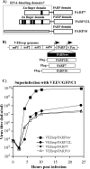
FIG 1
FIG 1
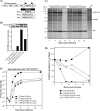
PARP7, PARP12L, and PARP10 fused with Flag tag efficiently inhibit VEEV replication. (A) Schematic representation of the PARP7, PARP12L, and PARP10 proteins. The amino-terminal, putative RNA-binding, and PARP domains are indicated. (B) Schematic representation of VEEV replicons encoding different PARPs under the control of the subgenomic promoter. The second promoter drives expression of the Pac gene. (C) BHK-21 cells were infected with the indicated packaged replicons at an MOI of 20 infectious units per cell. At 1 h postinfection, they were superinfected with VEEV/GFP/C1 virus (see Materials and Methods for details) at an MOI of 1 PFU/cell. At the indicated times after the second infection, medium was replaced, and titers of the released virus were determined by plaque assay on BHK-21 cells.
FIG 2
FIG 2
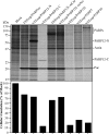
Expression of PARP7, PARP10, and PARP12L strongly affects cellular translation. (A) BHK-21 cells in 6-well Costar plates (5 × 10⁵ cells/well) were infected at an MOI of 20 infectious units per cell with packaged VEEV replicons encoding PARP7, PARP10, and PARP12L fused with Flag tag (see Materials and Methods for details) or VEErep, encoding PARP12L in reverse orientation. At the indicated times postinfection, proteins were metabolically pulse-labeled with [35S]methionine. Cell lysates were analyzed by SDS-PAGE followed by autoradiography. (B) Results of analysis of the same gels on a Storm phosphorimager. The radioactivity in the gel fragments containing cellular proteins of 15 to 42 kDa was measured and normalized to the levels from the same fragments in mock-infected cells. (C) Quantitative Western blot analysis of PARP7, PARP10, and PARP12L accumulation at 4 and 8 h postinfection (PI) by replicons. Protein levels were analyzed using a Flag-specific monoclonal antibody (MAb) and secondary infrared dye-labeled Abs. Images were acquired and processed on a Li-Cor imager.
FIG 3
FIG 3
The amino-terminal (PARP12-N) and carboxy-terminal (PARP12-C) domains, representing the natural short isoform of PARP12 and the PARP domain, respectively, are less efficient inhibitors of cellular translation. (A) Schematic representation of VEEV replicons encoding different fragments of PARP12 and PARP12L in reverse orientation. (B) Quantitative analysis of expression of different forms of PARP12. Cell lysates were prepared at 8 h postinfection with replicons. Protein levels were analyzed using a Flag-specific MAb and secondary infrared dye-labeled Abs. Images were acquired and processed on a Li-Cor imager. (C) BHK-21 cells in 6-well Costar plates (5 × 10⁵ cells/well) were infected with packaged VEEV replicons encoding PARP12-N, PARP12-C, and PARP12L fused with Flag tag (see Materials and Methods for details) or VEErep, encoding PARP12L in reverse orientation, at an MOI of 20 infectious units per cell. At the indicated times postinfection, proteins were metabolically pulse-labeled with [35S]methionine. Cell lysates were analyzed by SDS-PAGE followed by autoradiography. (D) Results of analysis of the same gels on a Storm phosphorimager. The radioactivity in gel fragments containing cellular proteins of 15 to 42 kDa was measured and normalized to the levels from the same fragments in mock-infected cells. (E) BHK-21 cells were infected with the indicated packaged replicons at an MOI of 20 infectious units per cell. At 1 h postinfection, they were superinfected with VEEV/GFP/C1 virus at an MOI of 1 PFU/cell. At the indicated times after the second infection, medium was replaced, and titers of the released virus were determined by plaque assay on BHK-21 cells.
FIG 4
FIG 4
Ribosylation of PARP12L is required for its translation inhibition functions. (A) Schematic representation of VEEV replicons encoding PARP12L in reverse orientation, Flag-PARP12L, or Flag-PARP12L with an H574A mutation in the catalytic site of the PARP domain (PARP12L-mCat variant) or an E569A mutation in its acceptor site (PARP12L-mAcc variant). (B) Quantitative analysis of expression of the indicated PARP12L mutants. Cell lysates were prepared at 8 h postinfection with replicons. The protein level was analyzed using a Flag-specific MAb and secondary infrared dye-labeled Abs. Images were acquired and processed on a Li-Cor imager. (C) BHK-21 cells in 6-well Costar plates (5 × 10⁵ cells/well) were infected with packaged VEEV replicons encoding Flag-PARP12-mCat and Flag-PARP12L-mAcc at an MOI of 20 infectious units per cell. At the indicated times postinfection, proteins were metabolically pulse-labeled with [35S]methionine. Cell lysates were analyzed by SDS-PAGE followed by autoradiography. (D) Results of analysis of the same gels on a Storm phosphorimager. The radioactivity in gel fragments containing cellular proteins of 15 to 42 kDa was measured and normalized to the levels from the same fragments in mock-infected cells. (E) BHK-21 cells were infected with the indicated packaged replicons at an MOI of 20 infectious units per cell. At 1 h postinfection, they were superinfected with VEEV/GFP/C1 virus at an MOI of 1 PFU/cell. At the indicated times after the second infection, medium was replaced, and titers of the released virus were determined by plaque assay on BHK-21 cells.
FIG 5
FIG 5
PARP proteins and their mutants cause translational shutoff in NIH 3T3 cells with efficiencies similar to those detected in BHK-21 cells. NIH 3T3 cells in 6-well Costar plates (5 × 10⁵ cells/well) were infected with the indicated packaged VEEV replicons at an MOI of 20 infectious units per cell. At 7 h postinfection, proteins were metabolically pulse-labeled with [35S]methionine. Cell lysates were analyzed by SDS-PAGE followed by autoradiography. The radioactivity in gel fragments containing cellular proteins of 15 to 42 kDa was measured on a Storm phosphorimager and normalized to the levels from the same fragments in mock-infected cells.
FIG 6
FIG 6
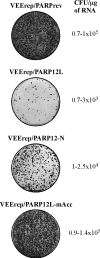
PARP12L variants demonstrate different levels of cytotoxicity. Equal amounts of in vitro-synthesized RNAs representing VEEV replicons encoding PARP12 in reverse orientation, Flag-PARP12L, Flag-PARP12-N, and Flag-PARP12L-mAcc, were electroporated into BHK-21 cells. Different numbers of cells were then seeded into dishes, and colonies of Purr cells were selected. Stained colonies in the dishes, which initially contained equal numbers of electroporated cells, are presented. The efficiency of formation of Purr colonies was measured in CFU per μg of RNA used for electroporation.
FIG 7
FIG 7
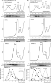
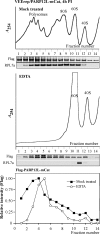
Upon expression in BHK-21 cells, the long isoform of PARP12 (PARP12L) forms two types of complexes. (A) BHK-21 cells were infected with packaged replicon VEErep/PARP12L at an MOI of 20 infectious units per cell. At 4 h postinfection, cells were harvested and lysed with NP-40. After pelleting the nuclei, the lysates were either directly analyzed by ultracentrifugation in the sucrose gradients and fractionated as described in Materials and Methods or additionally incubated prior to ultracentrifugation in the presence of 10 mM EDTA to disassemble ribosomes. Ribosomes and their subunits and other protein complexes were pelleted by another round of ultracentrifugation and further analyzed by quantitative Western blotting using Flag-specific MAbs and antibodies specific to the ribosomal RPL7a protein, which is located in the 60S ribosomal subunit. Images were acquired and processed using a Li-Cor imager. The lower panel presents the results of quantitative analysis of Flag-PARP12L distribution in the sucrose gradient. The intensity of the signal in each fraction was normalized to the highest concentration of Flag-PARP12L. (B) Cells were infected and lysates were analyzed as described above except that before ultracentrifugation, they were treated with 4 μg/ml of RNase A to destroy polysomes. The lower panel presents the results of quantitative analysis of PARP12L and 60S ribosomal subunit distribution in the sucrose gradients. The intensity of the signal in each fraction was normalized to the signal detected in the fractions containing highest concentration of Flag-PARP12L or RPL7a. These experiments were repeated twice with essentially the same results.
FIG 8
FIG 8
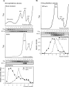
Expression of PARP12L, but not replication of VEErep itself, leads to destruction of polysomes. (A) BHK-21 cells were infected with packaged replicon VEErep/PARP12L at an MOI of 20 infectious units per cell. At 12 h postinfection, cells were harvested and lysed with NP-40, and the lysate was analyzed without additional treatment by ultracentrifugation in a sucrose gradient. Ribosomes, their subunits, and other protein complexes were pelleted from the fractions by additional ultracentrifugation and further analyzed by quantitative Western blotting using Flag-specific MAbs and antibodies specific to ribosomal RPL7a protein. Images were acquired and processed on a Li-Cor imager. The lower panel presents the results of quantitative analysis of Flag-PARP12L and 60S ribosomal subunit distribution in the sucrose gradient. The data were normalized to the signals detected in the fractions containing the highest concentrations of Flag-PARP12L and 60S subunit. (B) BHK-21 cells were infected with packaged VEErep/PARPrev replicon at an MOI of 20 infectious units per cell. At 4 and 12 h postinfection, cells were harvested and lysed with NP-40, and the lysates were analyzed without additional treatment by ultracentrifugation as described above for panel A.
FIG 9
FIG 9
The amino-terminal (PARP12-N) and the carboxy-terminal (PARP-C) domains of PARP12L form different complexes. BHK-21 cells were infected with packaged replicon VEErep/PARP12-C (A) or VEErep/PARP12-N (B) at an MOI of 20 infectious units per cell. At 4 h postinfection, cells were harvested and lysed with NP-40. After pelleting the nuclei, the lysates were either directly analyzed by ultracentrifugation in the sucrose gradients and fractionated as described in Materials and Methods or additionally incubated in the presence of 10 mM EDTA to disassemble ribosomes or with RNase A at concentration of 4 μg/ml for 15 min on ice to degrade polysomes. Ribosomes and their subunits and other protein complexes were pelleted by another round of ultracentrifugation as described in Materials and Methods and further analyzed by quantitative Western blotting using Flag-specific MAbs and antibodies specific to ribosomal RPL7a protein. Images were acquired and processed using a Li-Cor imager. The lower panels present the results of quantitative analysis of Flag-PARP12-C and Flag-PARP12-N distribution in the sucrose gradient. The intensity of the signal in each fraction was normalized to the highest concentration of Flag-PARP12L.
FIG 10
FIG 10
Mutation in PARP12L catalytic site does not abrogate formation of protein complexes. BHK-21 cells were infected with packaged VEErep/PARP12L-mCat replicon at an MOI of 20 infectious units per cell. At 4 h postinfection, cells were harvested and lysed with NP-40. After pelleting the nuclei, the lysates were either directly analyzed by ultracentrifugation in sucrose gradients and fractionated as described in Materials and Methods or additionally incubated in the presence of 10 mM EDTA to disassemble ribosomes. Ribosomes and their subunits and other protein complexes were pelleted by another round of ultracentrifugation as described in Materials and Methods and further analyzed by quantitative Western blotting using Flag-specific MAbs and antibodies specific to ribosomal RPL7a protein. Images were acquired and processed using a Li-COR imager. The lower panel presents the results of quantitative analysis of Flag-PARP12L-mCat distribution in the sucrose gradient. The intensity of the signal in each fraction was normalized to the highest concentration of Flag-PARP12L-mCat.
FIG 11
FIG 11
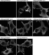
PARPs and their mutants demonstrate different intracellular distributions. BHK-21 cells seeded in Ibidi 8-well μ-slides were infected with the indicated packaged replicons at an MOI of 20 infectious units per cell. At 6 h postinfection, cells were fixed, permeabilized, and stained with a Flag-specific MAb and an Alexa Fluor 555-labeled secondary antibody. Images are presented as maximum-intensity projections of 6 optical sections. Bars correspond to 10 μm.

References

    1. Ramos HJ, Gale M., Jr 2011. RIG-I like receptors and their signaling crosstalk in the regulation of antiviral immunity. Curr. Opin. Virol. 1:167–176. 10.1016/j.coviro.2011.04.004 - DOI - PMC - PubMed
    1. Der SD, Yang YL, Weissmann C, Williams BR. 1997. A double-stranded RNA-activated protein kinase-dependent pathway mediating stress-induced apoptosis. Proc. Natl. Acad. Sci. U. S. A. 94:3279–3283. 10.1073/pnas.94.7.3279 - DOI - PMC - PubMed
    1. Der SD, Zhou A, Williams BR, Silverman RH. 1998. Identification of genes differentially regulated by interferon alpha, beta, or gamma using oligonucleotide arrays. Proc. Natl. Acad. Sci. U. S. A. 95:15623–15628. 10.1073/pnas.95.26.15623 - DOI - PMC - PubMed
    1. Lenschow DJ, Giannakopoulos NV, Gunn LJ, Johnston C, O'Guin AK, Schmidt RE, Levine B, Virgin HW., IV 2005. Identification of interferon-stimulated gene 15 as an antiviral molecule during Sindbis virus infection in vivo. J. Virol. 79:13974–13983. 10.1128/JVI.79.22.13974-13983.2005 - DOI - PMC - PubMed
    1. Liu SY, Sanchez DJ, Aliyari R, Lu S, Cheng G. 2012. Systematic identification of type I and type II interferon-induced antiviral factors. Proc. Natl. Acad. Sci. U. S. A. 109:4239–4244. 10.1073/pnas.1114981109 - DOI - PMC - PubMed

Publication types

MeSH terms