. 2014 Feb;88(4):2291-300.
doi: 10.1128/JVI.02829-13. Epub 2013 Dec 11.

STAT1 interaction with E3-14.7K in monocytes affects the efficacy of oncolytic adenovirus

Affiliations

STAT1 interaction with E3-14.7K in monocytes affects the efficacy of oncolytic adenovirus

Emma Spurrell et al. J Virol. 2014 Feb.

Erratum in

Abstract

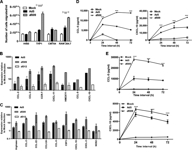
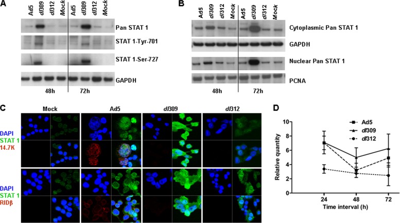
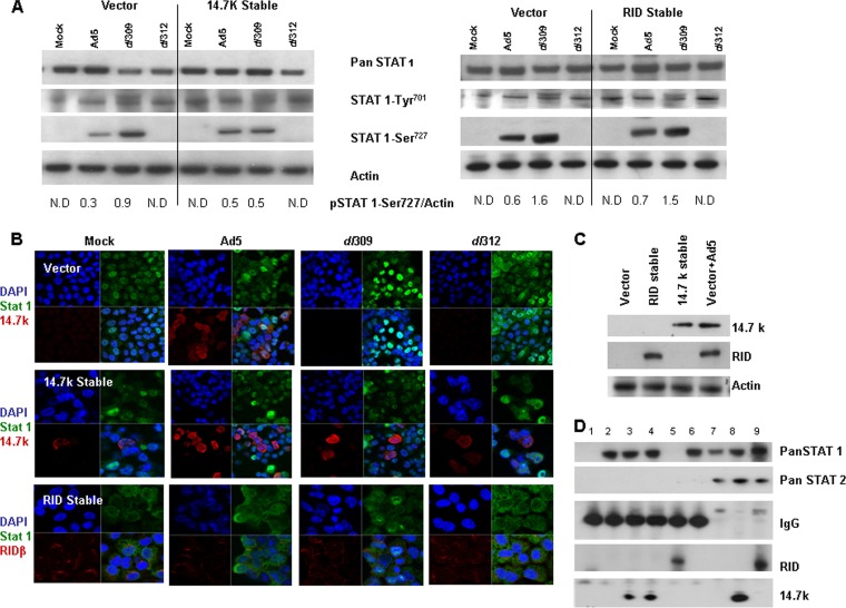
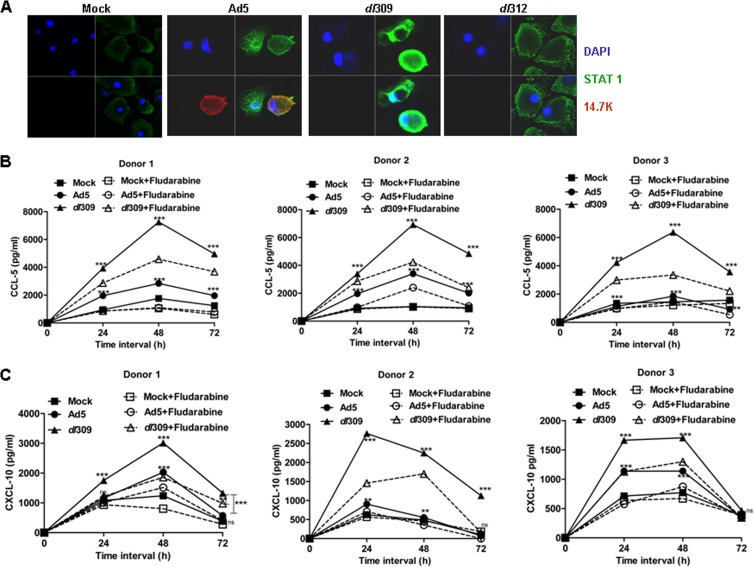
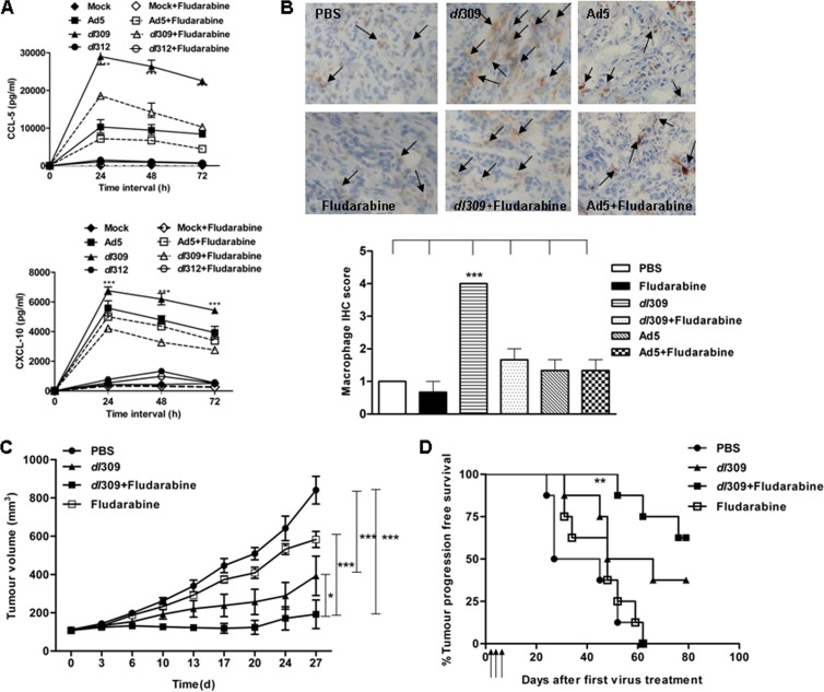
Oncolytic viruses based on adenovirus type 5 (Ad5) have been developed as a new class of therapeutic agents for cancers that are resistant to conventional therapies. Clinical experience shows that these agents are safe, but virotherapy alone has not achieved long-term cure in cancer patients. The vast majority of oncolytic adenoviruses used in clinical trials to date have deletion of the E3B genes. It has been demonstrated that the antitumor potency of the E3B-deleted mutant (dl309) is inferior to adenovirus with E3B genes intact. Tumors treated with dl309 show markedly greater macrophage infiltration than E3B-intact adenovirus. However, the functional mechanisms for this were not previously known. Here, we demonstrate that deletion of E3B genes increases production of chemokines by monocytes after adenovirus infection and increases monocyte migration. The E3B 14,700-Da protein (E3B-14.7K) inhibits STAT1 function by preventing its phosphorylation and nuclear translocation. The STAT1 inhibitor, fludarabine, rescues the effect of E3B-14.7K deletion by downregulating target chemokine expression in human and murine monocytes and results in an enhanced antitumor efficacy with dl309 in vivo. These findings have important implications for clinical use of E3B-deleted oncolytic adenovirus and other E3B-deleted adenovirus vector-based therapy.


Figures

FIG 1
FIG 1
The effect of E3B protein expression on the ability to chemoattract human and murine macrophage cell lines. (A) Quantitative analysis of monocytic cell line migration toward supernatants from epithelial and monocytic cells collected 72 h after adenoviral infection. The numbers of human THP-1 cells that had migrated toward the supernatants collected from human H460 and THP-1 cells and of murine RAW 264.7 cells that had migrated toward the supernatants collected from murine CMT-64 and RAW 264.7 cells were counted. At the indicated MOI, 70% of monocytes (THP-1 and RAW 264.7 cells) and 50% of tumor cells (H460 and CMT-64) were infected by adenovirus and expressed viral proteins. The samples were assayed in triplicates, and the experiment was carried out in duplicate. (B) Quantitative PCR estimation of chemokine mRNA in human THP-1 cells following Ad5, dl309, or dl312 infection at 48 h postinfection (MOI of 5,000). (C) Quantitative PCR estimation of cytokine and chemokine mRNAs in murine RAW 264.7 cells following Ad5, dl309, or dl312 infection at 48 h postinfection (MOI of 5,000). (D) Expression of CCL-5 and CXCL-10 by ELISA in THP-1 cells following mock, Ad5, or dl309 infection at an MOI of 5,000. (E) Expression of CCL-5 and CXCL-10 by ELISA in RAW 264.7 cells following mock, Ad5, or dl309 infection at an MOI of 5,000. Biological duplicate experiments were performed. *, P < 0.05; **, P < 0.01; ***, P < 0.001.
FIG 2
FIG 2
Expression of STAT1 in THP-1 cells after infection with different adenoviruses. (A) The expression of total (pan-STAT1) and phosphorylated STAT1 in THP-1 cells infected with Ad5, dl309, of dl312 or mock infected. Total protein from THP-1 cells was extracted at 48 h and 72 h after viral or mock infection and assayed by Western blotting. (B) STAT1 protein expression in nuclear and cytoplasmic compartments of THP-1 cell extracts infected with Ad5, dl309, or dl312 or mock infected. The results shown in panels A and B are from biological duplicate experiments. (C) STAT1 and E3B protein expression in THP-1 cells by confocal microscopy. Representative images showing STAT1 and E3B protein distribution 48 h following adenoviral or mock infection. The experiment was carried out in duplicate. (D) STAT1 mRNA expression in THP-1 cells mock infected or infected with Ad5, dl309, or dl312 over the time course indicated. GAPDH, glyceraldehyde-3-phosphate dehydrogenase.
FIG 3
FIG 3
Expression of STAT 1 protein in H460 cells primed with 100 ng/ml IFN-γ after adenovirus infection. (A) The expression of total and phosphorylated STAT1 in human lung carcinoma H460 cells primed with 100 ng/ml IFN-γ for 24 h prior to infection with Ad5, dl309, or dl312 at an MOI of 100 or mock infected. The total protein from the H460 cells was extracted at 48 h or 72 h after viral or mock infection and assayed by Western blotting. (B) STAT1 protein expression in nuclear and cytoplasmic compartments of H460 cell extracts infected with Ad5, dl309, or dl312 or mock infected. The data shown in panels A and B are from biological duplicate experiments. (C) STAT1 and E3B protein expression in THP-1 cells by confocal microscopy. Representative confocal images of H460 cells showing STAT 1 and E3B protein distribution 48 h following adenoviral or mock infection. Experiments were performed in duplicate.
FIG 4
FIG 4
Identification of interaction of E3B-14.7K with STAT1. (A) STAT1 protein expression in whole-cell extracts from E3B-14.7K, RID, or empty vector stably transfected cell lines primed with IFN-γ for 24 h and subsequently infected with Ad5, dl309, or dl312 at an MOI of 100 or mock infected. The specific bands for STAT1 and actin were quantified. The ratio of phospho-STAT1 (pSTAT1)-Ser 727 to actin is shown under the blots. (B) Confocal images showing STAT1 and E3B protein distribution in E3B-14.7K or RIDβ stably transfected cell lines and empty vector-transfected cells infected with Ad5, dl309, or dl312. (C) Western blot confirming the expression of E3B-14.7K and RID in stable H460 cell lines expressing E3B-14.7K or RID or transfected with the vector control or Ad5-infected vector control (48 h postinfection at an MOI of 100). (D) Direct interaction of STAT1 and E3B-14.7K proteins. The lysates from the human stable cell lines H460-pcDNA3.1 and H460 expressing E3B-14.7K and E3B RIDα/β were immunoprecipitated with anti-STAT1 antibody and probed with E3B-14.7K or RID or vice versa. The experiment was duplicated. Lane 1, H460-pcDNA3.1 extract incubated with nonspecific rabbit IgG; lane 2, H460-pcDNA3.1 cell extract immunoprecipitated with STAT1 antibody; lane 3, E3B-14.7K stable transfectant cell extract immunoprecipitated with E3B-14.7K antibody; lane 4, E3B-14.7K stable transfectant cell extract immunoprecipitated with STAT1 antibody; lane 5, RID stable transfectant cell extract immunoprecipitated with RID antibody; lane 6, RID stable transfectant cell extract immunoprecipitated with STAT1 antibody; lanes 7, 8, and 9, input of lysates from H460-pcDNA3.1, E3B-14.7K, and E3B RIDα/β, respectively.
FIG 5
FIG 5
Expression of STAT1, adenovirus E3B-14.7K protein, and chemokines in primary human monocytes and the effects of fludarabine on chemokine expression. (A) Confocal images showing STAT1 and E3B-14.7K protein distribution in human primary monocytes 48 h after infection with Ad5, dl309, or dl312 or mock infection. (B) CCL-5 secretion by human monocytes from three donors. Monocytes were pretreated with 10 μM fludarabine or vehicle buffer for 24 h and then infected with Ad5 or dl309 at an MOI of 5,000 or mock infected. The supernatants from the cell cultures were harvested, and CCL-5 was detected by ELISA 48 h after viral infection. (C) CXCL-10 secretion by human monocytes from three donors; monocytes were pretreated with 10 μM fludarabine or vehicle buffer for 24 h and then infected with Ad5 or dl309 at an MOI of 5,000 or mock infected. The supernatants from the cell cultures were harvested, and CXCL-10 was detected by ELISA 48 h after viral infection. Experiments were conducted individually on human monocytes taken from the three donors. The ELISA was performed in duplicate. *, P < 0.05; **, P < 0.01; ***, P < 0.001.
FIG 6
FIG 6
Effects of STAT1 inhibitor (fludarabine) on chemokine expression, macrophage infiltration, and antitumor efficacy by E3B-deleted adenovirus. (A) Expression of CCL-5 and CXCL-10 was assayed by ELISA on RAW 264.7 cells untreated or pretreated with 10 μM fludarabine for 24 h and infected with Ad5, dl309, or dl312 at an MOI of 5,000 or mock-infected. (B) Macrophage infiltration and quantification within murine CMT-93 tumors as determined by IHC after treatment with the different agents. Representative positive cells are indicated (arrows). Original magnification, ×200. (C) CMT-93 tumor growth curves after different treatments as described in Materials and Methods. (D) Kaplan-Meier survival analysis of mice bearing CMT-93 tumors after different treatments (n = 8/group). *, P < 0.05; **, P < 0.01; ***, P < 0.001.

References

    1. Garber K. 2006. China approves world's first oncolytic virus therapy for cancer treatment. J. Natl. Cancer Inst. 98:298–300. 10.1093/jnci/djj111 - DOI - PubMed
    1. Wong HH, Lemoine NR, Wang Y. 2010. Oncolytic viruses for cancer therapy: overcoming the obstacles. Viruses 2:78–106. 10.3390/v2010078 - DOI - PMC - PubMed
    1. Prestwich RJ, Harrington KJ, Pandha HS, Vile RG, Melcher AA, Errington F. 2008. Oncolytic viruses: a novel form of immunotherapy. Expert Rev. Anticancer Ther. 8:1581–1588. 10.1586/14737140.8.10.1581 - DOI - PMC - PubMed
    1. Ikeda K, Wakimoto H, Ichikawa T, Jhung S, Hochberg FH, Louis DN, Chiocca EA. 2000. Complement depletion facilitates the infection of multiple brain tumors by an intravascular, replication-conditional herpes simplex virus mutant. J. Virol. 74:4765–4775. 10.1128/JVI.74.10.4765-4775.2000 - DOI - PMC - PubMed
    1. Ikeda K, Ichikawa T, Wakimoto H, Silver JS, Deisboeck TS, Finkelstein D, Harsh GR, IV, Louis DN, Bartus RT, Hochberg FH, Chiocca EA. 1999. Oncolytic virus therapy of multiple tumors in the brain requires suppression of innate and elicited antiviral responses. Nat. Med. 5:881–887. 10.1038/11320 - DOI - PubMed

Publication types

MeSH terms

LinkOut - more resources