. 2014 Apr;69(4):1005-16.
doi: 10.1093/jac/dkt486. Epub 2013 Dec 12.

Rapid kill of malaria parasites by artemisinin and semi-synthetic endoperoxides involves ROS-dependent depolarization of the membrane potential

Affiliations

Rapid kill of malaria parasites by artemisinin and semi-synthetic endoperoxides involves ROS-dependent depolarization of the membrane potential

Thomas Antoine et al. J Antimicrob Chemother. 2014 Apr.

Abstract

Objectives: Artemisinin and artemisinin semi-synthetic derivatives (collectively known as endoperoxides) are first-line antimalarials for the treatment of uncomplicated and severe malaria. Endoperoxides display very fast killing rates and are generally recalcitrant to parasite resistance development. These key pharmacodynamic features are a result of a complex mechanism of action, the details of which lack consensus. Here, we report on the primary physiological events leading to parasite death.

Methods: Parasite mitochondrial (ΔΨm) and plasma membrane (ΔΨp) electrochemical potentials were measured using real-time single-cell imaging following exposure to pharmacologically relevant concentrations of endoperoxides (artemisinin, dihydroartemisinin, artesunate and the synthetic tetraoxane RKA182). In addition, mitochondrial electron transport chain components NADH:quinone oxidoreductase (alternative complex I), bc1 (complex III) and cytochrome oxidase (complex IV) were investigated to determine their functional sensitivity to the various endoperoxides.

Results: Parasite exposure to endoperoxides resulted in rapid depolarization of parasite ΔΨm and ΔΨp. The rate of depolarization was decreased in the presence of a reactive oxygen species (ROS) scavenger and Fe(3+) chelators. Depolarization of ΔΨm by endoperoxides is not believed to be through the inhibition of mitochondrial electron transport chain components, owing to the lack of significant inhibition when assayed directly.

Conclusions: The depolarization of ΔΨm and ΔΨp is shown to be mediated via the generation of ROS that are initiated by iron bioactivation of endoperoxides and/or catalysed by iron-dependent oxidative stress. These data are discussed in the context of current hypotheses concerning the mode of action of endoperoxides.

Keywords: Plasmodium; free radicals; haem; iron; lipid peroxidation; mitochondria; oxidative damage.


Figures

Figure 1.
Figure 1.
Chemical structures of endoperoxides, their corresponding P. falciparum growth inhibition values. Chemical structures of (a) artemisinin and its semi-synthetic derivatives, (b) deoxyartemisinin and (c) the synthetic 1,2,4,5-tetraoxane drug-development candidate RKA182 used in this study. The endoperoxide bridge is highlighted in grey. (d) P. falciparum growth inhibition IC50 values of endoperoxide antimalarials. Values are the mean ± SEM of results from three independent experiments.
Figure 2.
Figure 2.
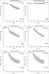
Effect of endoperoxide antimalarials on both the plasma and mitochondrial membrane potential of P. falciparum. Time course of TMRE-dependent fluorescence of P. falciparum-infected erythrocytes after the addition of (a) 100 nM atovaquone, (b) 100 nM deoxyartemisinin, (c) 100 nM artemisinin, (d) 100 nM tetraoxane (compound RKA182), (e) 100 nM artesunate and (f) 100 nM dihydroartemisinin. The data were normalized to 100% in untreated cells and to 0% in FCCP (10 μM)-treated cells. Graphs show means from experiments performed independently ± standard errors (n ≥ 7).
Figure 3.
Figure 3.
Effect of atovaquone, artemisinin and tetraoxane (RKA182) on fluorescent mitochondria from P. falciparum trophozoites. Bright-field fluorescence (a) and TMRE fluorescence images of concanamycin-pretreated infected erythrocytes before induction (b) and 300 s after induction (c) with 100 nM atovaquone (1), 100 nM artemisinin (2) and 100 nM tetraoxane (3). The green in these images is a pseudocolour. ‘M’ indicates the parasite mitochondrion and ‘FV’ indicates the food vacuole.
Figure 4.
Figure 4.
Effect of artemisinin and tetraoxane on mitochondrial membrane potential only. Plasma membrane potential is depolarized by treatment of P. falciparum-infected erythrocytes with 200 nM concanamycin A before addition of inhibitors. Time course of TMRE-plasma independent fluorescence is followed after addition of (a) 10 nM atovaquone, (b) 100 nM artemisinin and (c) 100 nM tetraoxane. Data were normalized to 100% in untreated cells and to 0% in FCCP (10 μM)-treated cells. Graphs show means from experiments performed independently ± standard errors (n ≥ 7).
Figure 5.
Figure 5.
Effect of artemisinin and tetraoxane on plasma membrane potential only. Mitochondrial membrane potential is depolarized by treatment of P. falciparum-infected erythrocytes with 100 nM atovaquone before addition of inhibitors. Time course of TMRE-mitochondrial independent fluorescence is followed after addition of (a) 200 nM concanamycin A, (b) 100 nM artemisinin and (c) 100 nM tetraoxane. Data were normalized to 100% in untreated cells and to 0% in FCCP (10 μM)-treated cells. Graphs show means from experiments performed independently ± standard errors (n ≥ 7).
Figure 6.
Figure 6.
Effect of artemisinin and tetraoxane on the membrane potential in the presence of iron chelators. Time course of TMRE-dependent fluorescence of P. falciparum-infected erythrocytes after addition of (a) 100 nM artemisinin and (b) 100 nM tetraoxane. Cells are not treated (open circles) or subjected to iron chelator treatment with 100 μM DFO (filled circles) or 100 μM DFP (filled diamonds). Data were normalized to 100% in untreated cells and to 0% in FCCP (10 μM)-treated cells. Graphs show means from experiments performed independently ± standard errors (n ≥ 7).
Figure 7.
Figure 7.
Effect of artemisinin and tetraoxane on the membrane potential in the presence of ROS scavenger. Time course of TMRE-dependent fluorescence of P. falciparum-infected erythrocytes after addition of (a) 100 nM artemisinin and (b) 100 nM tetraoxane. Cells are not treated (open circles) or subjected to ROS scavenger treatment with 100 μM Tiron (filled circles). Data were normalized to 100% in untreated cells and to 0% in FCCP (10 μM)-treated cells. Graphs show means from experiments performed independently ± standard errors (n ≥ 7).

References

    1. Klayman DL. Qinghaosu (artemisinin): an antimalarial drug from China. Science. 1985;228:1049–55. - PubMed
    1. Charman SA, Arbe-Barnes S, Bathurst IC, et al. Synthetic ozonide drug candidate OZ439 offers new hope for a single-dose cure of uncomplicated malaria. Proc Natl Acad Sci USA. 2011;108:4400–5. - PMC - PubMed
    1. O'Neill PM, Amewu RK, Nixon GL, et al. Identification of a 1,2,4,5-tetraoxane antimalarial drug-development candidate (RKA 182) with superior properties to the semisynthetic artemisinins. Angew Chem Int Ed Engl. 2010;49:5693–7. - PubMed
    1. O'Neill PM, Barton VE, Ward SA. The molecular mechanism of action of artemisinin—the debate continues. Molecules. 2010;15:1705–21. - PMC - PubMed
    1. Meshnick SR, Yang YZ, Lima V, et al. Iron-dependent free radical generation from the antimalarial agent artemisinin (qinghaosu) Antimicrob Agents Chemother. 1993;37:1108–14. - PMC - PubMed

Publication types

MeSH terms