. 2014 Mar 6;123(10):1596-603.
doi: 10.1182/blood-2013-08-523860. Epub 2013 Dec 13.

Clot contraction: compression of erythrocytes into tightly packed polyhedra and redistribution of platelets and fibrin

Affiliations

Clot contraction: compression of erythrocytes into tightly packed polyhedra and redistribution of platelets and fibrin

Douglas B Cines et al. Blood. .

Abstract

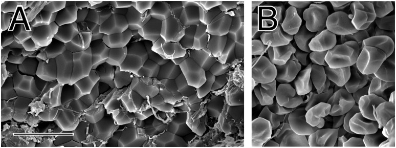
Contraction of blood clots is necessary for hemostasis and wound healing and to restore flow past obstructive thrombi, but little is known about the structure of contracted clots or the role of erythrocytes in contraction. We found that contracted blood clots develop a remarkable structure, with a meshwork of fibrin and platelet aggregates on the exterior of the clot and a close-packed, tessellated array of compressed polyhedral erythrocytes within. The same results were obtained after initiation of clotting with various activators and also with clots from reconstituted human blood and mouse blood. Such close-packed arrays of polyhedral erythrocytes, or polyhedrocytes, were also observed in human arterial thrombi taken from patients. The mechanical nature of this shape change was confirmed by polyhedrocyte formation from the forces of centrifugation of blood without clotting. Platelets (with their cytoskeletal motility proteins) and fibrin(ogen) (as the substrate bridging platelets for contraction) are required to generate the forces necessary to segregate platelets/fibrin from erythrocytes and to compress erythrocytes into a tightly packed array. These results demonstrate how contracted clots form an impermeable barrier important for hemostasis and wound healing and help explain how fibrinolysis is greatly retarded as clots contract.


Figures

Figure 1
Figure 1
The structure of contracted human whole blood clots. Scanning electron micrographs of whole-blood clots activated by thrombin following recalcification. (A,C) Inside of contracted clots, revealing close-packed polyhedra. (B,D) Outside of contracted clots, showing a thick meshwork of fibrin and platelet aggregates. Magnification bar = 10 µm.
Figure 2
Figure 2
The structure of erythrocytes from centrifuged unclotted human blood. Scanning electron micrograph showing the shape change of erythrocytes as a result of the forces of centrifugation at 6000g. Polyhedrocytes were observed with centrifugation of blood at forces of 1000g or greater, but not at lower forces. From the weight of a single erythrocyte of ∼1 pN or 10−7 dyn force applied to half of its surface area, the minimal stress required to induce polyhedrocyte formation and tight packing is estimated to be ∼75 to 150 dyn/cm2, well within the range of stress generated by platelets during contraction. Magnification bar = 10 µm.
Figure 3
Figure 3
The structure of contracted human whole blood clots by confocal light microscopy. (A) Confocal light micrograph of contracted whole-blood clot activated by thrombin following recalcification. Erythrocyte membranes of 30% of cells were fluorescently labeled with DiD. (B) Whole blood before clotting. Leukocytes and platelets were labeled with calcein (green); larger cells are leukocytes and smaller ones are platelets. Fibrinogen was labeled with Alexa 564 but is not visible, because it is not polymerized. (C) Three-dimensional reconstruction of contracted whole-blood clot. The outside of the clot is on the top, where there are platelets (green), fibrin (red), and biconcave erythrocytes (white). Erythrocytes deeper in the clot are polyhedral. Magnification bar = 25 µm.
Figure 4
Figure 4
The structure of clots made from reconstituted human whole blood activated with kaolin and mouse whole blood. (A-C) Scanning electron micrographs of clots made from reconstituted human whole blood consisting of 2 mg/mL fibrinogen, 167 000 platelets/μL, 37% hematocrit, activated by kaolin following recalcification. (A-B) Inside of these contracted clots, revealing close-packed polyhedra. (C) Outside of contracted clot, showing a thick meshwork of fibrin and platelet aggregates. (D) Scanning electron micrograph of the inside of a mouse whole-blood clot. Magnification bar = 20 µm (A) and 10 µm (B-D).
Figure 5
Figure 5
Polyhedral erythrocytes in human coronary artery thrombi. (A-B) Scanning electron micrographs of portions of thrombi aspirated from the coronary arteries of patients with ST-elevation myocardial infarction. Erythrocytes make up only about 11% of the volume of these thrombi, but some are polyhedral. Magnification bar = 10 µm.

Comment in

References

    1. Marder VJ, Aird WC, Bennett JS, Schulman S, White GC. Hemostasis and Thrombosis: Basic Principles and Clinical Practice. 6th ed. Philadelphia, PA: Lippincott Williams and Wilkins; 2013.
    1. Undas A, Ariëns RA. Fibrin clot structure and function: a role in the pathophysiology of arterial and venous thromboembolic diseases. Arterioscler Thromb Vasc Biol. 2011;31(12):e88–e99. - PubMed
    1. Coller BS. Historical perspective and future directions in platelet research. J Thromb Haemost. 2011;9(Suppl 1):374–395. - PMC - PubMed
    1. Lane DA, Grant PJ. Role of hemostatic gene polymorphisms in venous and arterial thrombotic disease. Blood. 2000;95(5):1517–1532. - PubMed
    1. Jen CJ, McIntire LV. The structural properties and contractile force of a clot. Cell Motil. 1982;2(5):445–455. - PubMed

Publication types

LinkOut - more resources