Dihydroartemisinin induces apoptosis of cervical cancer cells via upregulation of RKIP and downregulation of bcl-2
- PMID: 24335512
- PMCID: PMC3974829
- DOI: 10.4161/cbt.27223
Dihydroartemisinin induces apoptosis of cervical cancer cells via upregulation of RKIP and downregulation of bcl-2
Abstract
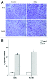
Treatment of recurrent and metastatic cervical cancer remains a challenge, especially in developing countries, which lack efficient screening programs. In recent years, artemisinin and its derivatives, such as dihydroartemisinin (DHA), which were traditionally used as anti-malarial agent, have been shown to inhibit tumor growth with low toxicity to normal cells. In this study, we investigated mechanisms underlying the anti-tumor effect of DHA in cervical cancer. We evaluated the role of DHA on the expression of bcl-2 and Raf kinase inhibitor protein (RKIP), which is a suppressor of metastasis. The MTT assay was used to compare the proliferation of untreated and DHA-treated Hela and Caski cervical cancer cells. Flow cytometry was used to determine the percentage of cells at each stage of the cell cycle in untreated and DHA-treated cells. We used RT-PCR and western blots to determine the expression of bcl-2 and RKIP mRNA and proteins. We evaluated the effect of DHA treatment in nude mice bearing Hela or Caski tumors. DHA-treated cells showed a time- and dose-dependent inhibition of proliferation and a significant increase in apoptosis. The expression of RKIP was significantly upregulated and the expression of bcl-2 was significantly downregulated in DHA-treated cells compared with control cells. DHA treatment caused (1) a significant inhibition of tumor growth and (2) a significant increase in the apoptotic index in nude mice bearing Hela or Caski tumors. Our data suggest that DHA inhibits cervical cancer growth via upregulation of RKIP and downregulation of bcl-2.
Keywords: Raf kinase inhibitor protein; apoptosis; bcl-2; cervival cancer; dihydroartemisinin; metastasis; tumor.
Figures



Similar articles
-
Anti-cancer activity of DHA on gastric cancer--an in vitro and in vivo study.Tumour Biol. 2013 Dec;34(6):3791-800. doi: 10.1007/s13277-013-0963-0. Epub 2013 Aug 2. Tumour Biol. 2013. PMID: 23907579
-
Dihydroartemisinin inhibits growth of pancreatic cancer cells in vitro and in vivo.Anticancer Drugs. 2009 Feb;20(2):131-40. doi: 10.1097/CAD.0b013e3283212ade. Anticancer Drugs. 2009. PMID: 19209030
-
Oxymatrine inhibits the proliferation of CaSki cells via downregulating HPV16E7 expression.Oncol Rep. 2016 Jul;36(1):291-8. doi: 10.3892/or.2016.4800. Epub 2016 May 10. Oncol Rep. 2016. PMID: 27176229
-
Exploring combinations of dihydroartemisinin for cancer therapy: A comprehensive review.Biochem Biophys Res Commun. 2025 Jun 8;765:151854. doi: 10.1016/j.bbrc.2025.151854. Epub 2025 Apr 18. Biochem Biophys Res Commun. 2025. PMID: 40262468 Review.
-
Dihydroartemisinin: A Potential Natural Anticancer Drug.Int J Biol Sci. 2021 Jan 16;17(2):603-622. doi: 10.7150/ijbs.50364. eCollection 2021. Int J Biol Sci. 2021. PMID: 33613116 Free PMC article. Review.
Cited by
-
Co-delivery of dihydroartemisinin and docetaxel in pH-sensitive nanoparticles for treating metastatic breast cancer via the NF-κB/MMP-2 signal pathway.RSC Adv. 2018 Jun 13;8(39):21735-21744. doi: 10.1039/c8ra02833h. eCollection 2018 Jun 13. RSC Adv. 2018. PMID: 35541720 Free PMC article.
-
The biological complexity of RKIP signaling in human cancers.Exp Mol Med. 2015 Sep 25;47(9):e185. doi: 10.1038/emm.2015.70. Exp Mol Med. 2015. PMID: 26403261 Free PMC article. Review.
-
Modulation of Navitoclax Sensitivity by Dihydroartemisinin-Mediated MCL-1 Repression in BCR-ABL+ B-Lineage Acute Lymphoblastic Leukemia.Clin Cancer Res. 2017 Dec 15;23(24):7558-7568. doi: 10.1158/1078-0432.CCR-17-1231. Epub 2017 Oct 3. Clin Cancer Res. 2017. PMID: 28974549 Free PMC article.
-
Protein kinase C-mediated phosphorylation of RKIP regulates inhibition of Na-alanine cotransport by leukotriene D(4) in intestinal epithelial cells.Am J Physiol Cell Physiol. 2014 Dec 1;307(11):C1010-6. doi: 10.1152/ajpcell.00284.2014. Epub 2014 Sep 17. Am J Physiol Cell Physiol. 2014. PMID: 25231108 Free PMC article.
-
Development of nanoscale drug delivery systems of dihydroartemisinin for cancer therapy: A review.Asian J Pharm Sci. 2022 Jul;17(4):475-490. doi: 10.1016/j.ajps.2022.04.005. Epub 2022 May 14. Asian J Pharm Sci. 2022. PMID: 36105316 Free PMC article. Review.
References
-
- Peralta-Zaragoza O, Deas J, Gómez-Cerón C, García-Suastegui WA, Fierros-Zárate GdelS, Jacobo-Herrera NJ. HPV-Based Screening, Triage, Treatment, and Followup Strategies in the Management of Cervical Intraepithelial Neoplasia. Obstet Gynecol Int. 2013;2013:912780. doi: 10.1155/2013/912780. - DOI - PMC - PubMed
-
- Mathew A, George PS. Trends in incidence and mortality rates of squamous cell carcinoma and adenocarcinoma of cervix--worldwide. Asian Pac J Cancer Prev. 2009;10:645–50. - PubMed
-
- Iida K, Nakayama K, Rahman MT, Rahman M, Ishikawa M, Katagiri A, Yeasmin S, Otsuki Y, Kobayashi H, Nakayama S, et al. EGFR gene amplification is related to adverse clinical outcomes in cervical squamous cell carcinoma, making the EGFR pathway a novel therapeutic target. Br J Cancer. 2011;105:420–7. doi: 10.1038/bjc.2011.222. - DOI - PMC - PubMed
MeSH terms
Substances
LinkOut - more resources
Full Text Sources
Other Literature Sources
Medical
Research Materials
Miscellaneous
