

Blockade of SDF-1 after irradiation inhibits tumor recurrences of autochthonous brain tumors in rats
- PMID: 24335554
- PMCID: PMC3870826
- DOI: 10.1093/neuonc/not149
Blockade of SDF-1 after irradiation inhibits tumor recurrences of autochthonous brain tumors in rats
Abstract
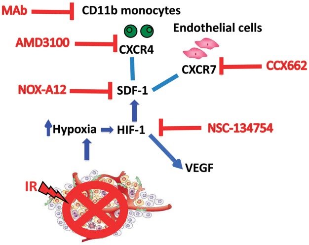
Background: Tumor irradiation blocks local angiogenesis, forcing any recurrent tumor to form new vessels from circulating cells. We have previously demonstrated that the post-irradiation recurrence of human glioblastomas in the brains of nude mice can be delayed or prevented by inhibiting circulating blood vessel-forming cells by blocking the interaction of CXCR4 with its ligand stromal cell-derived factor (SDF)-1 (CXCL12). In the present study we test this strategy by directly neutralizing SDF-1 in a clinically relevant model using autochthonous brain tumors in immune competent hosts.
Methods: We used NOX-A12, an l-enantiomeric RNA oligonucleotide that binds and inhibits SDF-1 with high affinity. We tested the effect of this inhibitor on the response to irradiation of brain tumors in rat induced by n-ethyl-N-nitrosourea.
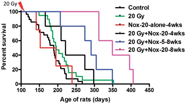
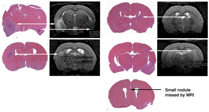
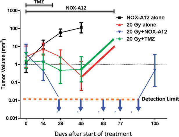
Results: Rats treated in utero with N-ethyl-N-nitrosourea began to die of brain tumors from approximately 120 days of age. We delivered a single dose of whole brain irradiation (20 Gy) on day 115 of age, began treatment with NOX-A12 immediately following irradiation, and continued with either 5 or 20 mg/kg for 4 or 8 weeks, doses and times equivalent to well-tolerated human exposures. We found a marked prolongation of rat life span that was dependent on both drug dose and duration of treatment. In addition we treated tumors only when they were visible by MRI and demonstrated complete regression of the tumors that was not achieved by irradiation alone or with the addition of temozolomide.
Conclusions: Inhibition of SDF-1 following tumor irradiation is a powerful way of improving tumor response of glioblastoma multiforme.
Keywords: CXCL12; CXCR4; CXCR7; ENU-induced tumors; NOX-A12; SDF-1; angiogenesis; glioblastoma; irradiation; vasculogenesis.
Figures

Comment in
-
Getting more out of radiation therapy in glioblastoma.Neuro Oncol. 2014 Jan;16(1):4-6. doi: 10.1093/neuonc/not233. Neuro Oncol. 2014. PMID: 24366974 Free PMC article. No abstract available.
Similar articles
-
Inhibition of vasculogenesis, but not angiogenesis, prevents the recurrence of glioblastoma after irradiation in mice.J Clin Invest. 2010 Mar;120(3):694-705. doi: 10.1172/JCI40283. Epub 2010 Feb 22. J Clin Invest. 2010. PMID: 20179352 Free PMC article.
-
SDF-1/CXCR4/CXCR7 is pivotal for vascular smooth muscle cell proliferation and chronic allograft vasculopathy.Transpl Int. 2015 Dec;28(12):1426-35. doi: 10.1111/tri.12651. Epub 2015 Sep 1. Transpl Int. 2015. PMID: 26265085
-
Inhibition of CXCR7 extends survival following irradiation of brain tumours in mice and rats.Br J Cancer. 2014 Mar 4;110(5):1179-88. doi: 10.1038/bjc.2013.830. Epub 2014 Jan 14. Br J Cancer. 2014. PMID: 24423923 Free PMC article.
-
Targeting SDF-1/CXCR4 to inhibit tumour vasculature for treatment of glioblastomas.Br J Cancer. 2011 Jun 7;104(12):1805-9. doi: 10.1038/bjc.2011.169. Epub 2011 May 17. Br J Cancer. 2011. PMID: 21587260 Free PMC article. Review.
-
Stromal cell-derived factor-1 (SDF-1) as a target in liver diseases.Am J Physiol Gastrointest Liver Physiol. 2016 Aug 1;311(2):G203-9. doi: 10.1152/ajpgi.00193.2016. Epub 2016 Jun 16. Am J Physiol Gastrointest Liver Physiol. 2016. PMID: 27313175 Review.
Cited by
-
Vasculogenesis: a crucial player in the resistance of solid tumours to radiotherapy.Br J Radiol. 2014 Mar;87(1035):20130686. doi: 10.1259/bjr.20130686. Br J Radiol. 2014. PMID: 24338942 Free PMC article. Review.
-
Targeting CXCL12/CXCR4 and myeloid cells to improve the therapeutic ratio in patient-derived cervical cancer models treated with radio-chemotherapy.Br J Cancer. 2019 Jul;121(3):249-256. doi: 10.1038/s41416-019-0497-3. Epub 2019 Jun 26. Br J Cancer. 2019. PMID: 31239542 Free PMC article.
-
Imposing Phase II and Phase III Clinical Trials of Targeted Drugs for Glioblastoma: Current Status and Progress.Front Oncol. 2021 Sep 9;11:719623. doi: 10.3389/fonc.2021.719623. eCollection 2021. Front Oncol. 2021. PMID: 34568049 Free PMC article. Review.
-
CXCR4 increases in-vivo glioma perivascular invasion, and reduces radiation induced apoptosis: A genetic knockdown study.Oncotarget. 2016 Dec 13;7(50):83701-83719. doi: 10.18632/oncotarget.13295. Oncotarget. 2016. PMID: 27863376 Free PMC article.
-
Potential Role of CXCR4 Targeting in the Context of Radiotherapy and Immunotherapy of Cancer.Front Immunol. 2018 Dec 21;9:3018. doi: 10.3389/fimmu.2018.03018. eCollection 2018. Front Immunol. 2018. PMID: 30622535 Free PMC article. Review.
References
-
- Hochberg FH, Pruitt A. Assumptions in the radiotherapy of glioblastoma. Neurology. 1980;30(9):907–911. - PubMed
-
- Liang BC, Thornton AF, Jr., Sandler HM, Greenberg HS. Malignant astrocytomas: focal tumor recurrence after focal external beam radiation therapy. J Neurosurg. 1991;75(4):559–563. - PubMed
-
- Sneed PK, Gutin PH, Larson DA, et al. Patterns of recurrence of glioblastoma multiforme after external irradiation followed by implant boost. Int J Radiat Oncol Biol Phys. 1994;29(4):719–727. - PubMed
-
- McDonald MW, Shu HK, Curran WJ, Jr., Crocker IR. Pattern of failure after limited margin radiotherapy and temozolomide for glioblastoma. Int J Radiat Oncol Biol Phys. 2011;79(1):130–136. - PubMed
Publication types
MeSH terms
Substances
Grants and funding
LinkOut - more resources
Full Text Sources
Other Literature Sources
Medical
