Ginsenoside Rg1 enhances the resistance of hematopoietic stem/progenitor cells to radiation-induced aging in mice
- PMID: 24335839
- PMCID: PMC4075748
- DOI: 10.1038/aps.2013.136
Ginsenoside Rg1 enhances the resistance of hematopoietic stem/progenitor cells to radiation-induced aging in mice
Abstract
Aim: To investigate the effects of ginsenoside Rg1 on the radiation-induced aging of hematopoietic stem/progenitor cells (HSC/HPCs) in mice and the underlying mechanisms.
Methods: Male C57BL/6 mice were treated with ginsenoside Rg1 (20 mg·kg(-1)·d(-1), ip) or normal saline (NS) for 7 d, followed by exposure to 6.5 Gy X-ray total body irradiation. A sham-irradiated group was treated with NS but without irradiation. Sca-1(+) HSC/HPCs were isolated and purified from their bone marrow using MACS. DNA damage was detected on d 1. The changes of anti-oxidative activities, senescence-related markers senescence-associated β-galactosidase (SA-β-gal) and mixed colony-forming unit (CFU-mix), P16(INK4a) and P21(Cip1/Waf1) expression on d 7, and cell cycle were examined on d 1, d 3, and d 7.
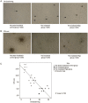
Results: The irradiation caused dramatic reduction in the number of Sca-1(+) HSC/HPCs on d 1 and the number barely recovered until d 7 compared to the sham-irradiated group. The irradiation significantly decreased SOD activity, increased MDA contents and caused DNA damage in Sca-1(+) HSC/HPCs. Moreover, the irradiation significantly increased SA-β-gal staining, reduced CFU-mix forming, increased the expression of P16(INK4a) and P21(Cip1/Waf1) in the core positions of the cellular senescence signaling pathways and caused G1 phase arrest of Sca-1(+) HSC/HPCs. Administration of ginsenoside Rg1 caused small, but significant recovery in the number of Sca-1(+) HSC/HPCs on d 3 and d 7. Furthermore, ginsenoside Rg1 significantly attenuated all the irradiation-induced changes in Sca-1(+) HSC/HPCs, including oxidative stress reaction, DNA damage, senescence-related markers and cellular senescence signaling pathways and cell cycle, etc.
Conclusion: Administration of ginsenoside Rg1 enhances the resistance of HSC/HPCs to ionizing radiation-induced senescence in mice by inhibiting the oxidative stress reaction, reducing DNA damage, and regulating the cell cycle.
Figures


Similar articles
-
Ginsenoside Rg1 protects against Sca-1+ HSC/HPC cell aging by regulating the SIRT1-FOXO3 and SIRT3-SOD2 signaling pathways in a γ-ray irradiation-induced aging mice model.Exp Ther Med. 2020 Aug;20(2):1245-1252. doi: 10.3892/etm.2020.8810. Epub 2020 May 28. Exp Ther Med. 2020. PMID: 32765665 Free PMC article.
-
Protective Effect of Ginsenoside Rg1 on Hematopoietic Stem/Progenitor Cells through Attenuating Oxidative Stress and the Wnt/β-Catenin Signaling Pathway in a Mouse Model of d-Galactose-induced Aging.Int J Mol Sci. 2016 Jun 9;17(6):849. doi: 10.3390/ijms17060849. Int J Mol Sci. 2016. PMID: 27294914 Free PMC article.
-
[Experimental study of relationship between effect of ginsenoside Rg1 to delay hematopoietic stem cell senescence and expression of p16(INK4a)].Zhongguo Zhong Yao Za Zhi. 2011 Mar;36(5):608-13. Zhongguo Zhong Yao Za Zhi. 2011. PMID: 21657082 Chinese.
-
Inhibitory effects and molecular mechanisms of ginsenoside Rg1 on the senescence of hematopoietic stem cells.Fundam Clin Pharmacol. 2023 Jun;37(3):509-517. doi: 10.1111/fcp.12863. Epub 2023 Jan 7. Fundam Clin Pharmacol. 2023. PMID: 36582074 Review.
-
Ginsenoside Rg1 as a Potential Regulator of Hematopoietic Stem/Progenitor Cells.Stem Cells Int. 2021 Dec 31;2021:4633270. doi: 10.1155/2021/4633270. eCollection 2021. Stem Cells Int. 2021. PMID: 35003268 Free PMC article. Review.
Cited by
-
Dynamic Protective Effect of Chinese Herbal Prescription, Yiqi Jiedu Decoction, on Testis in Mice with Acute Radiation Injury.Evid Based Complement Alternat Med. 2021 Jun 1;2021:6644093. doi: 10.1155/2021/6644093. eCollection 2021. Evid Based Complement Alternat Med. 2021. PMID: 34122603 Free PMC article.
-
Research on the anti-aging mechanisms of Panax ginseng extract in mice: a gut microbiome and metabolomics approach.Front Pharmacol. 2024 Jun 20;15:1415844. doi: 10.3389/fphar.2024.1415844. eCollection 2024. Front Pharmacol. 2024. PMID: 38966558 Free PMC article.
-
Ginsenoside Rg1 can restore hematopoietic function by inhibiting Bax translocation-mediated mitochondrial apoptosis in aplastic anemia.Sci Rep. 2021 Jun 17;11(1):12742. doi: 10.1038/s41598-021-91471-1. Sci Rep. 2021. PMID: 34140535 Free PMC article.
-
Ginsenoside RG1 enhances the paracrine effects of bone marrow-derived mesenchymal stem cells on radiation induced intestinal injury.Aging (Albany NY). 2020 Dec 3;13(1):1132-1152. doi: 10.18632/aging.202241. Epub 2020 Dec 3. Aging (Albany NY). 2020. PMID: 33293477 Free PMC article.
-
Ginsenoside Rg1 protects against Sca-1+ HSC/HPC cell aging by regulating the SIRT1-FOXO3 and SIRT3-SOD2 signaling pathways in a γ-ray irradiation-induced aging mice model.Exp Ther Med. 2020 Aug;20(2):1245-1252. doi: 10.3892/etm.2020.8810. Epub 2020 May 28. Exp Ther Med. 2020. PMID: 32765665 Free PMC article.
References
-
- Gazit R, Weissman H, Rossl DJ. Hematopoietic stem cells and the aging hematopoietic system. Strain Hematol. 2008;45:218–23. - PubMed
-
- Chambers SM, Goodell MA. Hematopoietic stem cell aging: wrinkles in stem cell potential. Stem Cell Rev. 2007;3:201–11. - PubMed
-
- Cheng Y, Shen LH, Zhang JT. Anti-amnestic and anti-aging effects of ginsenoside Rg1 and Rb1 and its mechanism of action. Acta Pharmacol Sin. 2005;26:143–9. - PubMed
Publication types
MeSH terms
Substances
LinkOut - more resources
Full Text Sources
Other Literature Sources
Medical
Research Materials
Miscellaneous

