

Cancer-associated fibroblasts induce epithelial-mesenchymal transition of breast cancer cells through paracrine TGF-β signalling
- PMID: 24335925
- PMCID: PMC3915130
- DOI: 10.1038/bjc.2013.768
Cancer-associated fibroblasts induce epithelial-mesenchymal transition of breast cancer cells through paracrine TGF-β signalling
Abstract
Background: Cancer-associated fibroblasts (CAFs) activated by tumour cells are the predominant type of stromal cells in breast cancer tissue. The reciprocal effect of CAFs on breast cancer cells and the underlying molecular mechanisms are not fully characterised.
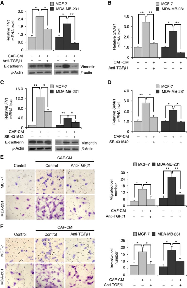
Methods: Stromal fibroblasts were isolated from invasive breast cancer tissues and the conditioned medium of cultured CAFs (CAF-CM) was collected to culture the breast cancer cell lines MCF-7, T47D and MDA-MB-231. Neutralising antibody and small-molecule inhibitor were used to block the transforming growth factor-β (TGF-β) signalling derived from CAF-CM, which effect on breast cancer cells.
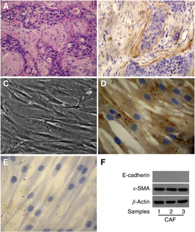
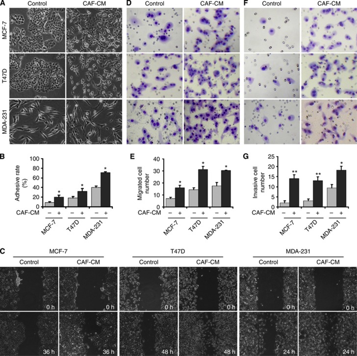
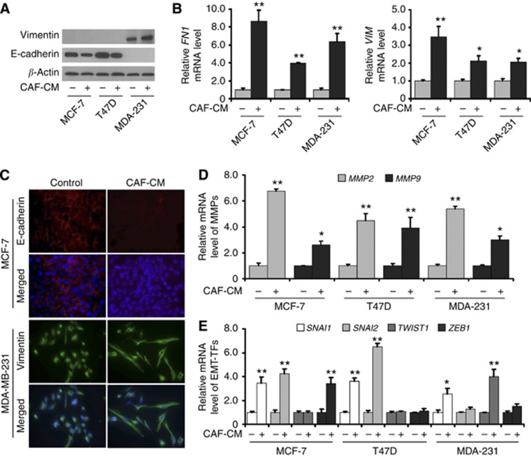
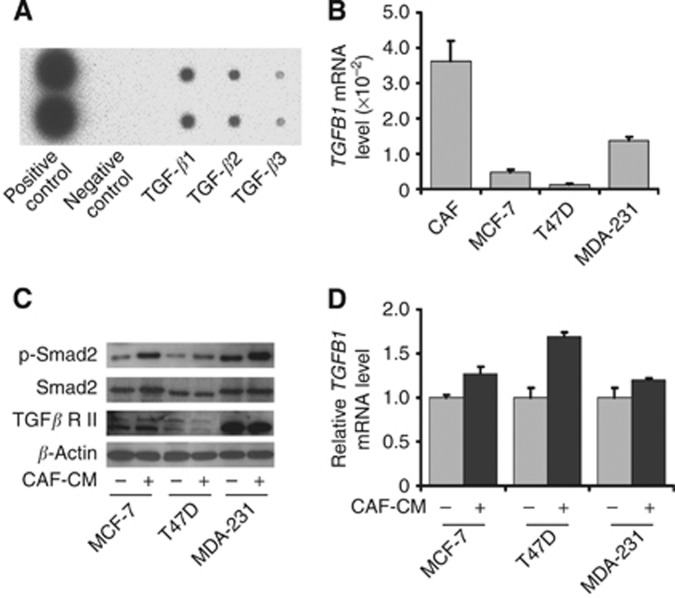
Results: The stromal fibroblasts isolated from breast cancer tissues showed CAF characteristics with high expression levels of α-smooth muscle actin and SDF1/CXCL12. The CAF-CM transformed breast cancer cell lines into more aggressive phenotypes, including enhanced cell-extracellular matrix adhesion, migration and invasion, and promoted epithelial-mesenchymal transition (EMT). Cancer-associated fibroblasts secreted more TGF-β1 than TGF-β2 and TGF-β3, and activated the TGF-β/Smad signalling pathway in breast cancer cells. The EMT phenotype of breast cancer cells induced by CAF-CM was reversed by blocking TGF-β1 signalling.
Conclusion: Cancer-associated fibroblasts promoted aggressive phenotypes of breast cancer cells through EMT induced by paracrine TGF-β1. This might be a common mechanism for acquiring metastatic potential in breast cancer cells with different biological characteristics.
Figures
Similar articles
-
Paracrine and epigenetic control of CAF-induced metastasis: the role of HOTAIR stimulated by TGF-ß1 secretion.Mol Cancer. 2018 Jan 11;17(1):5. doi: 10.1186/s12943-018-0758-4. Mol Cancer. 2018. PMID: 29325547 Free PMC article.
-
Stearoyl-CoA desaturase 1 and paracrine diffusible signals have a major role in the promotion of breast cancer cell migration induced by cancer-associated fibroblasts.Br J Cancer. 2015 May 12;112(10):1675-86. doi: 10.1038/bjc.2015.135. Epub 2015 Apr 16. Br J Cancer. 2015. PMID: 25880005 Free PMC article.
-
Cancer-associated fibroblasts induce epithelial-mesenchymal transition of bladder cancer cells through paracrine IL-6 signalling.BMC Cancer. 2019 Feb 11;19(1):137. doi: 10.1186/s12885-019-5353-6. BMC Cancer. 2019. PMID: 30744595 Free PMC article.
-
Overexpression of matrix metalloproteinase-9 in breast cancer cell lines remarkably increases the cell malignancy largely via activation of transforming growth factor beta/SMAD signalling.Cell Prolif. 2019 Sep;52(5):e12633. doi: 10.1111/cpr.12633. Epub 2019 Jul 2. Cell Prolif. 2019. PMID: 31264317 Free PMC article. Review.
-
Adipose-Derived Stromal Cells and Cancer-Associated Fibroblasts: Interactions and Implications in Tumor Progression.Int J Mol Sci. 2024 Oct 28;25(21):11558. doi: 10.3390/ijms252111558. Int J Mol Sci. 2024. PMID: 39519109 Free PMC article. Review.
Cited by
-
Role of Microenvironmental Components in Head and Neck Squamous Cell Carcinoma.J Pers Med. 2023 Nov 17;13(11):1616. doi: 10.3390/jpm13111616. J Pers Med. 2023. PMID: 38003931 Free PMC article. Review.
-
Fibroblasts Promote Resistance to KRAS Silencing in Colorectal Cancer Cells.Cancers (Basel). 2024 Jul 20;16(14):2595. doi: 10.3390/cancers16142595. Cancers (Basel). 2024. PMID: 39061234 Free PMC article.
-
Lung cancer-associated mesenchymal stem cells promote tumor metastasis and tumorigenesis by induction of epithelial-mesenchymal transition and stem-like reprogram.Aging (Albany NY). 2021 Mar 19;13(7):9780-9800. doi: 10.18632/aging.202732. Epub 2021 Mar 19. Aging (Albany NY). 2021. PMID: 33744858 Free PMC article.
-
CXCL12/CXCR4: a symbiotic bridge linking cancer cells and their stromal neighbors in oncogenic communication networks.Oncogene. 2016 Feb 18;35(7):816-26. doi: 10.1038/onc.2015.139. Epub 2015 May 11. Oncogene. 2016. PMID: 25961926 Review.
-
Characterization of gastric cancer-stimulated signaling pathways and function of CTGF in cancer-associated fibroblasts.Cell Commun Signal. 2024 Jan 2;22(1):8. doi: 10.1186/s12964-023-01396-7. Cell Commun Signal. 2024. PMID: 38167009 Free PMC article.
References
-
- Ackland ML, Michalczyk A, Whitehead RH. PMC42, a novel model for the differentiated human breast. Exp Cell Res. 2001;263 (1:14–22. - PubMed
-
- Ao M, Franco OE, Park D, Raman D, Williams K, Hayward SW. Cross-talk between paracrine-acting cytokine and chemokine pathways promotes malignancy in benign human prostatic epithelium. Cancer Res. 2007;67 (9:4244–4253. - PubMed
-
- Du X, Li XQ, Li L, Xu YY, Feng YM. The detection of ESR1/PGR/ERBB2 mRNA levels by RT-QPCR: a better approach for subtyping breast cancer and predicting prognosis. Breast Cancer Res Treat. 2013;138 (1:59–67. - PubMed
-
- Erez N, Truitt M, Olson P, Arron ST, Hanahan D. Cancer-associated fibroblasts are activated in incipient neoplasia to orchestrate tumor-promoting inflammation in an NF-kappaB-dependent manner. Cancer Cell. 2010;17 (2:135–147. - PubMed
Publication types
MeSH terms
Substances
LinkOut - more resources
Full Text Sources
Other Literature Sources
Medical
Miscellaneous
