Review
. 2014 Jan;14(1):24-35.
doi: 10.1038/nri3567. Epub 2013 Dec 13.

Human memory T cells: generation, compartmentalization and homeostasis

Affiliations
Review

Human memory T cells: generation, compartmentalization and homeostasis

Donna L Farber et al. Nat Rev Immunol. 2014 Jan.

Abstract

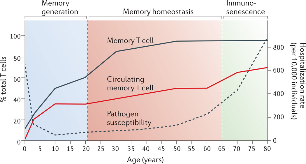
Memory T cells constitute the most abundant lymphocyte population in the body for the majority of a person's lifetime; however, our understanding of memory T cell generation, function and maintenance mainly derives from mouse studies, which cannot recapitulate the exposure to multiple pathogens that occurs over many decades in humans. In this Review, we discuss studies focused on human memory T cells that reveal key properties of these cells, including subset heterogeneity and diverse tissue residence in multiple mucosal and lymphoid tissue sites. We also review how the function and the adaptability of human memory T cells depend on spatial and temporal compartmentalization.


Figures

Figure 1
Figure 1. Memory T cell frequency, pathogen susceptibility and mortality throughout human life
Memory T cells pass through three distinct phases: memory generation, memory homeostasis and immunosenescence. Memory T cells are mostly generated following antigen exposure during infancy, youth and young adulthood (ages 0–20). Their levels subsequently plateau and are maintained through homeostasis throughout adulthood (ages 30–65), after which they enter the third stage and exhibit senescent changes (ages 65 and up). Previous studies have shown that there is an increase in the frequency of memory T cells in the blood (red line) over time,. In the whole body, which includes the blood, intestines, lungs, skin, liver, brain and lymphoid tissues, the overall frequency of memory T cells (black line) also increases with age. The increase in memory T cell frequency throughout the body inversely correlates to a decrease pathogen susceptibility (dashed line) calculated from infectious disease hospitalization rates (per 10,000 people) recorded from 1998–2006 in the US based on 40,085,978 total hospitalizations.
Figure 2
Figure 2. Model for the generation of human memory T cell subsets
A schematic model for differentiation of circulating and tissue-resident memory T cell subsets. Progressive differentiation of the three major circulating subsets — stem cell memory T cells (TSCM cells), central memory T cells (TCM cells) and effector memory T cells (TEM cells) — from activated naïve T cells is shown relative to the extent of antigen exposure. Effector T cells (TEff cells) represent terminally differentiated cells, and death is one outcome of increased antigen exposure and proliferation. Naïve, TSCM and TCM cells circulate and migrate to lymphoid tissue, whereas TEM and TEff cells are the subsets with the capacity to traffic to peripheral tissues. Tissueresident memory T cells (TRM cells) in peripheral tissue sites may derive from either TEM or TEff cells that migrate to these sites via tissue-specific influences. It is possible that TCM cells could develop into TRM cells in lymphoid sites (dotted line). TRM cells in peripheral compartments are likely terminally differentiated since they do not circulate or convert to other memory T cell subsets.
Figure 3
Figure 3. Schematic of memory T cell heterogeneity in peripheral blood and tissues
A Diagram shows the tissue distribution and migration patterns of the major human memory T cell subsets, including three major circulating populations —, stem cell memory T cells (TSCM cells), central memory T cells (TCM cells) and effector memory T cells (TEM cells) — and TRM cells in multiple sites, as well as CD8+ TRM cells that are defined by expression of CD103 and are associated with mucosal sites and skin. The individual sites are defined in terms of circulation (red), lymphoid origin (grey) or peripheral tissues (yellow). Circulating TSCM, TCM and TEM cell subsets migrate from the blood and circulate through the spleen and lungs, where they can be primed to migrate to intestines. They also migrate via the lymphatics and efferent vessels to lymph nodes. TRM cells predominate in skin, lungs, bone marrow and intestines, but may also be present within the CD69+TEM subsets in the spleen and lymph nodes. Mucosal sites and the skin also contain specific CD103+ TRM cells. The expression of certain chemokine receptors and/or integrins is associated with T cell migration and/or residence in lymph nodes (CCR7), skin (CCR4, CLA and CCR10), intestines (CCR9 and integrin α4β7), lungs (CCR6) and bone marrow (integrin α2β1). B Key phenotypic and functional properties of circulating and resident subsets are shown, with CD45RA and CCR7 distinguishing circulating memory T cell subsets and CD69 (and CD103) expression delineating TRM cells from circulating subsets. Memory subsets can produce similar types of recall cytokines, such as interleukin-2 (IL-2), interferon-γ (IFNγ) and tumour necrosis factor (TNF), but differ in the extent and quality of these responses.
Figure 4
Figure 4. Model for compartmentalization of antigen-specific memory T cell subsets in space and time
This schematic shows the relative naïve and memory T cell subset frequencies in the circulation and peripheral sites and at different life stages. The schematic also shows the biased specificity for microbial antigens derived from pathogens that has been observed in specific tissue sites, including cytomegalovirus (CMV) in the blood; Epstein-Barr virus (EBV) in the spleen; vaccinia virus in lymph nodes; influenza virus in the lungs; rotavirus in the intestine and herpes simplex virus (HSV) and varicella zoster virus (VZV) in the skin. In addition, the specificities of some memory T cells at mucosal sites (lungs, intestine and skin) are biased for antigens from the microflora. The relative frequencies of each T cell subset in each tissue site for youths through adults are compiled from Ref. 11, and are extrapolated for infants based on Refs. , . At birth, there is a preponderance of naïve T cells in the circulation, and an abundance of mucosal microbial antigens are encountered during infancy, resulting in seeding of mucosal sites with TEM cells specific for mucosal pathogens, which could develop into TRM cells in situ. Infant skin has few, if any T cells (R. Clark, personal communication) and therefore estimates for memory T cell content in skin begin during youth. During childhood, exposure to the ubiquitous pathogenic and non-pathogenic microbial species in each site occurs and new memory T cells are formed which are partitioned as TRM cells in skin and mucosal sites, and as TCM cells in lymphoid tissues. This basic partitioning of antigen-specific memory T cell subsets in tissues is maintained during adulthood, with more TEM and TCM cell subsets gradually accumulating in the circulation and lymph nodes that could potentially replenish and/or convert to TRM cells that are lost through attrition.

Similar articles

Cited by

References

    1. Remakus S, Sigal LJ. Memory CD8(+) T cell protection. Adv Exp Med Biol. 2013;785:77–86. - PubMed
    1. Wherry EJ, Ahmed R. Memory CD8 T-cell differentiation during viral infection. J Virol. 2004;78:5535–5545. - PMC - PubMed
    1. Teijaro JR, et al. Costimulation modulation uncouples protection from immunopathology in memory T cell responses to influenza virus. J Immunol. 2009;182:6834–6843. - PubMed
    1. Teijaro JR, et al. Cutting edge: tissue-retentive lung memory CD4 T cells mediate optimal protection to respiratory virus infection. J Immunol. 2011;187:5510–5514. This study identified retention of CD4+ TRM cells in mouse lungs and demonstrated superior protective capacity of lung TRM cells compared with circulating spleen memory CD4+ T cells to influenza virus infection.

    1. Teijaro JR, Verhoeven D, Page CA, Turner D, Farber DL. Memory CD4 T cells direct protective responses to influenza virus in the lungs through helper-independent mechanisms. J Virol. 2010;84:9217–9226. - PMC - PubMed

Publication types