. 2014 Feb;46(2):152-60.
doi: 10.1038/ng.2853. Epub 2013 Dec 15.

Therapeutic modulation of eIF2α phosphorylation rescues TDP-43 toxicity in amyotrophic lateral sclerosis disease models

Affiliations

Therapeutic modulation of eIF2α phosphorylation rescues TDP-43 toxicity in amyotrophic lateral sclerosis disease models

Hyung-Jun Kim et al. Nat Genet. 2014 Feb.

Abstract

Amyotrophic lateral sclerosis (ALS) is a fatal, late-onset neurodegenerative disease primarily affecting motor neurons. A unifying feature of many proteins associated with ALS, including TDP-43 and ataxin-2, is that they localize to stress granules. Unexpectedly, we found that genes that modulate stress granules are strong modifiers of TDP-43 toxicity in Saccharomyces cerevisiae and Drosophila melanogaster. eIF2α phosphorylation is upregulated by TDP-43 toxicity in flies, and TDP-43 interacts with a central stress granule component, polyA-binding protein (PABP). In human ALS spinal cord neurons, PABP accumulates abnormally, suggesting that prolonged stress granule dysfunction may contribute to pathogenesis. We investigated the efficacy of a small molecule inhibitor of eIF2α phosphorylation in ALS models. Treatment with this inhibitor mitigated TDP-43 toxicity in flies and mammalian neurons. These findings indicate that the dysfunction induced by prolonged stress granule formation might contribute directly to ALS and that compounds that mitigate this process may represent a novel therapeutic approach.


Figures

Figure 1
Figure 1. Yeast plasmid overexpression screen highlights the role of stress granules in TDP-43 toxicity
a) Histogram showing the functional categories of genes (GO term process) with the relative frequency of genes from the TDP-43 overexpression screen compared to the yeast genome and to the FUS overexpression screen . Both the TDP-43 and FUS screens were enriched for RNA metabolic process genes, while the TDP-43 screen was enriched for cell cycle, transport, and protein modification process genes. b) While the TDP-43 and FUS screens were both enriched for RNA metabolic process genes, most of the hits did not overlap with the exception of three genes. c) Stress granule genes identified in the screen enhance or suppress TDP-43 toxicity. Spotting assay showing that on the galactose plate (expression ON) the co-expression of HRP1, KEM1, or PBP1 with TDP-43 leads to enhanced toxicity (reduced growth) while the co-expression of TIS11 or VTS1 with TDP-43 leads to suppression of toxicity (increased growth). d) RNA-binding protein focused yeast interaction network for TDP-43 screen hits reveal connections to PAB1 and EIF2A homolog. Ten out of forty yeast genes that modified TDP-43 toxicity when overexpressed are annotated as RNA-binding proteins. These are displayed as black circles. GeneMANIA was used to search for interacting genes based physical and genetic interactions. Interacting genes are displayed as grey circles and network edges are colored based on the type of interaction (red=physical interaction and green=genetic interaction). This analysis revealed strong connections to PAB1 and YGR054W, which are highlighted in blue. YGR054W is the yeast homolog of a human translation initiation factor EIF2A. e) GFP-tagged ALS-linked disease gene TDP-43 forms aggregates when expressed in yeast that colocalize with the stress granule marker PUB1-mCherry. Scale bar, 5 μm.
Figure 2
Figure 2. Genes that impact stress granule formation modulate TDP-43 toxicity
a) TDP-43 expression increases eIF2α-phosphorylation levels. Genes were expressed in the nervous system in a drug-inducible manner with the elavGS driver. eIF2α- phosphorylation level of elavGS/UAS-YFP and elavGS, TDP-43/UAS-YFP flies fed as adults on RU486 (40μg/ml), and assessed at the indicated time-points. Genotypes: Control is elavGS/UAS-YFP. TDP-43 is elavGS, UAS-TDP-43(S)/UAS-YFP. Mean ± s.e.m., n=3 independent experiments. *p<0.05, *** p<0.001, n.s., not significant. (Student’s t-test). b) Altering the levels of genes that reduce stress granule formation mitigates TDP-43 toxicity, and that promote stress granule formation enhances TDP-43 toxicity. Mean ± 95% CI of four experiments. Genotypes: elavGS/UAS-YFP is elavGS/UAS-YFP. elavGS, TDP-43/UAS-YFP is elavGS, UAS-TDP-43(S)/UAS-YFP. elavGS, TDP-43/Gadd34 RNAi is elavGS, UAS-TDP-43(S)/UAS-Gadd34. RNAiHMS00811. elavGS, TDP-43/Rox8 RNAi is elavGS, UAS-TDP-43(S)/UAS-Rox8. RNAiHMS00472. elavGS, TDP-43/PEK RNAi is elavGS, UAS-TDP-43(S)/UAS-PEK. RNAiGL00030. All flies raised with RU486 (40μg/ml) (125 flies per genotype). ANOVA for significance, followed by Tukey’s multiple comparison test, **p<0.01, *** p<0.001, # p<0.0001, n.s., not significant. c) eIF2α-phosphorylation level of elavGS, TDP-43/UAS-YFP, elavGS, TDP-43/Rox8 RNAi, elavGS, TDP-43/Gadd34 RNAi and elavGS, TDP-43/PEK RNAi. Mean ± s.e.m., n=3 independent experiments. *p<0.05, n.s., not significant (Student’s t-test). d) Total, nuclear and cytosolic TDP-43 protein level in 10d fly heads. Genes predicted to increase stress granules formation increase cytoplasmic TDP-43 protein levels. Mean ± s.e.m., n=3 independent experiments. *** p<0.001, n.s., not significant (Student’s t-test).
Figure 3
Figure 3. The interaction between TDP-43 and Ataxin-2 is mediated through the polyA binding protein motif of Ataxin-2
a) Schematic representation of the versions of Ataxin-2 generated and tested for interactions with TDP-43. Colored boxes indicate the putative RNA-binding Lsm domain, the Lsm-associated domain LsmAD, the PABP-interacting motif PAM2 and the polyQ stretch. Deletion region of ΔLSM is aa277-346, and ΔPAM2 is aa920-932 of ATXN2-32Q. Both domains are well conserved from yeast to mammal. b) TDP-43 toxicity is not affected by co-expression of ATXN2 lacking the PAM2 motif. Whereas TDP-43 toxicity is enhanced with co-expression of ATXN2-32Q or ATXN2-32Q-ΔLSM (TDP-43+ATXN2-32Q or TDP-43+ΔLSM), there is no effect with co-expression of ATXN2-32Q-ΔPAM2 (TDP-43+ΔPAM2). Genotypes: YFP is gmr-GAL4(YH3)/UAS-YFP. TDP-43 is UAS-TDP-43(M)/+; gmr-GAL4(YH3)/+. TDP-43+ATXN2-32Q is UAS-TDP-43(M)/+; gmr-GAL4(YH3)/UAS-ATXN2-32Q. TDP-43+ΔPAM2 is UAS-TDP-43(M)/+; gmr-GAL4(YH3)/UAS-ATXN2-32Q.ΔPAM2. TDP-43+ΔLSM is UAS-TDP-43(M)/+; gmr-GAL4(YH3)/UAS- ATXN2-32Q.ΔLSM. Scale bar for eyes, 100 μm; for sections, 10 μm. c) Expression of TDP-43 in the nervous system reduces lifespan (green, compared with normal in brown). Expression of ATXN2-32Q greatly enhanced TDP-43 toxicity (blue), but the ATXN2-32Q.ΔPAM2 deletion mutant form did not (red). Genotypes: Elav/+ is elav3A-GAL4/+ (n=177 flies). Elav, TDP-43/+ is elav3A-GAL4, UAS-TDP-43(S)/+ (n=156). Elav, TDP-43/ATXN2-32Q is elav3A-GAL4, UAS-TDP-43(S)/UAS-ATXN2-32Q (n=152). Elav, TDP-43/ΔPAM2 is elav3A-GAL4, UAS-TDP-43(S)/UAS-ATXN2-32Q. ΔPAM2 (n=138). d) TDP-43 caused progressive loss of climbing ability when expressed in the adult nervous system (green) which is dramatically enhanced by co-expression of ATXN2-32Q (blue). However, the ΔPAM2 form of ATXN2-Q32 has little effect (red) compared to TDP-43 alone. Mean ± 95% CI of four experiments (n=125 flies per genotype). # p<0.0001, n.s., not significant (ANOVA for significance, followed by Tukey’s multiple comparison test). e) Total, nuclear and cytosolic TDP-43 protein levels in 10d fly heads. Cytosolic TDP-43 protein level is greatly increased by ATXN2-32Q; ΔPAM2 expression does not affect TDP-43 localization or level. Mean ± s.e.m., n=3-4. # p<0.0001, n.s., not significant (ANOVA for significance, followed by Tukey’s multiple comparison test). d–e Genotypes: elavGS, TDP-43/+ is elavGS, UAS-TDP-43(S)/+.elavGS, TDP-43/ATXN2-32Q is elavGS, UAS-TDP-43(S)/ UAS-ATXN2-32Q. elavGS, TDP-43/ΔPAM2 is elavGS, UAS-TDP-43(S)/ UAS-ATXN2-32Q.ΔPAM2. All flies raised with RU486 (20 μg/ml). *p<0.05, **p< 0.01, n.s. not significant. (ANOVA for significance, followed by Student’s t-test).
Figure 4
Figure 4. Poly(A) binding protein is required for TDP-43 toxicity
a) dPABP upregulation enhances TDP-43 and ATXN2-32Q toxicity. However, dPABP does not interact with the ΔPAM2 form of ATXN2-32Q. Genotype: TDP-43 is UAS-TDP-43(M)/+; gmr-GAL4(YH3)/+. TDP-43+dPABP is UAS-TDP-43(M)/+; gmr-GAL4(YH3)/UAS-dPABP. PABP is gmr-GAL4(YH3)/UAS-dPABP. ATXN2-32Q+dPABP is gmr-GAL4(YH3), UAS-dPABP/ UAS-ATXN2-32Q.ΔPAM2+dPABP is gmr-GAL4(YH3), UAS-dPABP/ UAS-ATXN2-32Q.ΔPAM2. Scale bar for eyes, 100 μm; for sections, 10 μm. b) TDP-43 loss of climbing ability is suppressed by downregulation of dPABP. Genotypes: elavGS, TDP-43/+ is elavGS, UAS-TDP-43(S)/+. elavGS, TDP-43/dPABP RNAi is elavGS, UAS-TDP-43(S)/UAS-dPABP.RNAiJF03104 (125 flies/genotype). Mean ± 95% CI of four experiments. *** p<0.001, n.s. not significant (Student’s t-test). c) Total, nuclear and cytosolic TDP-43 protein levels in 10d fly heads. PABP downregulation decreases cytosolic TDP-43 protein level. Genotype: elavGS, TDP-43/+ is elavGS, UAS-TDP-43(S)/+. elavGS, TDP-43/dPABP. RNAi is elavGS, UAS-TDP-43(S)/UAS-dPABP.RNAiJF03104. Mean ± s.e.m., n=3 independent experiments *** p<0.001, n.s. not significant (Student’s t-test).
Figure 5
Figure 5. PABPC1 is mislocalized in motor neurons from ALS patient tissue
a–d, Immunostaining for PABPC1 in the cervical spinal cord of control and ALS patients. a) PABPC1 is expressed in spinal cord motor neurons, and is localized throughout the cytoplasm in a diffuse pattern. b) Occasionally PABPC1 cytoplasmic accumulations were observed in normal motorneurons (arrow). c) In ALS patient motor neurons, PABPC1 is present in distinct cytoplasmic and dense accumulations (arrow). d) PABPC1 cytoplasmic accumulations were also observed in the ALS motorneurons where there was a clearing in the middle. Scale bars, 30 μm. e) The number of motor neurons containing cytoplasmic accumulation of PABPC1 in ALS patients was significantly greater (p=0.01, one-tailed Mann-Whitney test) than in motor neurons from controls. For quantification of accumulations of PABPC1 in individual normal versus ALS cases, see Supplementary Table 1. n=5 controls, 4 ALS patients, mean ± s.e.m.
Figure 6
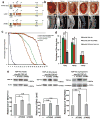
Figure 6. PERK inhibitor treatment rescues TDP-43 toxicity in Drosophila and primary rat neurons
a,b, PERK inhibitor reduces eIF2α-phosphorylation levels and mitigates TDP-43 toxicity to flies. a) Immunoblot of flies expressing TDP-43 in the nervous system. PERK inhibitor decreases eIF2α-phosphorylation levels, but not TDP-43 protein levels. Quantification performed on 3 independent experiments, normalized to tubulin. Mean ± s.e.m. **p<0.01, n.s., not significant (Student’s t-test). b) Flies expressing TDP-43 by elav were treated with DMSO or PERK inhibitor (GSK2606414,10μM). Genotypes: elav3A-GAL4, UAS-TDP-43(S)/UAS-YFP. Mean ± s.e.m. n=4 experiments. **p<0.01, n.s., not significant (Student’s t-test). c-f, PERK inhibitor (PERKi) reduces TDP43 toxicity in primary rat cortical neurons. c) Representative micrographs showing longitudinal imaging of primary cortical neurons transfected with mApple as morphology marker and TDP43-EGFP. White arrows show cells still counted as alive in survival analysis. Cells were transfected at 4 DIV; hr indicate imaging time post transfection. Scale bar, 20μM. d-e) Cumulative hazard plots for survival analysis performed on primary cortical neurons treated with PERKi and transfected with mApple and d) EGFP, e) TDP43-EGFP. PERKi treatment (500nM) of neurons expressing TDP43-EGFP decreased the risk of death by 14%, compared to neurons expressing TDP43-EGFP with vehicle (DMSO) only treatment. Three independent experiments were analyzed to determine statistical significance and cumulative hazard plots. Statistics was performed by Cox proportional hazards analysis; see Supplementary Table 2 for cells per condition and hazard ratios. *, p<0.05. **, p<0.01. ***, p<0.001. #, p<0.0001. n.s., not significant.

References

    1. Boillee S, Vande Velde C, Cleveland DW. ALS: a disease of motor neurons and their nonneuronal neighbors. Neuron. 2006;52:39–59. - PubMed
    1. Cleveland DW, Rothstein JD. From Charcot to Lou Gehrig: deciphering selective motor neuron death in ALS. Nat Rev Neurosci. 2001;2:806–19. - PubMed
    1. Al-Chalabi A, et al. The genetics and neuropathology of amyotrophic lateral sclerosis. Acta Neuropathol. 2012;124:339–52. - PubMed
    1. Couthouis J, et al. Evaluating the role of the FUS/TLS-related gene EWSR1 in amyotrophic lateral sclerosis. Hum Mol Genet. 2012;21:2899–911. - PMC - PubMed
    1. Couthouis J, et al. A yeast functional screen predicts new candidate ALS disease genes. Proc Natl Acad Sci U S A. 2011;108:20881–90. - PMC - PubMed

Publication types

MeSH terms

Substances