. 2014 Apr;22(4):702-12.
doi: 10.1038/mt.2013.282. Epub 2013 Dec 12.

Sustained correction of motoneuron histopathology following intramuscular delivery of AAV in pompe mice

Affiliations

Sustained correction of motoneuron histopathology following intramuscular delivery of AAV in pompe mice

Mai K Elmallah et al. Mol Ther. 2014 Apr.

Abstract

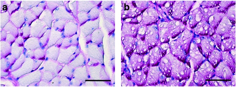
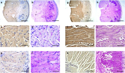
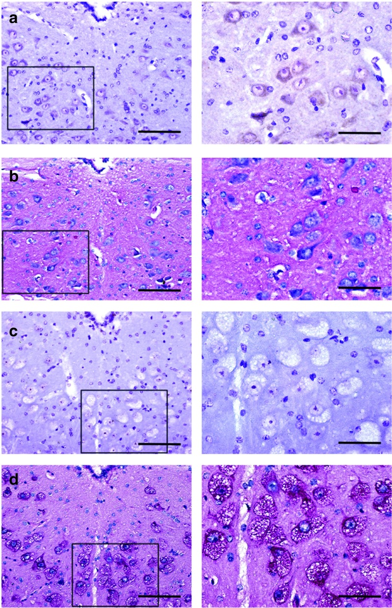
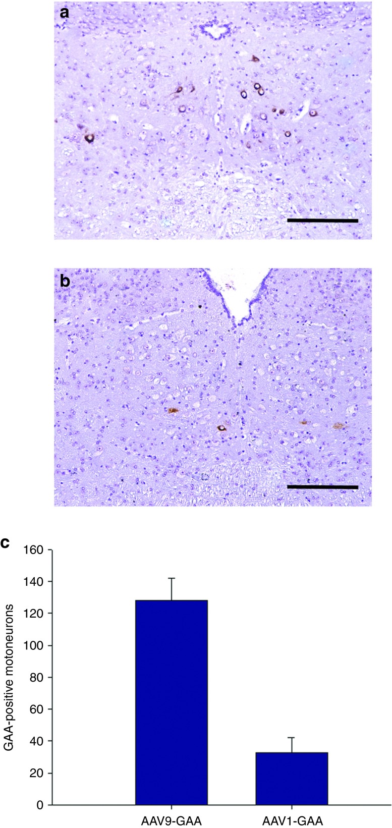
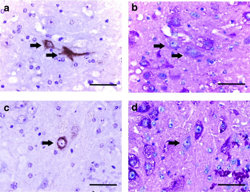
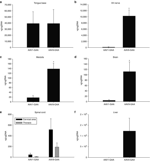
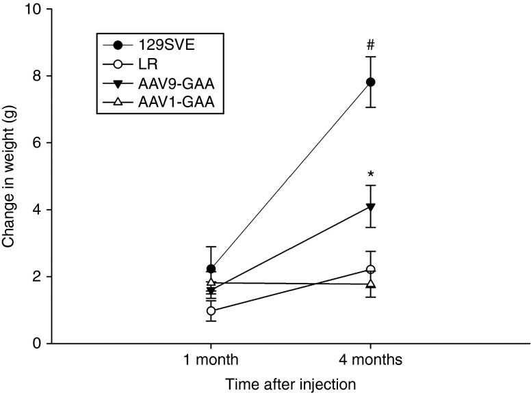
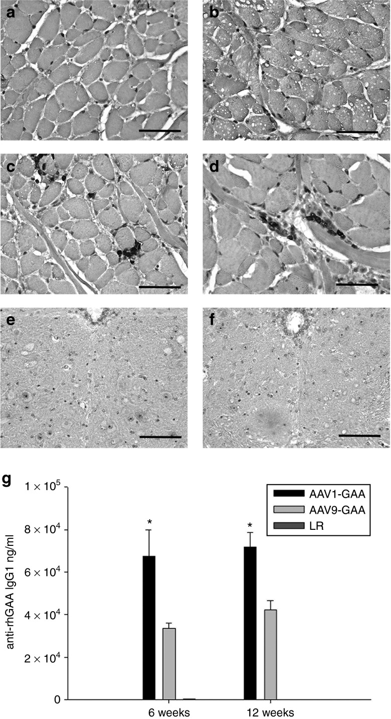
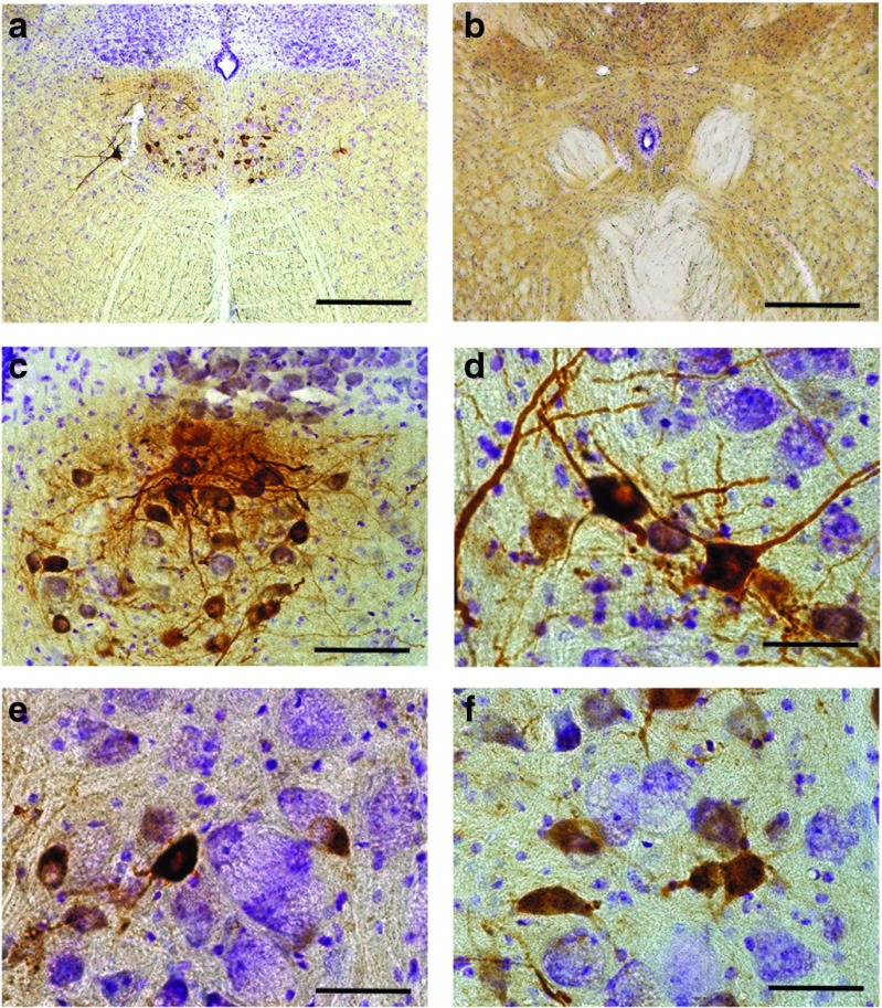
Pompe disease is an autosomal recessive disorder caused by mutations in the acid-α glucosidase (GAA) gene. Lingual dysfunction is prominent but does not respond to conventional enzyme replacement therapy (ERT). Using Pompe (Gaa(-/-)) mice, we tested the hypothesis that intralingual delivery of viral vectors encoding GAA results in GAA expression and glycogen clearance in both tongue myofibers and hypoglossal (XII) motoneurons. An intralingual injection of an adeno-associated virus (AAV) vector encoding GAA (serotypes 1 or 9; 1 × 10(11) vector genomes, CMV promoter) was performed in 2-month-old Gaa(-/-) mice, and tissues were harvested 4 months later. Both serotypes robustly transduced tongue myofibers with histological confirmation of GAA expression (immunochemistry) and glycogen clearance (Period acid-Schiff stain). Both vectors also led to medullary transgene expression. GAA-positive motoneurons did not show the histopathologic features which are typical in Pompe disease and animal models. Intralingual injection with the AAV9 vector resulted in approximately threefold more GAA-positive XII motoneurons (P < 0.02 versus AAV1); the AAV9 group also gained more body weight over the course of the study (P < 0.05 versus AAV1 and sham). We conclude that intralingual injection of AAV1 or AAV9 drives persistent GAA expression in tongue myofibers and motoneurons, but AAV9 may more effectively target motoneurons.


Figures

Figure 1
Figure 1
Representative examples of periodic acid Schiff (PAS) staining in genioglossus tissue sections from an adult (a) wild-type and (b) a sham-treated Gaa−/− mouse. The PAS reaction recognizes glycogen and is evident by the magenta coloring. Myofibers from the (b) sham-treated Gaa−/− mice are PAS-positive and have swollen vacuolar appearance with disruption of cellular architecture. Scale bars = 200 µm.
Figure 2
Figure 2
Immunohistochemical acid-α glucosidase (GAA) staining and periodic acid Schiff (PAS) staining of transverse lingual sections from the base of the tongue. Tissues were harvested at 4 months following intralingual injections with (a–f) AAV9-hGAA and (g–l) AAV1-hGAA. Alternating tissue sections were stained for GAA or PAS. The approximate site of the tongue AAV injection is indicated by the asterisk, and the sites marked c–f or i–l are shown in an expanded scale immediately below each top panel. Note that (c) GAA-positive myofibers are negative for (d) PAS. In contrast, the area negative for (e) GAA is also positive for (f) PAS. Similar results were seen in the AAV1-hGAA–injected lingual sections (panels g and h). Scale bars: 800 µm (a,b,g,h); 50 µm (c–f, i–l).
Figure 3
Figure 3
Immunohistochemical acid-α glucosidase (GAA) staining (panels a and c) and periodic acid Schiff (PAS) staining (panels b and d) of the XII motor nucleus in wild-type and sham-treated Gaa−/− tissues. The area highlighted by the box in the left panels is shown at a higher magnification in the right panels. (a) Positive GAA staining is evident in XII motoneurons with an absence of PAS staining (b) in wild-type tissues. In contrast, sham-treated Gaa−/− tissues show complete absence of (c) GAA immunostaining and (d) swollen PAS positive motoneurons. Scale bars = a–d left panels: 50 µm; right panels: 100 µm.
Figure 4
Figure 4
Persistent acid-α glucosidase (GAA) expression in Gaa−/− XII motoneurons after intralingual injection of AAV9-hGAA or AAV1-hGAA. All tissues were harvested 4 months following AAV delivery to tongue. (a) AAV9-hGAA–treated Gaa−/− mice had more robust GAA expression in the XII motor nucleus as compared to (b) AAV1-hGAA. The average number of GAA-positive cells in the XII nucleus is shown in Panel c (*P < 0.05 compared to AAV9). Scale bars = a,b: 50 µm.
Figure 5
Figure 5
Acid-α glucosidase (GAA)-positive XII motoneurons have no glycogen accumulation. Higher magnification views of XII motoneurons stained for GAA (brown) and periodic acid Schiff (PAS) (magenta) in alternate sections. The arrows indicate the same cell in consecutive histological sections. The juxtaposition of GAA-positive-GAA-negative cells allows direct comparison of the histological appearance. Note that GAA-positive cells lack PAS staining and do not have the vacuolar appearance typical of adjacent Gaa−/− motoneurons. Panels a and b: examples from AAV9-treated tissues; panels c and d: examples from AAV1-treated tissues. Scale bars = a–d: 200 µm.
Figure 6
Figure 6
Mean number of vector genome copies 4 months after intralingual injection. Vector genome copies in the tongue were similar between (a) AAV1-hGAA– and AAV9-hGAA–treated Gaa−/− mice, but were greater in the (b) XII nerve, (c) medulla, (d) brain, (e) spinal cord, and (f) liver of AAV9-hGAA– versus AAV1-hGAA–treated Gaa−/− mice.
Figure 7
Figure 7
Mean increase in body weight following intralingual adeno-associated virus (AAV) injection. All experimental groups gained weight similarly over the first month of the study, but by the fourth month differences emerged between groups. The AAV9-hGAA–treated Gaa−/− mice had a significant increase in weight gain (*) compared to both LR- and AAV1-hGAA–injected Gaa−/− mice. The LR-treated mice were housed as cage mates with the AAV1-hGAA and AAV9-hGAA groups.
Figure 8
Figure 8
CD3 staining in the genioglossus and XII motor nucleus. No T-cell infiltrate was observed in sham-injected genioglossal myofibers of (a) wild-type or (b) Gaa−/− mice. In contrast, a mild T-cell infiltrate was noted in the genioglossus of both (c) AAV1-hGAA– and (d) AAV9-hGAA–injected mice. However, no T-cell infiltrate was observed in the medulla or XII motor nucleus of (e) AAV9-hGAA– or (f) AAV1-hGAA–treated animals. An IgG response against acid-α glucosidase (GAA) was noted in both (g) the AAV1-hGAA– and the AAV9-hGAA–treated mice. Scale bars = 200 µm.
Figure 9
Figure 9
Representative examples of medullary acid-α glucosidase (GAA) immunostaining 12 months following intralingual injection with AAV1-hGAA in a Gaa−/− mouse. Positive immunostaining for GAA is restricted to the XII motor nucleus in the rostral medulla at 12 months following intralingual injection of (a) AAV1-hGAA. A representative section caudal to the XII motor nucleus with no GAA labeling is shown in (b). Panels c and d show examples of “clustering” of GAA-positive motoneurons which was typical at 12 months postinjection. The contrast in the histological appearance of GAA-positive versus negative cells is illustrated in (e–f). The GAA-positive motoneurons have a central nucleus without evidence of vacuoles. In contrast, cells not expressing GAA (*) in panels e and f have an eccentric nucleus with an enlarged vacuole filled soma. Scale bars = a: 50 µm; b: 200 µm.

References

    1. Fuller DD, ElMallah MK, Smith BK, Corti M, Falk DJ, Byrne BJ. Pompe disease and the respiratory system. Res Physiol Neurobiol. 2013;189:241–249. - PMC - PubMed
    1. Byrne BJ, Falk DJ, Pacak CA, et al. Pompe disease gene therapy. Hum Mol Genet. 2011;20 R1:R61–8. - PMC - PubMed
    1. Jones HN, Muller CW, Lin M, et al. Oropharyngeal dysphagia in infants and children with infantile Pompe disease. Dysphagia. 2010;25:277–83. - PubMed
    1. Margolis ML, Howlett P, Goldberg R, Eftychiadis A, Levine S. Obstructive sleep apnea syndrome in acid maltase deficiency. Chest. 1994;105:947–9. - PubMed
    1. Muller CW, Jones HN, OGrady G, Suarez AH, Heller JH, Kishnani PS. Language and speech function in children with infantile Pompe disease. J Ped Neurol. 2009;7:147–156.

Publication types

MeSH terms