

Review
doi: 10.1038/nrmicro3136.
Bacterial programmed cell death: making sense of a paradox
Affiliations
- PMID: 24336185
- PMCID: PMC4422510
- DOI: 10.1038/nrmicro3136
Item in Clipboard
Review
Bacterial programmed cell death: making sense of a paradox
Nat Rev Microbiol.
2014 Jan.
Abstract
Although the concept of programmed cell death (PCD) in bacteria has been met with scepticism, a growing body of evidence suggests that it can no longer be ignored. Several recent studies indicate that the phenotypic manifestations of apoptosis, which are processes that are associated with ordered cellular disassembly in eukaryotes, are conserved in bacteria. In this Opinion article, I propose a model for the coordinated control of potential bacterial PCD effectors and argue that the processes involved are functionally analogous to eukaryotic PCD systems.
Figures
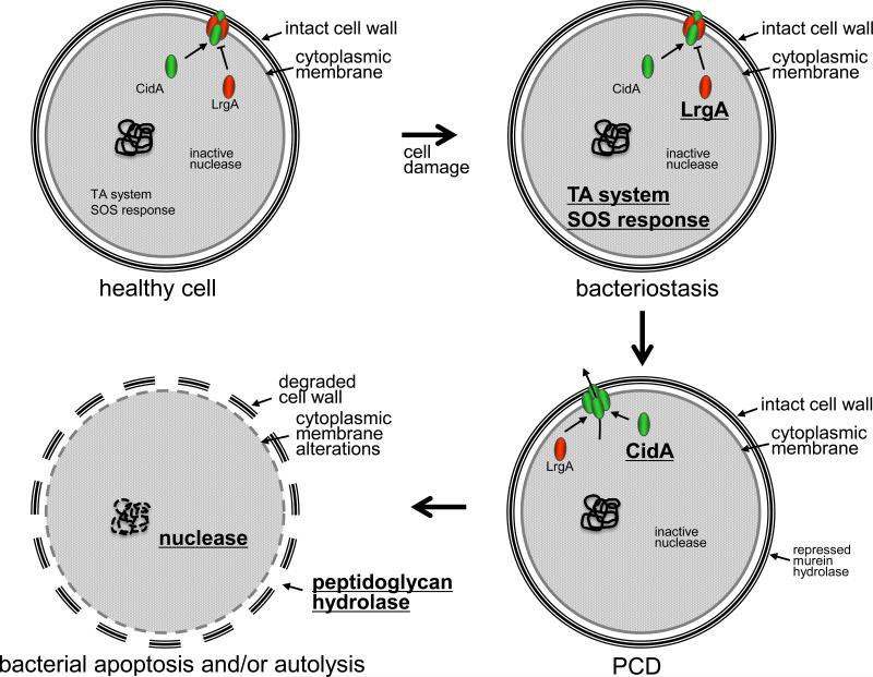
In this model, a healthy cell that has endured some type of damage (e.g. UV-induced mutations) will initially, and likely simultaneously, respond by inducing toxin-antitoxin (TA) systems (e.g. MazEF) and repair mechanisms (the SOS response). The Cid and Lrg proteins are present but are kept inactive at this point because of the inhibitory effect of the Lrg proteins on the Cid PCD effector proteins. The activity of the TA system, by virtue of its inhibitory effect on translation, results in the transition of the cell to a quiescent state (bacteriostasis) that maximizes the energy and resources needed to repair the damage. If the damage is beyond repair, a “point of no return” is reached and the inhibitory effect of the Lrg proteins on Cid is released by an unknown mechanism and cell death ensues. Finally, post-mortem signaling results in the activation of apoptosis-like processes and/or autolysis, including nuclease activation (such as BapE) and DNA fragmentation, membrane alterations, and peptidoglycan hydrolase-mediated cell wall degradation.
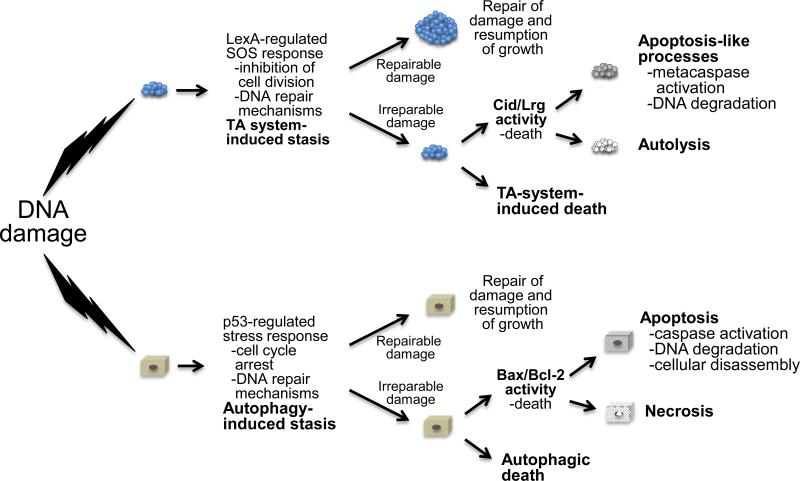
Cell stress, such as that elicited by DNA damaging agents, induces a stress response program that includes DNA repair mechanisms and cell death pathways. This response includes mechanisms to inhibit cell division, directing all available resources to repair the damage. If the levels of damage are minimal, the repair mechanisms will be sufficient to restore the cell to working order. Similar to the role of p53 in assessing the extent of damage to the cell, and then coordinating an appropriate response, it is envisioned that the LexA regulator of the SOS response serves a similar role in coordinating the response to DNA damage in bacteria. In both cases, this includes processes that result in the recycling of cytoplasmic components (TA systems and autophagy) in an attempt to fuel DNA repair. If the damage is irreparable, the repair mechanisms will be overwhelmed and PCD, either Cid-/Lrg-induced death (prokaryotes) or Bcl-2 protein family-induced death (eukaryotes), is induced. Alternatively, TA system-induced or autophagic death can also be induced. Finally, post-mortem events, such as those associated with intrinsic apoptosis and necrosis (eukaryotes), and apoptosis-like processes and autolysis (prokaryotes) are activated.
References
-
- Bayles KW. Are the molecular strategies that control apoptosis conserved in bacteria? Trends Microbiol. 2003;11:306–311. - PubMed
-
- Lappann M, et al. A dual role of extracellular DNA during biofilm formation of Neisseria meningitidis. Mol Microbiol. 2010;75:1355–1371. doi:10.1111/j.1365-2958.2010.07054.x. - PubMed
Publication types
MeSH terms
Substances
Grants and funding
LinkOut - more resources
Full Text Sources
Other Literature Sources
Miscellaneous
