. 2014 Jan;21(1):103-9.
doi: 10.1038/nsmb.2737. Epub 2013 Dec 15.

Dynamics of yeast histone H2A and H2B phosphorylation in response to a double-strand break

Affiliations

Dynamics of yeast histone H2A and H2B phosphorylation in response to a double-strand break

Cheng-Sheng Lee et al. Nat Struct Mol Biol. 2014 Jan.

Abstract

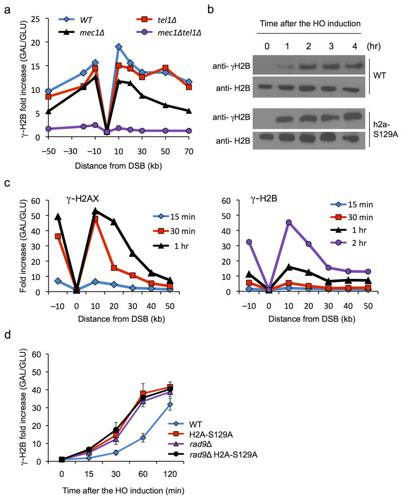
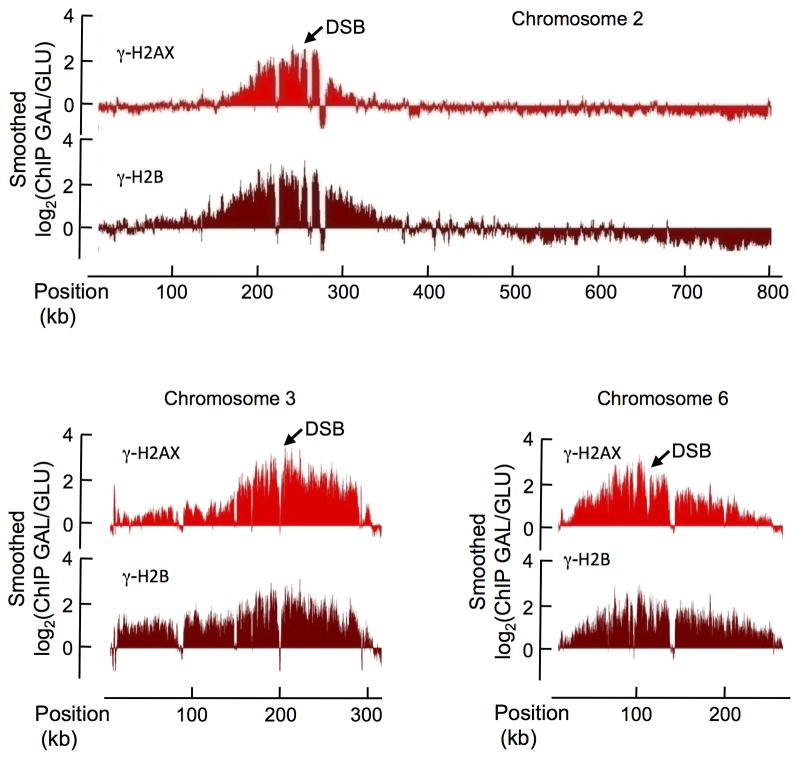
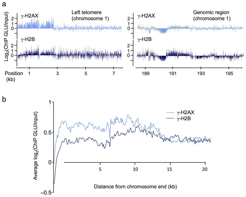
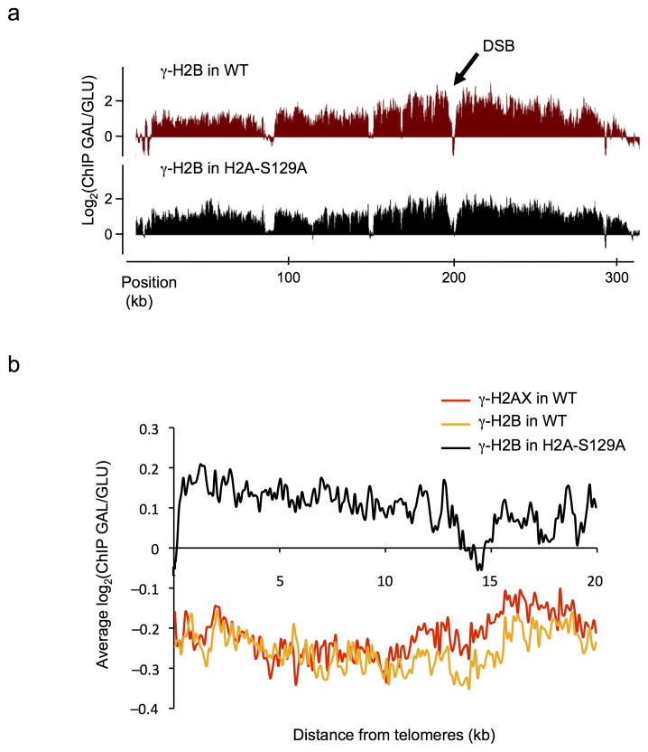
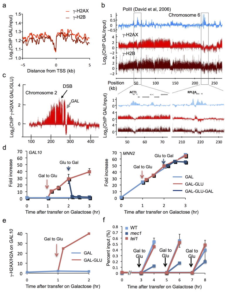
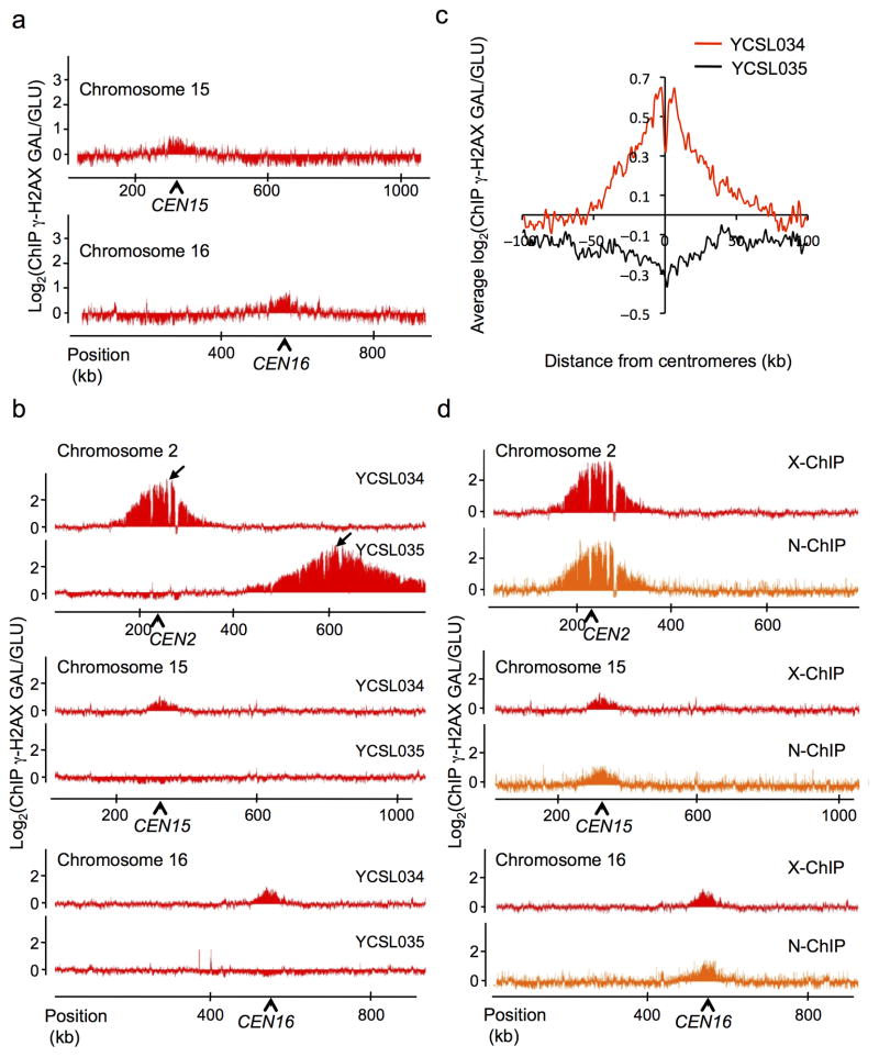
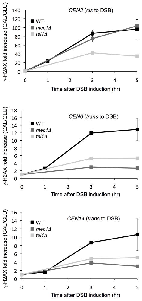
In budding yeast, a single double-strand break (DSB) triggers extensive Tel1 (ATM)- and Mec1 (ATR)-dependent phosphorylation of histone H2A around the DSB, to form γ-H2AX. We describe Mec1- and Tel1-dependent phosphorylation of histone H2B at T129. γ-H2B formation is impaired by γ-H2AX and its binding partner Rad9. High-density microarray analyses show similar γ-H2AX and γ-H2B distributions, but γ-H2B is absent near telomeres. Both γ-H2AX and γ-H2B are strongly diminished over highly transcribed regions. When transcription of GAL7, GAL10 and GAL1 genes is turned off, γ-H2AX is restored within 5 min, in a Mec1-dependent manner; after reinduction of these genes, γ-H2AX is rapidly lost. Moreover, when a DSB is induced near CEN2, γ-H2AX spreads to all other pericentromeric regions, again depending on Mec1. Our data provide new insights in the function and establishment of phosphorylation events occurring on chromatin after DSB induction.


Figures

Figure 1
Figure 1. Spreading of γ-H2AX and γ-H2B around an HO-induced DSB
a. γ-H2B spreads extensively around an unrepaired DSB at MATα in strain JKM179, as measured by ChIP 2 hr after HO endonuclease expression was induced. Data for cells lacking Mec1 and/or Tel1 are shown b. Western blot analysis shows that the kinetics of γ-H2B formation is more rapid in a h2a-S129A strain lacking γ-H2AX phosphorylation. Levels of phosphorylation are normalized to the amount of total H2B protein. c. Slow kinetics of γ-H2B formation compared to γ-H2AX are seen in ChIP around the MAT DSB. d. The slow formation of γ-H2B 10 kb distal to the DSB site is accelerated both by eliminating γ-H2AX and by deleting RAD9. For ChIP assays in a, c and d, the fold increase was calculated by normalization of IP values with non-IP (input) values and then further normalized with reference to the values prior to induction of the DSB (0 hr). Error bars represent S.D. from three independent experiments.
Figure 2
Figure 2. Profiles of γ-H2AX and γ-H2B in the three HO-cuts strain after DSB induction
ChIP-chip measurements were performed with antibodies against γ-H2AX (light red) and γ-H2B (dark red), using yeast grown on galactose containing media for 1 hour (DSB induction). The log2 ratio of ChIP-chip signals obtained on Galactose versus Glucose growth is shown (GAL/GLU), across the chromosomes 2, 3 and 6, carrying HO cuts sites (indicated by arrows).
Figure 3
Figure 3. Profiles of γ-H2AX and γ-H2B in undamaged cells
a. The profiles of γ-H2AX (light blue) and γ-H2B (dark blue) obtained at the left telomere of chromosome 1 (left panel) and on a genomic region farther on the same chromosome (right panel) are shown. Note that γ-H2B is less enriched than γ-H2AX at the telomere, while both signals are equivalent further away. b. γ-H2AX and γ-H2B ChIP-chip signals were averaged on 20 kb at all chromosomes ends (left and right arms combined). Note the higher level of γ-H2AX at telomeres compared to γ-H2B.
Figure 4
Figure 4. Differential spreading of γ-H2B in a H2A-S129A mutant strain
a. γ-H2B ChIP-chip experiments were performed in a WT (in red) or H2A-S129A mutant (in black) background, carrying a DSB at the MAT locus on chromosome 3 before and after DSB induction. The profile of the ratio γ–H2AX GAL/GLU is shown. b. The GAL/GLU ratio obtained for γ-H2AX (red), γ-H2B in WT (orange) and γ-H2B in H2A mutant strain (black) were averaged on 20 kb at telomeres.
Figure 5
Figure 5. Transcriptionally active units are refractory to γ-H2AX and γ-H2B
a. Averaged γ-H2AX (light red) and γ-H2B (dark red) ChIP-chip signals upon galactose growth around the TSS of genes encompassed in γ-H2AX domains (supplemental methods). b. γ-H2AX, γ-H2B (this study) and RNA-Pol II (previously reported ) profiles are shown across chromosome 6. Bottom panel shows a magnification of two regions carrying highly transcribed genes (ACT1 and RPL2A). c. γ-H2AX profile on a part of chromosome 2, one hour after HO induction. The GAL gene cluster (gray arrow), highly transcribed in this growth condition, is strongly depleted in γ-H2AX. d. Left panel: γ-H2AX levels (measured by ChIP-qPCR) within GAL10 are very low for 3 h DSB induction, but rise rapidly if cells are transferred to glucose that shuts off GAL10 transcription (red arrow). γ-H2AX levels rapidly drop when cells are again placed on galactose (blue arrow). Right panel: at the MNN2 gene (not regulated by galactose), γ-H2AX levels rise continuously after transfer to galactose. e. The ratio between γ-H2AX and H2A is shown at GAL10 under different growth conditions (measured by ChIP-qPCR). f. ChIP-qPCR were performed in wildtype, Tel1 and/or Mec1-deleted strains, and transfer on glucose was performed at different time points as indicated. The rapid increase in γ-H2AX in GAL10 is seen if cells are transferred on glucose at 3, 5 or 7 h after DSB induction. This increase can be carried out by either Tel1 or Mec1. Error bars represent the range from two independent experiments.
Figure 6
Figure 6. γ-H2AX can spread in trans on physically close loci
a. Profile of γ-H2AX around centromeres of chromosomes 15 and 16 (indicated by arrows), after one-hour DSB induction in the 3-HO cuts strain, shown as the log2 ratio of the signal obtained after galactose versus glucose growth (GAL/GLU). b. γ-H2AX ChIP-chip was performed before and after galactose growth, either in strain YCSL034 carrying a single HO cut site at 13.5 kb from CEN2 or in strain YCSL035 carrying a single HO cut site 387 kb from CEN2. Profiles (ratio of GAL/GLU) obtained on chromosome 2 (carrying the DSB) and on chromosomes 15 and 16 are shown. c. Averaged γ-H2AX signal obtained after DSB induction, around centromere of undamaged chromosomes (i.e. chromosome 2 is excluded), using the data obtained with YCSL034 (red line) or YCSL035 (black line) strain. d. γ-H2AX ChIP-chip was performed before and after DSB induction, either using a protocol involving a crosslinking agent (X-ChIP in red) or in native conditions (N-ChIP in orange). Profiles obtained on chromosome 2 (carrying the DSB) and on chromosomes 15 and 16 are shown.
Figure 7
Figure 7. Effect of Mec1 and Tel1 kinases on spreading in cis and trans from a DSB induced near CEN2
An HO-induced DSB was created by galactose-mediated induction at a site 13.5 kb from CEN2. Increases in γ-H2AX were measured at the times indicated for a site 17.7 kb distal to the DSB on the same chromosome (a), 9.9 kb from CEN6 (b) and overlapping CEN14 (c). Fold increase is calculated by normalizing IP value to input and then to 0hr. Note that in tel1Δ strains there is a lower level of spreading in both cis and trans, whereas the absence of MEC1 specifically affects spreading in trans. Error bars represent the range from two independent experiments.

References

    1. Shroff R, et al. Distribution and dynamics of chromatin modification induced by a defined DNA double-strand break. Curr Biol. 2004;14:1703–1711. - PMC - PubMed
    1. Rogakou EP, Boon C, Redon C, Bonner WM. Megabase chromatin domains involved in DNA double-strand breaks in vivo. J Cell Biol. 1999;146:905–916. - PMC - PubMed
    1. Harrison JC, Haber JE. Surviving the breakup: the DNA damage checkpoint. Annu Rev Genet. 2006;40:209–235. - PubMed
    1. Kim JA, Kruhlak M, Dotiwala F, Nussenzweig A, Haber JE. Heterochromatin is refractory to gamma-H2AX modification in yeast and mammals. J Cell Biol. 2007;178:209–218. - PMC - PubMed
    1. Szilard RK, et al. Systematic identification of fragile sites via genome-wide location analysis of gamma-H2AX. Nat Struct Mol Biol. 2010;17:299–305. - PMC - PubMed

References for Online Methods

    1. Lee SE, et al. Saccharomyces Ku70, mre11/rad50 and RPA proteins regulate adaptation to G2/M arrest after DNA damage. Cell. 1998;94:399–409. - PubMed
    1. Kim JA, Kruhlak M, Dotiwala F, Nussenzweig A, Haber JE. Heterochromatin is refractory to gamma-H2AX modification in yeast and mammals. J Cell Biol. 2007;178:209–218. - PMC - PubMed
    1. Sandell LL, Zakian VA. Loss of a yeast telomere: arrest, recovery, and chromosome loss. Cell. 1993;75:729–739. - PubMed
    1. Sun K, Coic E, Zhou Z, Durrens P, Haber JE. Saccharomyces forkhead protein Fkh1 regulates donor preference during mating-type switching through the recombination enhancer. Genes Dev. 2002;16:2085–2096. - PMC - PubMed
    1. Foster ER, Downs JA. Methylation of H3 K4 and K79 is not strictly dependent on H2B K123 ubiquitylation. J Cell Biol. 2009;184:631–638. - PMC - PubMed

Publication types