Review
. 2014 Feb;28(2):180-8.
doi: 10.1038/eye.2013.277. Epub 2013 Dec 13.

Visual regulation of refractive development: insights from animal studies

Affiliations
Review

Visual regulation of refractive development: insights from animal studies

E L Smith 3rd et al. Eye (Lond). 2014 Feb.

Abstract

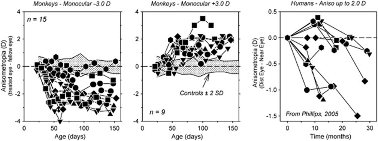
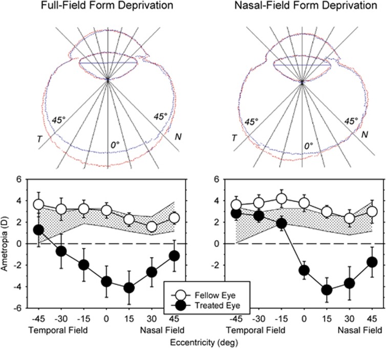
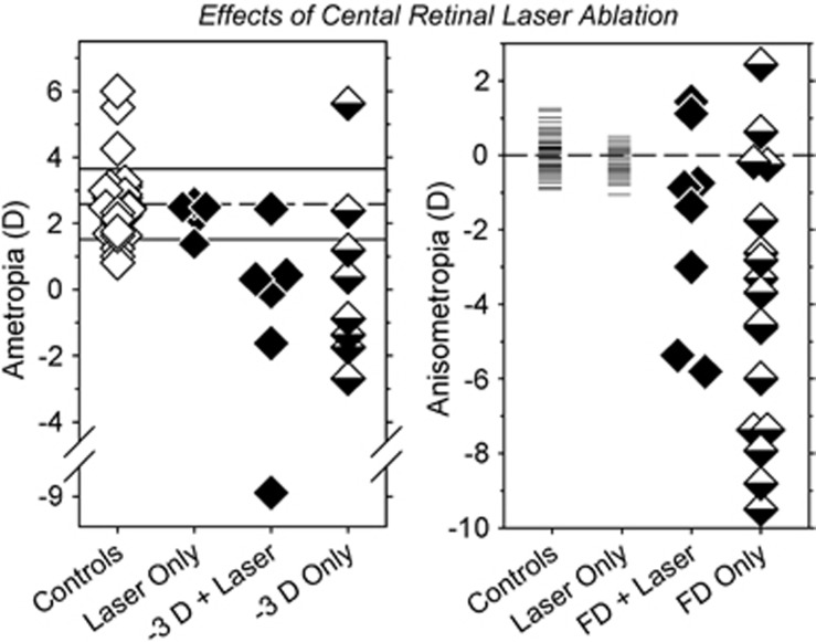
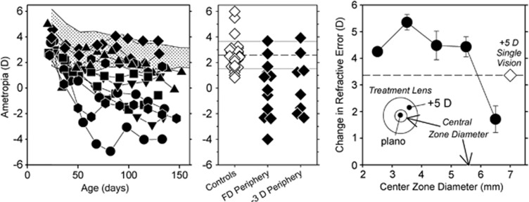
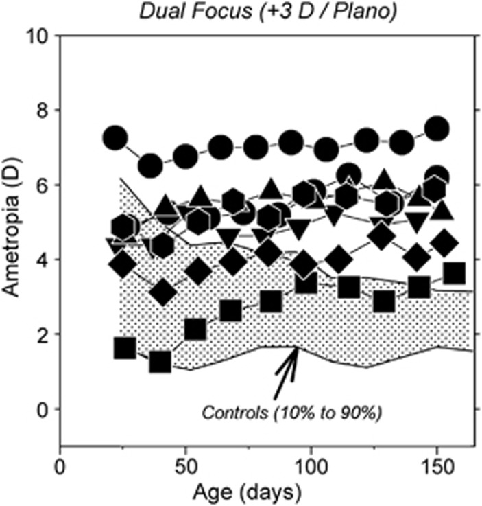
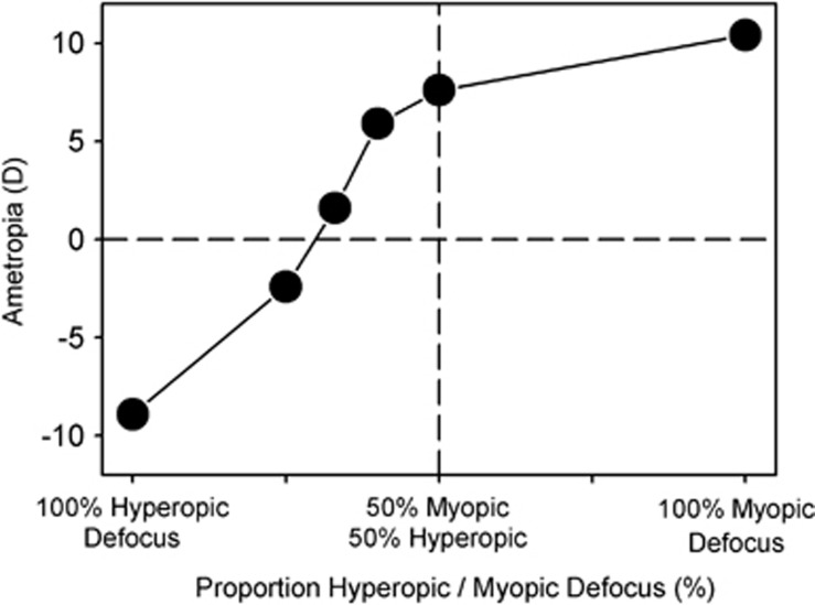
Investigations employing animal models have demonstrated that ocular growth and refractive development are regulated by visual feedback. In particular, lens compensation experiments in which treatment lenses are used to manipulate the eye's effective refractive state have shown that emmetropization is actively regulated by signals produced by optical defocus. These observations in animals are significant because they indicate that it should be possible to use optical treatment strategies to influence refractive development in children, specifically to slow the rate of myopia progression. This review highlights some of the optical performance properties of the vision-dependent mechanisms that regulate refractive error development, especially those that are likely to influence the efficacy of optical treatment strategies for myopia. In this respect, the results from animal studies have been very consistent across species; however, to facilitate extrapolation to clinical settings, results are presented primarily for nonhuman primates. In agreement with preliminary clinical trials, the experimental data show that imposed myopic defocus can slow ocular growth and that treatment strategies that influence visual signals over a large area of the retina are likely to be most effective.


Figures

Figure 1
Figure 1
Interocular differences in refractive error (treated eye correction−fellow eye correction) plotted as a function of age for individual monkeys reared with −3D (left panel) or +3D lenses over one eye (middle panel)., The fellow eyes viewed through zero-powered lenses. The first symbol represents the start of the lens-rearing period. The shaded regions indicate ±2 SD around the mean anisometropia for untreated control monkeys. The right panel illustrates the interocular difference in refractive error (distance corrected eye−near corrected eye) as a function of time for children corrected using a monovision strategy that imposed up to 2.0 D of relative hyperopia on the near corrected eye.
Figure 2
Figure 2
Top: outlines of horizontal magnetic resonance images for the treated (red) and fellow eyes (blue) of representative monkeys reared with monocular diffuser lenses that produced form deprivation over the entire visual field (left) or only over the nasal visual field (right). T, temporal; N, nasal. Bottom: mean (±SE) spherical-equivalent refractive corrections plotted as a function of eccentricity for the treated (filled circles) and fellow eyes (open circles) of monkeys reared monocular full-field (left) and nasal-field (right) form deprivation. The shade area represents ±2 SD from the mean for normal monkeys. Replotted from Smith et al.
Figure 3
Figure 3
Left: spherical-equivalent refractive corrections obtained at ages corresponding to the end of the lens-rearing period for the right or treated eyes of individual animals reared with unrestricted vision (controls), laser ablation of the treated eye fovea (laser only), foveal ablation and −3D lenses, or intact retinas and −3D treatment lenses. Right: interocular differences in refractive error (right or treated eye−left or fellow eye) for individual controls and animals reared with laser ablation of the fovea and unrestricted vision, foveal ablation and monocular form deprivation, or only monocular form deprivation.
Figure 4
Figure 4
Left: spherical-equivalent refractive corrections plotted as a function of age for monkeys reared with diffusers that had either 4 or 8 mm apertures centered over the pupils of both eyes. The treatment lenses provided unrestricted vision for the central 24 or 37 degrees of the retina; the rest of the retina experienced severe form deprivation. The shaded area represents the 10th to 90th percentile range of ametropias for control monkeys. Middle: ametropias measured at ages corresponding to the end of the lens-rearing period for the right eye of individual control monkeys (open symbols) and monkeys that were rearing with either peripheral form deprivation or peripheral hyperopic defocus. Right: changes in refractive error produced in chickens by treatment lenses with zero-powered central zones and +5D peripheral zones (filled circles) plotted as a function of the diameter of the central zone. The open diamond represents the changes in refraction produced by a +5D single-vision lens.
Figure 5
Figure 5
Spherical-equivalent refractive corrections plotted as a function of age for individual monkeys reared with dual-focus Fresnel lenses that had alternating refracting powers of 0D and +3D. The shaded area represents the 10th to 90th percentile range of ametropias for control monkeys.
Figure 6
Figure 6
Mean interocular differences in spherical-equivalent refractive corrections for lens-reared chickens plotted as a function of the proportion of the visual field that experienced hyperopic versus myopic defocus.

References

    1. Curtin BJ. Basic Science and Clinical Management. Harper & Row: Philadelphia; 1985. The Myopias.
    1. Rose L, Yinon U, Belkin M. Myopia induced in cats deprived of distance vision during development. Vision Res. 1974;14:1029–1032. - PubMed
    1. Young FA. The distribution of refractive errors in monkeys. Exp Eye Res. 1964;3:230–238. - PubMed
    1. Smith EL., IIIEnvironmentally induced refractive errors in animalsIn: Rosenfield M, Gilmartin B (eds).Myopia and Nearwork Butterworth-Heinemann: Oxford; 199857–90.
    1. Levinsohn FG. Zur frage der kunstlich erzeugten kurzsichtigkeit bei affen. Klinische Monatsblatter fur Augenheilkunde. 1919;61:794–803.

Publication types

LinkOut - more resources