. 2015 Jan 2;34(1):63-72.
doi: 10.1038/onc.2013.529. Epub 2013 Dec 16.

SALL4 is a new target in endometrial cancer

Affiliations

SALL4 is a new target in endometrial cancer

A Li et al. Oncogene. .

Abstract

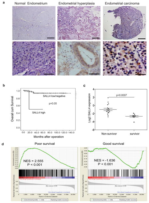
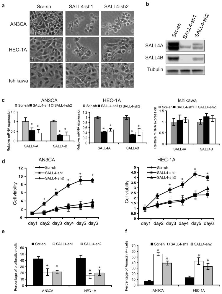
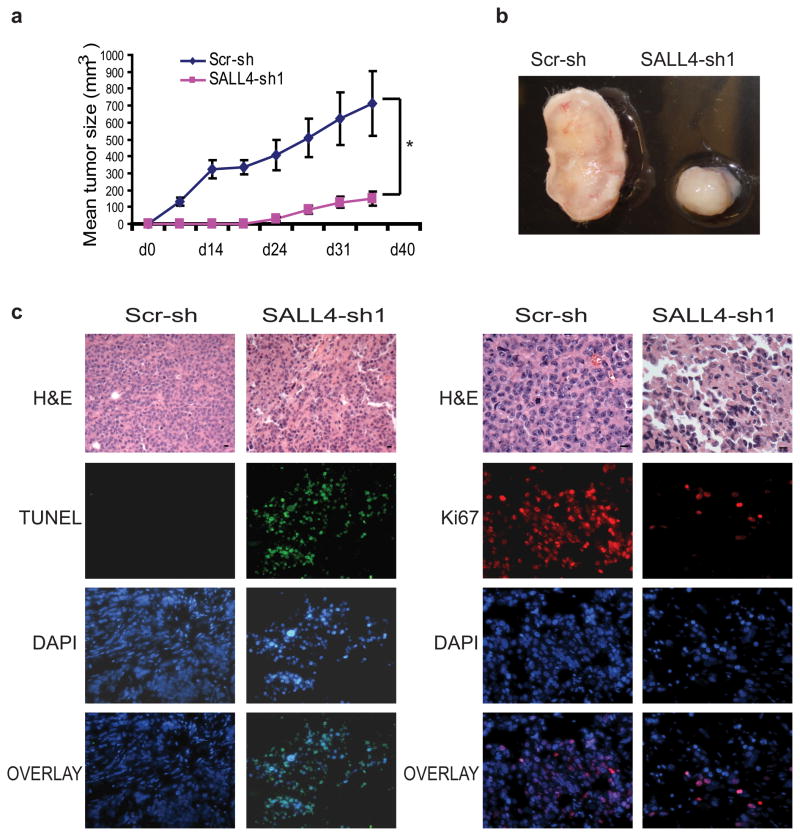
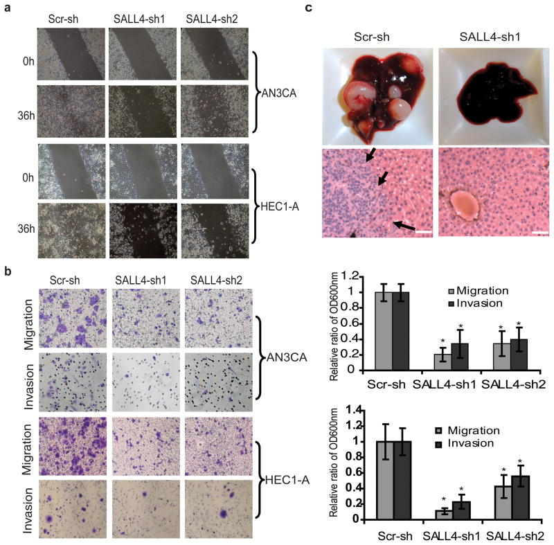
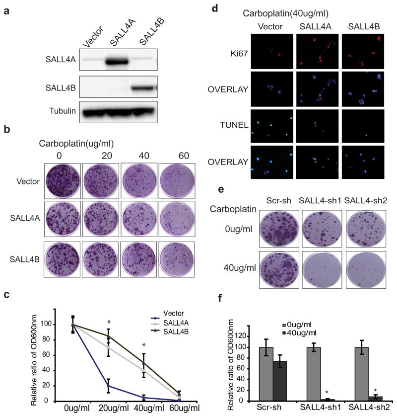
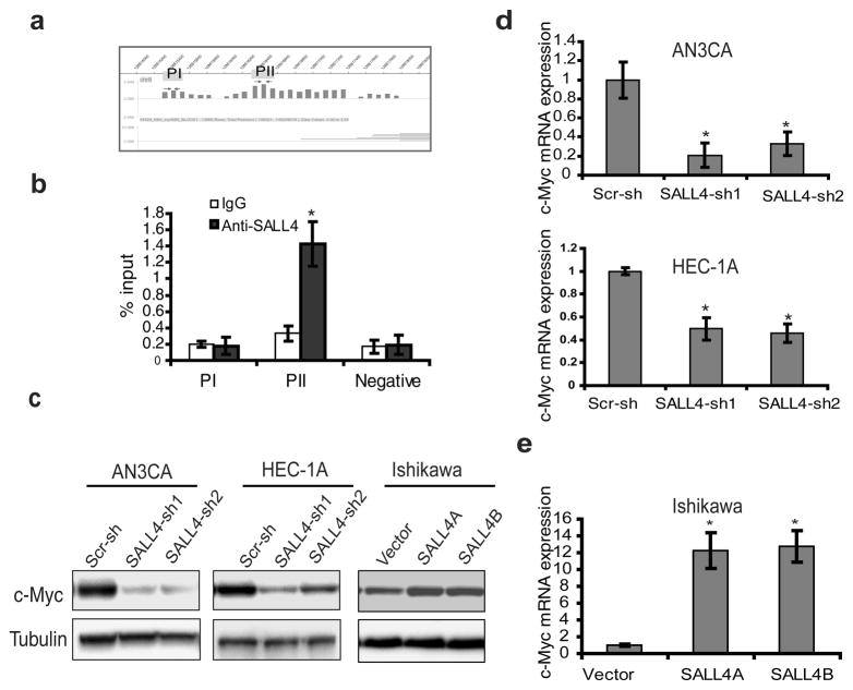
Aggressive cancers and embryonic stem (ES) cells share a common gene expression signature. Identifying the key factors/pathway(s) within this ES signature responsible for the aggressiveness of cancers can lead to a potential cure. In this study, we find that SALL4, a gene involved in the maintenance of ES cell self-renewal, is aberrantly expressed in 47.7% of primary human endometrial cancer samples. It is not expressed in normal or hyperplastic endometrial. More importantly, SALL4 expression is positively correlated with worse patient survival and aggressive features such as metastasis in endometrial carcinoma. Further functional studies have shown that loss of SALL4 inhibits endometrial cancer cell growth in vitro and tumorigenicity in vivo, as a result of inhibition of cell proliferation and increased apoptosis. In addition, downregulation of SALL4 significantly impedes the migration and invasion properties of endometrial cancer cells in vitro and their metastatic potential in vivo. Furthermore, manipulation of SALL4 expression can affect drug sensitivity of endometrial cancer cells to carboplatin. Moreover, we show that SALL4 specifically binds to the c-Myc promoter region in endometrial cancer cells. While downregulation of SALL4 leads to a decreased expression of c-Myc at both protein and mRNA levels, ectopic SALL4 overexpression causes increased c-Myc protein and mRNA expression, indicating that c-Myc is one of the SALL4 downstream targets in endometrial tumorigenesis. In summary, we are the first to demonstrate that SALL4 has functional role(s) in metastasis and drug resistance in aggressive endometrial cancer. As a consequence of its functional roles in cancer cell and absence in normal tissue, SALL4 is a potential novel therapeutic target for the high-risk endometrial cancer patient population.


Conflict of interest statement

Conflict of interest

The authors declare no conflict of interest.

Figures

Figure 1
Figure 1. SALL4 expression is associated with poor survival and metastasis in endometrial cancer patients
(a) Representative IHC images show positive SALL4 expression in endometrial cancer and absence of SALL4 in normal endometria and hyperplasia. Scale bars = 500μm (upper panels) and 50μm (lower panel). (b) Clinicopathological analysis demonstrates SALL4 expression is significantly correlated with worse survival of EC patients (p =0.05). SALL4 low/negative group includes IHC 0 and 1+, and SALL4 high group includes IHC 2+ or above. (c) Microarray analysis confirms that SALL4 expression was significantly higher in non-survivor compared to survivor of endometrial cancer. (d) Gene Set Enrichment Analysis (GSEA) reveals that in high SALL4-expressing endometrial carcinoma, there is an enrichment of gene sets upregulated in cancers with poor survival (left panel, p<0.001); On the contrary, gene sets that are enriched in cancers with good survival are enriched in SALL4-negative endometrial carcinoma (right panel, p<0.001).
Figure 2
Figure 2. SALL4 depletion decreases cell viability by inhibiting proliferation and triggering cell apoptosis in vitro
(a) Phase-contrast photomicrographs show decreased viable cells by morphology after SALL4 knockdown. (b) Western blot demonstrates downregulation of SALL4 expression in AN3CA cells as a result of lentiviral-mediated RNA interference. (c) Downregulation of SALL4 expression by shRNA targeting can lead to decreased SALL4 expression in AN3CA and HEC-1A cells, but not in Ishikawa cells, evaluated by qRT-PCR. (d) Down-regulation of SALL4 leads to decreased cell viability. Cells expressing SALL4 shRNAs or scrambled control shRNAs were plated in 96-well plates (2000 cells per well) and cell viability assayed at various time points using the MTS assay. (e) Down-regulation of SALL4 leads to decreased cell proliferation. Cells expressing SALL4 shRNAs (SALL4-sh1 and SALL4 sh2) or scrambled shRNAs (Scr-sh) were pulse-labeled with BrdU for 1hr, followed by analysis of DNA replication with anti-BrdU antibody and flow cytometry analysis to measure proliferating cells. (f) Down-regulation of SALL4 leads to increased apoptosis. Cells expressing SALL4 shRNAs or scrambled shRNAs were collected, stained with PI and Annexin V, and processed for analysis by flow cytometry to measure the apoptotic cells. Error bars indicate standard error of three replicates (n =3) (*p<0.05, Student’s t test).
Figure 3
Figure 3. SALL4 depletion inhibits tumorigenecitiy of AN3CA cells in vivo
(a) Graph shows tumor volume (mm3) measured at various time points after transplantation (n = 5). Error bars indicate standard error from five mice (*p<0.05, Student’s t test). Tumor volume (mm3) was calculated by the following formula: length×width× height/2. (b) Representative gross picture of tumors derived from subcutaneous injection of AN3CA cells expressing SALL4-sh1 or scrambled shRNA (Scr-sh). (c) AN3CA tumor sections treated with SALL4-sh1 shRNA have reduced proliferation and increased apoptosis as measured by IHC Ki-67 and TUNEL staining, respectively.
Figure 4
Figure 4. SALL4 depletion inhibits endometrial cancer cells migration, invasion and metastasis
(a) Phase contrast microscopy images demonstrate impaired AN3CA and HEC-1A cells wound closure rate as a result of loss of SALL4. (b) Left panel shows that representative transwell cell migration and matrigel cell invasion images. Right panel shows that migration and invasion capacities were quantified after the cells stained with crystal violet solution. Data are presented as OD600nm. Error bars indicate standard error of three replicates (n =3) (*p<0.05). (c) Liver tissues from mice retro-orbitally injected with SALL4-knockdown or scrambled control shRNA-treated AN3CA cells. Upper panel, gross liver tissue pictures; lower panel, representative H&E staining on liver tissues. Arrows indicate metastatic tumor region. Scale bar represents 50μm (* p<0.05).
Figure 5
Figure 5. Change of SALL4 expression leads to alterations in drug sensitivity of endometrial cancer cells to Carboplatin
(a) Western blot demonstrates ectopic overexpression of SALL4 in Ishikawa cells. (b) Clonogenic assays on Ishikawa cells overexpressing SALL4A or SALL4B treated with indicated carboplatin concentrations; images show colonies stained with crystal violet solution. (c) Quantification of the relative colony formation in empty vector control group versus SALL4 overexpression groups following drug treatments (* p<0.05). (d) Overexpression of SALL4 renders Ishikawa more resistant to carboplatin treatment (40μg/ml carboplatin treated for 72hrs) by promoting proliferation and inhibiting apoptosis, as measured by Ki-67 and TUNEL staining. (e) Knockdown of SALL4 sensitized carboplatin-resistant cell line HEC-1A to carboplatin treatment, as measured by decreased colony formation. (f) Quantification of relative colony formation in control versus SALL4 knockdown HEC-1A cells with and without 40μg/ml carboplatin treatment (* p<0.05).
Figure 6
Figure 6. C-Myc is a direct target of SALL4 in endometrial carcinoma
(a) ChIP-on-Chip analysis of SALL4 binding on c-Myc promoter region in NB4 cells. (b) ChIP-qPCR assay performed using SALL4-specific antibody in AN3CA cells to verify ChIP-on-Chip result. Immunoprecipitated DNA was analyzed by quantitative PCR using specific primers. qChIP values were expressed as percentage of input chromatin as reported by others. Data represent the average and standard deviation from three parallel experiments. (c) Western blot demonstrates that SALL4 depletion significantly down-regulated c-Myc expression in AN3CA and HEC-1A cell lines, and overexpression of SALL4 up-regulated c-Myc expression in Ishikawa cells. (d, e) Real time RT-PCR analysis reveals that the mRNA of c-Myc is affected by SALL4 in AN3CA, HEC-1A and Ishikawa ((*p<0.05).

References

    1. Amant F, Moerman P, Neven P, Timmerman D, Van Limbergen E, Vergote I. Endometrial Cancer. Lancet. 2005;366:491–505. - PubMed
    1. Saso S, Chatterjee J, Georgiou E, Ditri AM, Smith JR, Ghaem-Maghami S. Endometrial Cancer. BMJ. 2011;343:d3954. - PubMed
    1. You JS, Kang JK, Seo DW, Park JH, Park JW, Lee JC, et al. Depletion of Embryonic Stem Cell Signature by Histone Deacetylase Inhibitor in Nccit Cells: Involvement of Nanog Suppression. Cancer Res. 2009;69:5716–25. - PubMed
    1. Ben-Porath I, Thomson MW, Carey VJ, Ge R, Bell GW, Regev A, et al. An Embryonic Stem Cell-Like Gene Expression Signature in Poorly Differentiated Aggressive Human Tumors. Nat Genet. 2008;40:499–507. - PMC - PubMed
    1. Elling U, Klasen C, Eisenberger T, Anlag K, Treier M. Murine Inner Cell Mass-Derived Lineages Depend on Sall4 Function. Proc Natl Acad Sci USA. 2006;103:16319–24. - PMC - PubMed

Publication types

MeSH terms

LinkOut - more resources