. 2013 Dec 16:3:3510.
doi: 10.1038/srep03510.

Structural basis of a nucleosome containing histone H2A.B/H2A.Bbd that transiently associates with reorganized chromatin

Affiliations

Structural basis of a nucleosome containing histone H2A.B/H2A.Bbd that transiently associates with reorganized chromatin

Yasuhiro Arimura et al. Sci Rep. .

Erratum in

Abstract

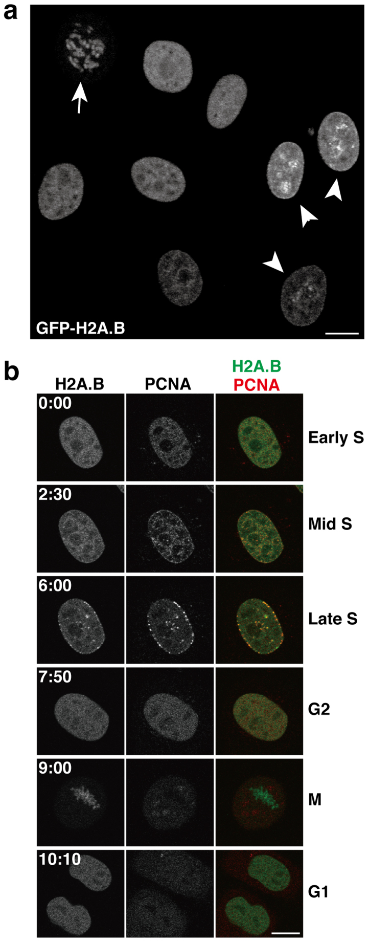
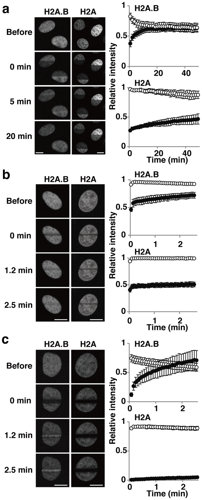
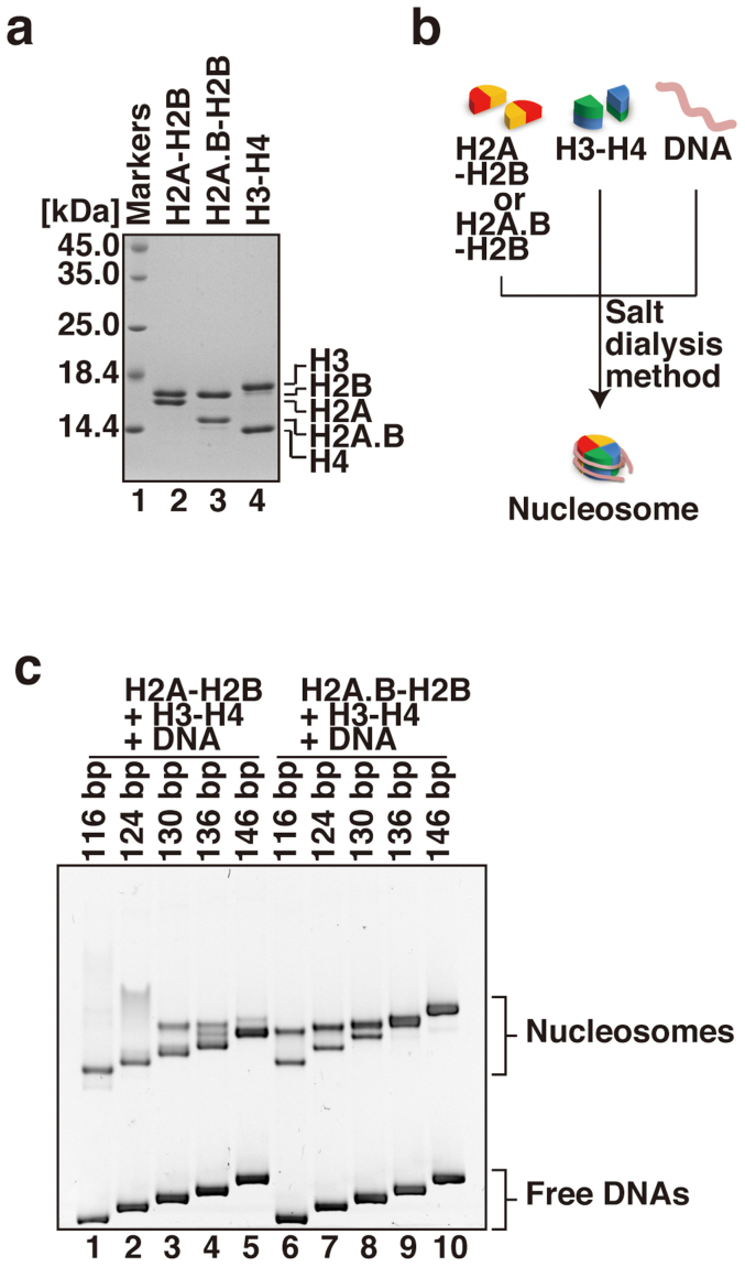
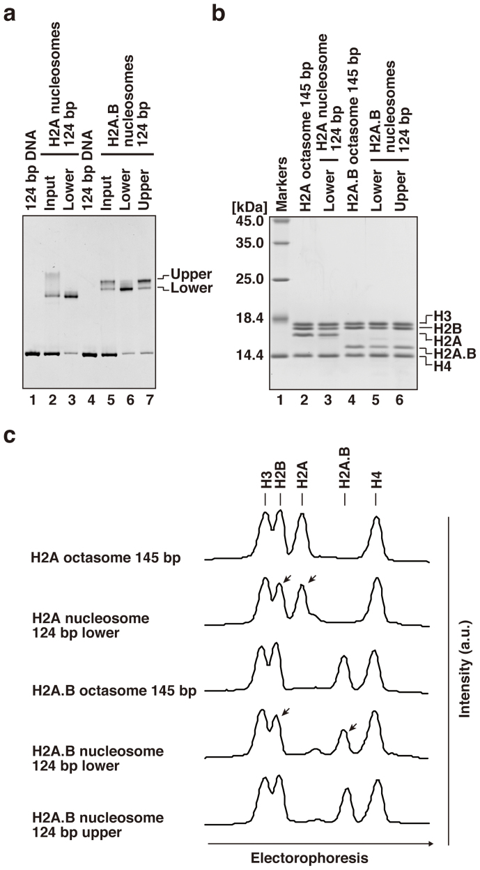
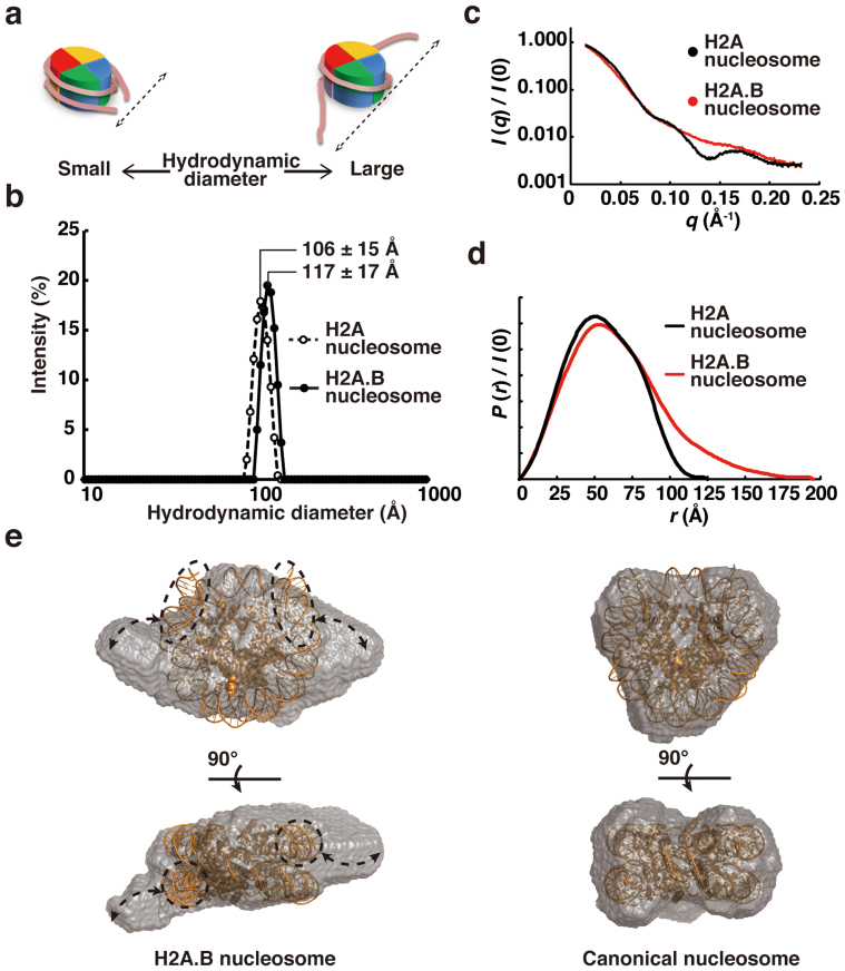
Human histone H2A.B (formerly H2A.Bbd), a non-allelic H2A variant, exchanges rapidly as compared to canonical H2A, and preferentially associates with actively transcribed genes. We found that H2A.B transiently accumulated at DNA replication and repair foci in living cells. To explore the biochemical function of H2A.B, we performed nucleosome reconstitution analyses using various lengths of DNA. Two types of H2A.B nucleosomes, octasome and hexasome, were formed with 116, 124, or 130 base pairs (bp) of DNA, and only the octasome was formed with 136 or 146 bp DNA. In contrast, only hexasome formation was observed by canonical H2A with 116 or 124 bp DNA. A small-angle X-ray scattering analysis revealed that the H2A.B octasome is more extended, due to the flexible detachment of the DNA regions at the entry/exit sites from the histone surface. These results suggested that H2A.B rapidly and transiently forms nucleosomes with short DNA segments during chromatin reorganization.


Figures

Figure 1
Figure 1. Localization of GFP-H2A.B in living cells.
(a) GFP-H2A.B distribution in living HeLa cells imaged by confocal microscopy. GFP-H2A.B is localized to interphase nuclei and mitotic chromosomes (arrow). In some nuclei, GFP-H2A.B is concentrated in foci (arrowheads). (b) GFP-H2A.B is concentrated in replication foci during S phase. HeLa cells expressing both GFP-H2A.B and PCNA-mCherry were grown on a glass-bottom dish under a confocal microscope. A cell exhibiting the early replication pattern of PCNA-mCherry was chosen for time-lapse imaging. Confocal images of GFP-H2A.B and PCNA-mCherry were obtained every 5 min (see Supplementary Movie S1). The elapsed time (hh:mm) from the start of recording is shown on each panel. During mid- to late-S phase, GFP-H2A.B becomes clearly concentrated in PCNA foci, where DNA replication occurs. Bars, 10 μm.
Figure 2
Figure 2. Kinetics of GFP-H2A.B in living cells.
The kinetics of GFP-H2A.B and GFP-H2A were analyzed by bleaching with a 488-nm laser (a and b), and by irradiation with a 405-nm laser to induce DNA damage (c). Fluorescence intensities of bleached or damaged (closed circles) and unbleached or undamaged (open circles) areas are plotted as relative values to the initial intensity of the same area before bleaching or damaging (averages with standard deviations; N = 14–24). (a) One-half of the nucleus was bleached using a 488-nm laser for ~38 s, and fluorescence images were obtained for 50 min. The first post-bleach image was acquired at ~45 s after the beginning of bleaching. (b) A 2 μm strip was bleached using a 488-nm laser for 110 ms, and fluorescence images were obtained for 2.5 min. The first post-bleach image was acquired at 1.8 s after the beginning of bleaching. (c) A 2 μm strip was irradiated using a 405-nm laser for 3.6 s, and fluorescence images were obtained for 2.5 min. The first post-bleach image was acquired at 4.9 s after the beginning of irradiation. Bars, 10 μm.
Figure 3
Figure 3. Reconstitution of the H2A.B and H2A nucleosomes with various lengths of DNAs.
(a) Purified human recombinant histone complexes were analyzed by 18% SDS-PAGE with Coomassie Brilliant Blue staining. (b) Schematic representation of the nucleosome reconstitution experiments by the salt dialysis method. (c) Nucleosomes reconstituted with 116 bp (lanes 1 and 6), 124 bp (lanes 2 and 7), 130 bp (lanes 3 and 8), 136 bp (lanes 4 and 9), and 146 bp (lanes 5 and 10) DNAs were analyzed by non-denaturing 6% PAGE, and the gel was stained with ethidium bromide after PAGE. Lanes 1–5 indicate the nucleosomes reconstituted with H2A–H2B and H3–H4, and lanes 6–10 indicate the nucleosomes reconstituted with H2A.B–H2B and H3–H4. The 146 bp DNA was the palindromic satellite DNA derivative.
Figure 4
Figure 4. H2A.B forms both an octasome and a hexasome with a 124 bp DNA fragment.
(a) Nucleosomes reconstituted with a 124 bp DNA were purified using a Prep Cell apparatus, and were analyzed by non-denaturing 6% PAGE, and the gel was stained with ethidium bromide after PAGE. Lanes 1 and 4 indicate the 124 bp DNA used in the nucleosome reconstitution. Lanes 2 and 3 indicate the H2A nucleosome samples before (input) and after (lower band) Prep Cell purification, respectively. Lane 5 indicates the H2A.B nucleosome sample before (input) purification, and lanes 6 (lower band) and 7 (upper band) indicate the H2A.B nucleosomes after Prep Cell purification. (b) The histone compositions of the purified nucleosomes were analyzed by 18% SDS-PAGE with Coomassie Brilliant Blue staining. Lane 1 indicates molecular mass markers. Lanes 2 and 4 represent the histones included in the purified fractions of the H2A and H2A.B octasomes reconstituted with a 145 bp 601 DNA fragment, respectively. Lane 3 depicts the histones included in the purified fraction of the H2A nucleosome with a 124 bp DNA fragment, as shown in panel a, lane 3. Lanes 5 and 6 show the histones included in the purified fractions of the H2A.B nucleosomes with a 124 bp DNA fragment. Lanes 5 and 6 indicate the lower and upper band fractions, as shown in panel a, lanes 6 and 7, respectively. (c) Line intensity profiles (a.u.) of the gel shown in (b). Arrowheads indicate bands with decreased intensity, due to the lack of a single H2A–H2B or H2A.B–H2B dimer in the hexasome.
Figure 5
Figure 5. Solution structure of the H2A.B octasome.
(a) Schematic representations of the relationship between the hydrodynamic diameter in the DLS analysis and the nucleosome structure. (b) Particle size distribution profiles in the DLS analysis of the H2A and H2A.B octasomes with a 145 bp 601 DNA fragment. Closed and open circles represent the particle size distributions of the H2A.B and H2A octasomes, respectively. The nucleosome concentration was 1.0 mg DNA/ml. The hydrodynamic diameters (Z-average size) of the H2A and H2A.B octasomes were 10.598 nm (error: 1.4658 nm) and 11.682 nm (error: 1.6588 nm), respectively. Both octasomes were detected as monodisperse. (c) The SAXS intensity curves of H2A and H2A.B octasomes with a 145 bp DNA fragment. (d) Distance distribution functions P(r) of the H2A and H2A.B octasomes. (e) Dummy atom models of the H2A.B octasome (left) and the H2A octasome (right). Side and top views are presented in the upper and lower panels, respectively. The crystal structure of the canonical nucleosome (PDB ID: 3AFA) is superimposed on the solution structures of the H2A and H2A.B octasomes. Putative flexible DNA segments are enclosed by dashed circles. Arrows indicate the extra volumes, which may be the mobile areas of the flexible DNA segments.

Similar articles

Cited by

References

    1. Luger K., Mader A. W., Richmond R. K., Sargent D. F. & Richmond T. J. Crystal structure of the nucleosome core particle at 2.8 Å resolution. Nature 389, 251–260 (1997). - PubMed
    1. Davey C. A., Sargent D. F., Luger K., Maeder A. W. & Richmond T. J. Solvent mediated interactions in the structure of the nucleosome core particle at 1.9 Å resolution. J. Mol. Biol. 319, 1097–1113 (2002). - PubMed
    1. Makde R. D., England J. R., Yennawar H. P. & Tan S. Structure of RCC1 chromatin factor bound to the nucleosome core particle. Nature 467, 562–566 (2010). - PMC - PubMed
    1. Kimura H. Histone dynamics in living cells revealed by photobleaching. DNA Repair 4, 939–950 (2005). - PubMed
    1. Park Y. J. & Luger K. Histone chaperones in nucleosome eviction and histone exchange. Curr. Opin. Struct. Biol. 18, 282–289 (2008). - PMC - PubMed

Publication types

MeSH terms

LinkOut - more resources