. 2013 Dec 11;33(50):19470-9.
doi: 10.1523/JNEUROSCI.2283-13.2013.

The Coffin-Lowry syndrome-associated protein RSK2 regulates neurite outgrowth through phosphorylation of phospholipase D1 (PLD1) and synthesis of phosphatidic acid

Affiliations

The Coffin-Lowry syndrome-associated protein RSK2 regulates neurite outgrowth through phosphorylation of phospholipase D1 (PLD1) and synthesis of phosphatidic acid

Mohamed-Raafet Ammar et al. J Neurosci. .

Abstract

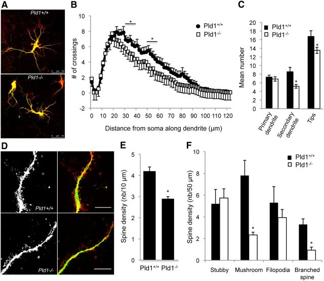
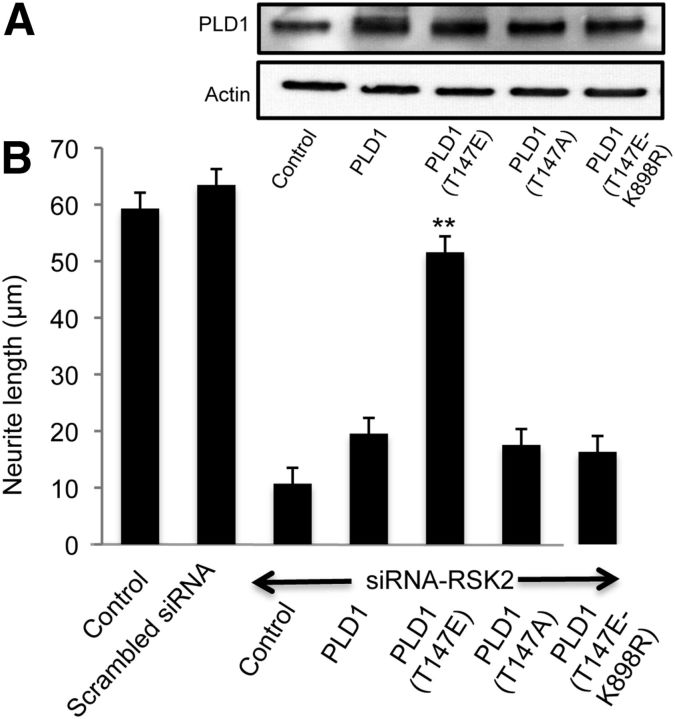
More than 80 human X-linked genes have been associated with mental retardation and deficits in learning and memory. However, most of the identified mutations induce limited morphological alterations in brain organization and the molecular bases underlying neuronal clinical features remain elusive. We show here that neurons cultured from mice lacking ribosomal S6 kinase 2 (Rsk2), a model for the Coffin-Lowry syndrome (CLS), exhibit a significant delay in growth in a similar way to that shown by neurons cultured from phospholipase D1 (Pld1) knock-out mice. We found that gene silencing of Pld1 or Rsk2 as well as acute pharmacological inhibition of PLD1 or RSK2 in PC12 cells strongly impaired neuronal growth factor (NGF)-induced neurite outgrowth. Expression of a phosphomimetic PLD1 mutant rescued the inhibition of neurite outgrowth in PC12 cells silenced for RSK2, revealing that PLD1 is a major target for RSK2 in neurite formation. NGF-triggered RSK2-dependent phosphorylation of PLD1 led to its activation and the synthesis of phosphatidic acid at sites of neurite growth. Additionally, total internal reflection fluorescence microscopy experiments revealed that RSK2 and PLD1 positively control fusion of tetanus neurotoxin insensitive vesicle-associated membrane protein (TiVAMP)/VAMP-7 vesicles at sites of neurite outgrowth. We propose that the loss of function mutations in RSK2 that leads to CLS and neuronal deficits are related to defects in neuronal growth due to impaired RSK2-dependent PLD1 activity resulting in a reduced vesicle fusion rate and membrane supply.


Figures

Figure 1.
Figure 1.
Involvement of RSK2 and PLD1 in neurite outgrowth. A, Cortical neurons from Rsk2−/−, Pld1−/−, or WT mice were stained for F-actin (red), β tubulin (green), and the DNA dye DRAQ5 (blue) at various stages of development. Scale bars, 10 μm. B, Percentage of cells at Stage 1, 2, or 3 counted after 24, 48, or 72 h in culture. At least 150 cells were counted for each condition. Similar observations were obtained with ≥3 different cell cultures. Statistical analyses were performed between the corresponding KO and WT pairs for each day of culture tested. Asterisks are placed above the corresponding error bars.
Figure 2.
Figure 2.
PLD1 KO affects neuronal development. A, Typical neurons from Pld1+/+ and Pld1−/− mice at 10 DIV. Scale bar, 50 μm. B, Sholl analysis revealed a significantly reduced number of crossings in Pld1−/− (n > 25 cells in each condition). C, Quantification of primary and secondary dendrites and neurite tips (n > 20 cells in each condition). D, Typical actin (red) and MAP2 (green) staining neurons from Pld1+/+ and Pld1−/− mice at 10 DIV. Scale bar, 10 μm. E, Quantification of PLD1 KO effect on the spine linear density. F, Quantification of Pld1 KO effect on spine morphology by counting the number of stubby, mushroom, filopodia, and branched spine. Similar observations were obtained with ≥3 different cell cultures (n > 25 cells in each condition).
Figure 3.
Figure 3.
RSK2 and PLD1 play a role in NGF-induced neurite outgrowth in PC12 cells. A, Insets depict the expression level of RSK2 and PLD1 3 d after PC12 cells were transfected with the indicated siRNA constructs and/or rescue plasmids. Actin staining serves as loading control. Neurite length from PC12 cells silenced for RSK2 or PLD1 expression after 3 d of NGF treatment. Coexpression of siRNA-resistant construct (human-RSK2 or human-PLD1) rescued the phenotype. B, Neurite length from PC12 cells exposed for 30 min to the RSK2 inhibitor BI-D1870 and/or to the PLD1 inhibitor (FIPI or CAY-93) before NGF addition for 3 d. Similar observations were obtained with ≥3 different cell cultures (n > 25 cells in each condition).
Figure 4.
Figure 4.
NFG stimulation triggers RSK2 and PLD1 phosphorylation in PC12 cells. Cells were stimulated with NGF (50 ng/ml) for 5–60 min and the levels of phosphoproteins and total RSK2 and PLD1 proteins were examined by Western blotting and quantified. A 30 min pretreatment with the RSK2 inhibitor BI-D1870 inhibited NGF-induced PLD1 phosphorylation in a dose-dependent manner (pPLD1/PLD1 levels were normalized to the control condition in absence of BI-D1870). Quantifications were performed on three independent experiments.
Figure 5.
Figure 5.
RSK2 stimulates PLD1 activity in NGF-stimulated PC12 cells. A, Distribution of RSK2 and PLD1 in resting and NGF-stimulated PC12 cells. NGF treatment induced a partial colocalization of RSK2-GFP and PLD1-mcherry at the cell periphery. Scale bars, 10 μm. B, RSK2 and PLD1 are present in the same protein complex in NGF-stimulated PC cells. After NGF treatment, immunoprecipitation (IP) of endogenous RSK2 coprecipitated endogenous PLD1 as revealed by immunoblot (IB). Conversely, immunoprecipitation of endogenous PLD1 coprecipitated endogenous RSK2. GAPDH is used as a negative control. C, Effect of PLD1 or RSK2 inhibitors on NGF-induced PLD activity in PC12 cells. FIPI, PLD inhibitor; CAY-10593, PLD1 inhibitor; BI-D1870, RSK2 inhibitor. Similar observations were obtained with ≥3 different cell cultures.
Figure 6.
Figure 6.
Phosphorylation of PLD1 is important for neurite outgrowth. A, Expression level of PLD1 and the various PLD1 mutants in RSK2-silenced cells. Actin staining serves as a loading control. B, Neurite length after NGF treatment in PC12 cells silenced for RSK2 expression and coexpressing either WT PLD1, the phosphorylation deficient mutant PLD1(T147A), the phosphomimetic PLD1(T147E) mutant, or the phosphomimetic and kinase dead PLD1(T147E-K898R) mutant. Expression of the phosphomimetic PLD1(T147E) rescued NGF-induced neurite outgrowth in cells with reduced RSK2 levels. Results were pooled from three independent experiments.
Figure 7.
Figure 7.
NGF-induced PA synthesis at the cell periphery is prevented by RSK2 and PLD1 inhibitors. A, Distribution of the PA sensor Spo20p-GFP in resting and NGF-stimulated PC12 cells. Thirty minute exposure to NGF triggers the recruitment of Spo20p-GFP to the cell periphery, revealing PA synthesis at the plasma membrane. The fluorescence distribution of Spo20p-GFP and the plasma membrane marker SNAP25 along the line drawn on the pictures is represented in arbitrary units. Pre-exposure to PLD1 or RSK2 inhibitors prevented the recruitment of Spo20p to the cell periphery in NGF-stimulated cells. Scale bars, 10 μm. B, Quantification of the Spo20p-GFP/SNAP25 colocalization. C, Control, Spo20p, or Spo20p(L67P)-expressing cells were grown in presence of NGF and the number and length of neurites were estimated. Similar observations were obtained with ≥3 different cell cultures (n > 25 cells in each condition).
Figure 8.
Figure 8.
PLD1 is associated with VAMP-7-positive vesicles. A, PC12 cells expressing PLD1-GFP were treated with NGF for 24 h and imaged by time-lapse video microscopy. A typical image extracted from a movie shows the vesicular pattern of PLD1-GFP, especially at the tip of the neurite (scale bar, 1 μm). PLD1-positive vesicles moved in both directions within the neurite (green and blue tracks) and toward the plasma membrane in the growth cone (red track). B, Colocalization of PLD1 and GFP-VAMP-2, GFP-VAMP-4, or RFP-VAMP-7 in PC12 cells. Selected areas were zoomed. C, NGF treatment induced a partial colocalization of PLD1 and VAMP-7 at neurite tips (arrows). Scale bars, 10 μm. Similar observations were obtained with ≥3 different cell cultures.
Figure 9.
Figure 9.
RSK2 and PLD1 regulate VAMP-7 vesicle fusion rate at the ends of growing neurites. A, Portion of a PC12 neurite tip expressing VAMP-7-pHluorine imaged by TIRF microscopy for 3 min. Twelve consecutive images from a 1.44 Hz movie are shown. Time is marked on each image. Arrows show the appearance and disappearance of two VAMP-7-pHluorine signals reflecting vesicle fusion and a shift of pHluorine signal due to the neutralization of the intravesicular pH. Scale bar, 1 μm. B, C, PC12 cells expressing VAMP-7-pHluorin were incubated overnight with NGF. Before VAMP-7 vesicle recording by TIRF microscopy, cells were preincubated with PLD inhibitors (FIPI or CAY-93) or RSK inhibitor (BI-D1870) for 30 min. The intervals between successive events (in seconds) were determined with ImageJ and their frequency plotted. The red dotted lines indicate the distribution of intervals between VAMP-7-pHluorine vesicles fusion events. Significance was determined by Wilcoxon signed-rank test.

Similar articles

Cited by

References

    1. Bader MF, Vitale N. Phospholipase D in calcium-regulated exocytosis: lessons from chromaffin cells. Biochim Biophys Acta. 2009;1791:936–941. doi: 10.1016/j.bbalip.2009.02.016. - DOI - PubMed
    1. Bader MF, Doussau F, Chasserot-Golaz S, Vitale N, Gasman S. Coupling actin and membrane dynamics during calcium-regulated exocytosis: a role for Rho and ARF GTPases. Biochim Biophys Acta. 2004;1742:37–49. doi: 10.1016/j.bbamcr.2004.09.028. - DOI - PubMed
    1. Béglé A, Tryoen-Tóth P, de Barry J, Bader MF, Vitale N. ARF6 regulates the synthesis of fusogenic lipids for calcium-regulated exocytosis in neuroendocrine cells. J Biol Chem. 2009;284:4836–4845. doi: 10.1074/jbc.M806894200. - DOI - PubMed
    1. Burgo A, Proux-Gillardeaux V, Sotirakis E, Bun P, Casano A, Verraes A, Liem RK, Formstecher E, Coppey-Moisan M, Galli T. A molecular network for the transport of the TI-VAMP/VAMP7 vesicles from cell center to periph. Dev Cell. 2012;23:166–180. doi: 10.1016/j.devcel.2012.04.019. - DOI - PubMed
    1. Cai D, Zhong M, Wang R, Netzer WJ, Shields D, Zheng H, Sisodia SS, Foster DA, Gorelick FS, Xu H, Greengard P. Phospholipase D1 corrects impaired betaAPP trafficking and neurite outgrowth in familial Alzheimer's disease-linked presenilin-1 mutant neurons. Proc Natl Acad Sci U S A. 2006;103:1936–1940. doi: 10.1073/pnas.0510710103. - DOI - PMC - PubMed

Publication types

Substances