. 2013 Dec 11;33(50):19480-91.
doi: 10.1523/JNEUROSCI.4067-13.2013.

Motor axon regeneration and muscle reinnervation in young adult and aged animals

Affiliations

Motor axon regeneration and muscle reinnervation in young adult and aged animals

Hyuno Kang et al. J Neurosci. .

Abstract

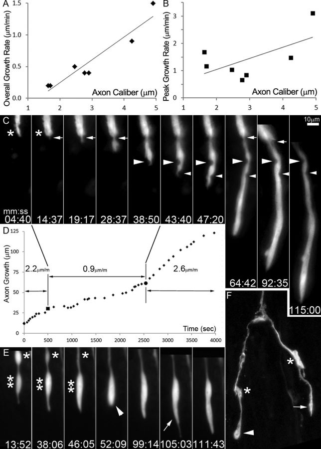
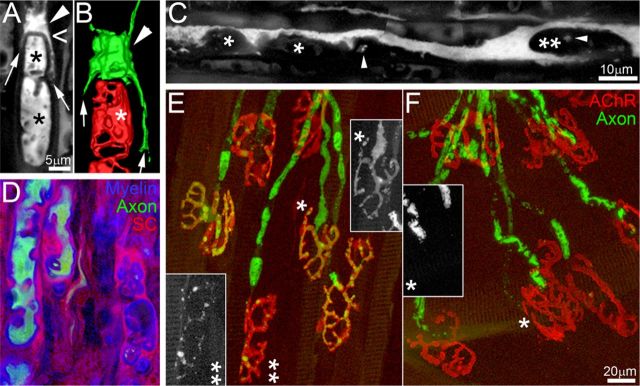
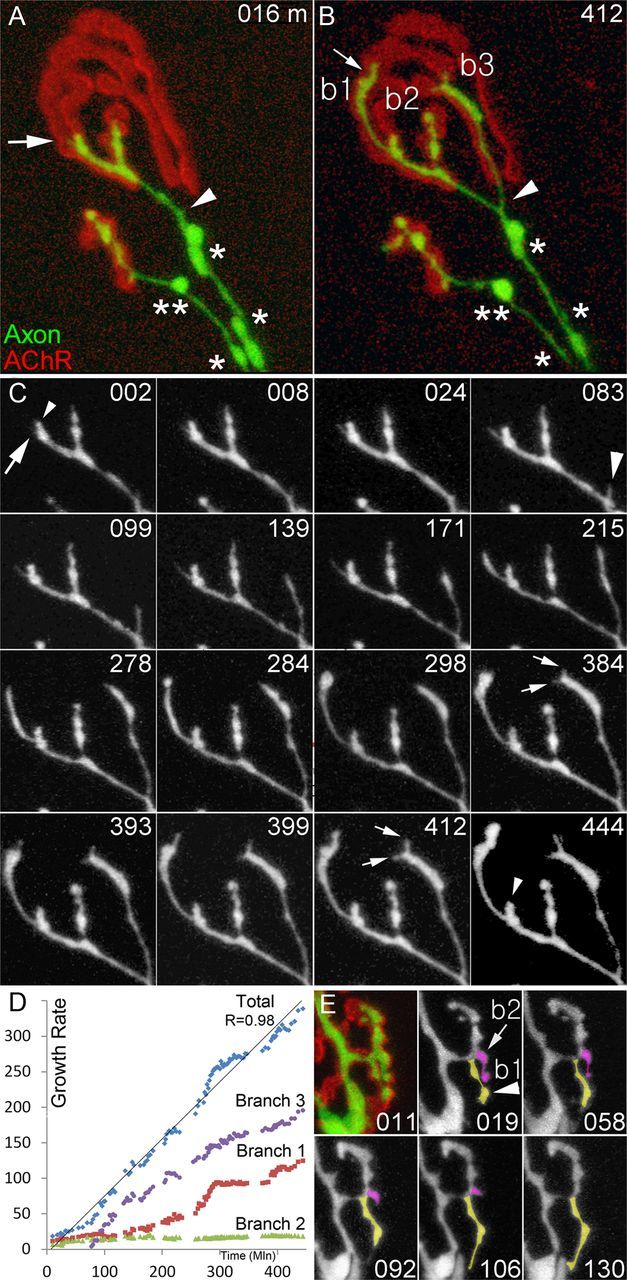
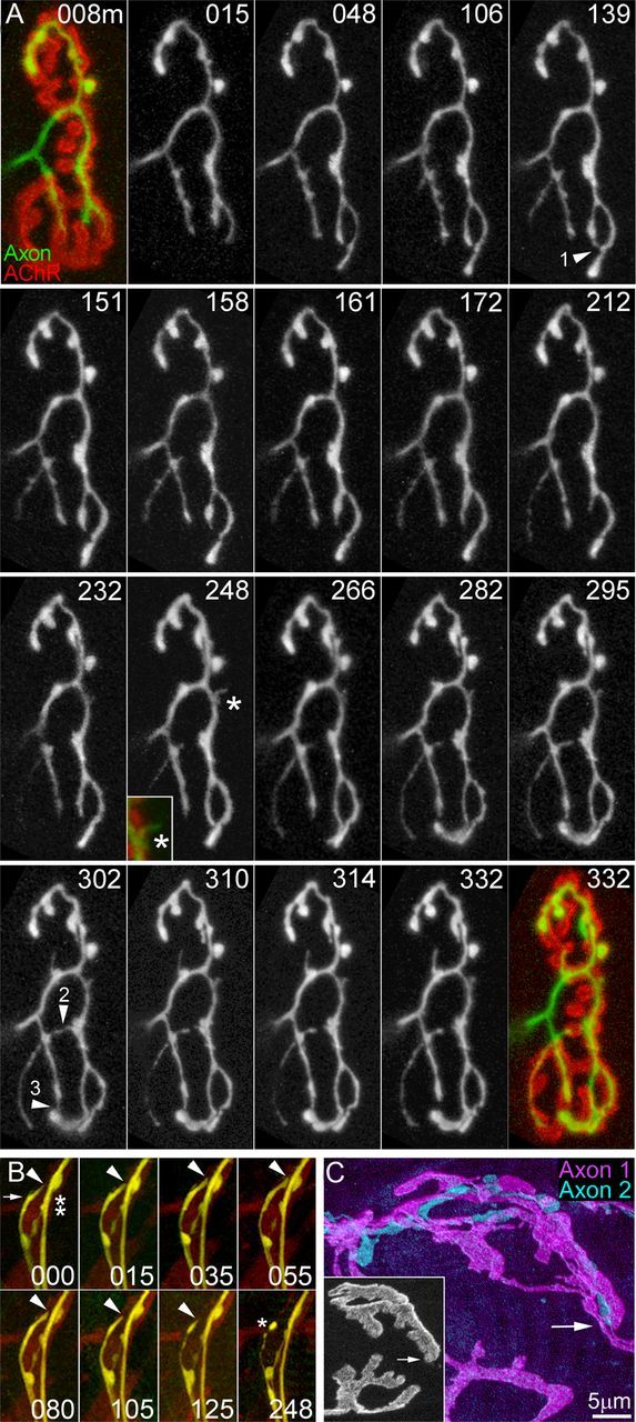
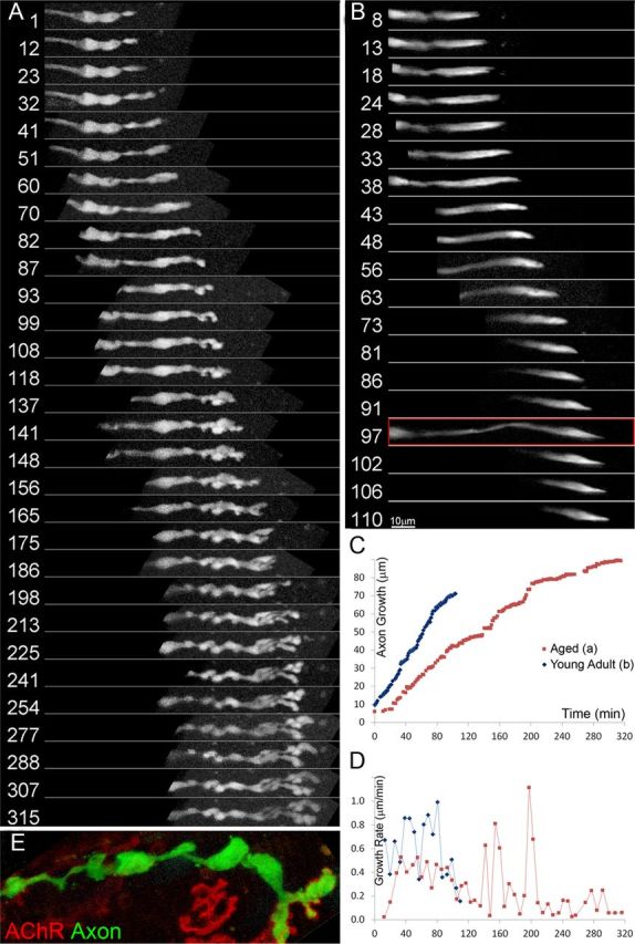
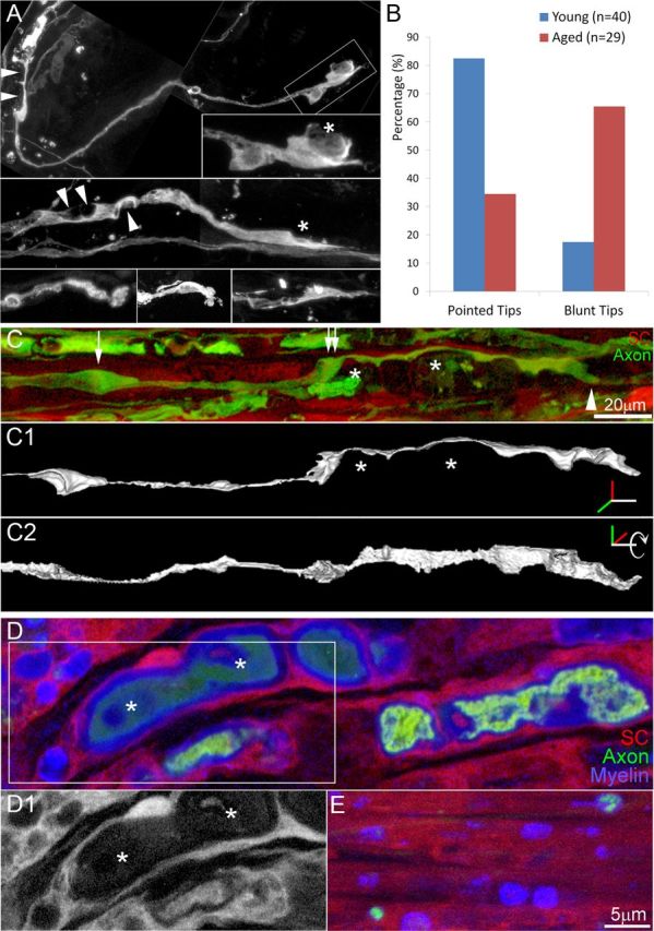
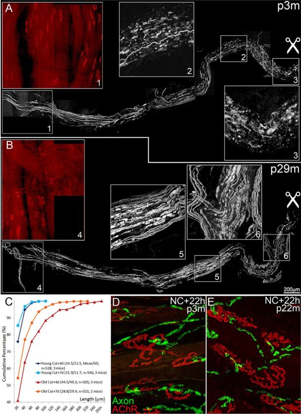
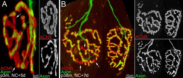
Injuries to peripheral nerves can cause paralysis and sensory disturbances, but such functional impairments are often short lived because of efficient regeneration of damaged axons. The time required for functional recovery, however, increases with advancing age (Verdú et al., 2000; Kawabuchi et al., 2011). Incomplete or delayed recovery after peripheral nerve damage is a major health concern in the aging population because it can severely restrict a person's mobility and independence. A variety of possible causes have been suggested to explain why nervous systems in aged individuals recover more slowly from nerve damage. Potential causes include age-related declines in the regenerative potential of peripheral axons and decreases in the supply or responsivity to trophic and/or tropic factors. However, there have been few direct analyses of age-related axon regeneration. Our aim here was to observe axons directly in young and old mice as they regenerate and ultimately reoccupy denervated neuromuscular synaptic sites to learn what changes in this process are age related. We find that damaged nerves in aged animals clear debris more slowly than nerves in young animals and that the greater number of obstructions regenerating axons encounter in the endoneurial tubes of old animals give rise to slower regeneration. Surprisingly, however, axons from aged animals regenerate quickly when not confronted by debris and reoccupy neuromuscular junction sites efficiently. These results imply that facilitating clearance of axon debris might be a good target for the treatment of nerve injury in the aged.

Keywords: aging; axon regeneration; in vivo imaging; neuromuscular system; self-avoidance; synapse reinnervation.


Figures

Figure 1.
Figure 1.
Regenerating axons in young animals slow when passing by obstacles. A, B, Graph showing that axon caliber correlates better to overall growth rate of regeneration (A) than peak growth rate (B). Caliber of individual regenerating axons (x-axis) and, on the y-axis, their overall regeneration rates and peak rates are plotted (n = 7). Correlation coefficient (R) is 0.79 for overall and 0.61 for peak rates. C, A regenerating axon was monitored for 2 h in an explanted triangularis sterni muscle from a 3-month-old mouse 5 d after a crush injury. The axon decelerated, accelerated, and even paused while growing >100 μm. Deceleration and pause were associated with axonal indentations along the axon shaft (arrows and arrowheads). Because the indentations are maintained until the end of imaging session, we think the growth delays are associated with the axon attempting to pass by multiple space-occupying obstacles. After these delays, the axon grew rapidly and showed no additional indentations. See the Results for details. The overall growth rate was 1.5 μm/min and the axon caliber near the growth cone was ∼4.9 μm. D, Graph showing a change in axon extension rate associated with obstacles in the path of the axon shown in C. Two vertical lines mark what we believe are the beginning and the end of the obstacle zone, where the axon was indented and its growth rate slowed. E, As axons regenerate, the axoplasm redistributes from a swelling into the distal compartment. An axonal varicosity (asterisk) just proximal to the growth cone (double asterisks) disappeared at the same time the growth cone added volume, suggesting that the axoplasm inside of the varicosity moved anterogradely into the growing tip. Also note that when the tip is blunt (arrowhead), its forward progress is less than when it becomes more pointed (arrow). The growth rate of this axon was 0.2 μm/min and the axon caliber near the growth cone was ∼1.6 μm. F, The blunt tips are not an artifact of in vivo time lapse imaging because acutely fixed preparations the same structures were observed. Shown is a regenerating axon with two branches: one with a pointed tip (arrow) and the other with a blunt tip (arrowhead). Varicosities (asterisks) were also found in the same axon.
Figure 2.
Figure 2.
Degenerating axon and myelin debris impedes axon regeneration. A, B, Confocal imaging reveals the debris near the growth tip of a regenerating axon just distal to the site of nerve damage (nerve crush occurred 3 d previously). A is a confocal section and B is a 3D rendering of the entire confocal stack of the same axon. See Results for details. C, Regenerating axons were also obstructed by nonfluorescent (i.e., non-neuronal) debris. Shown is a regenerating axon growing from the left of the figure, the shape of which was distorted by objects that are for the most part nonfluorescent (asterisks). As in the example shown in A and B, this axon is sending small branches around a large obstruction (double asterisks). The small dots of fluorescence (arrowheads) in the dark objects may indicate that there is some axonal debris intermixed with non-neural debris. D, Some of the nonfluorescent material in endoneurial tubes after nerve crush is glial. Transgenically labeled Schwann cells (red) and axonal debris (green) and immunolabeled myelin (blue) are all found densely packed in endoneurial tubes. E, F, Axonal debris disappears earlier at nerve terminal sites than in more proximal axonal pathways. Approximately 1 d after nerve damage, we noted axon fragmentation for the first time occurring asynchronously in different axons and terminals. For example, one junction in the same field of view still showed an unfragmented axon (asterisk in E), whereas other axons and their junctions were in various stage of degeneration (double asterisk in E). The debris was removed earlier from the nerve terminals than the more proximal nerve branches, as seen by the preservation of some fluorescent fragments in the axon when no fragments were visible at the junction sites (asterisk in F). Insets in E and F show the axon-only view.
Figure 3.
Figure 3.
Regenerating axons reoccupy former neuromuscular junction sites in a piecemeal way. AC, A regenerating axon at a neuromuscular junction site 4 d after nerve crush (A) and 7 h later (B). This neuromuscular junction was imaged repeatedly in vivo for 7+ h in a 3-month-old animal. The reinnervating axon (green) added a collateral branch (b3) that originated from the axon shaft (arrowheads) just proximal to a large cluster of AChRs (red). Notice the shrinkage (asterisks) and swellings (double asterisks) in the caliber and size of varicosities of the axon. C, Images taken every 3–5 min reveal the incremental pattern of synaptic regrowth. Although some branches were growing, others were quiescent. For example, branch b2 (B) grew very little over the 7 h, whereas branches 1 and 3 grew rapidly. The same phenomenon was seen at some sites where axon branches formed. Sometimes, one axon branch would grow and the other would not. For example, only one of the side branches of branch b1 grew extensively during this imaging session (large arrow vs small arrowhead). Conversely, branch b3 bifurcated and both of its daughter branches grew at similar rates, at least over the duration of this imaging session (small arrows). D, Graph showing that the cumulative growth rates of regenerating branches of the same axon are not necessarily the same. Branch 2 (green, b2 in B) grew only slightly during the 7+ h imaging session. However, branch 3 (purple) grew linearly from the moment of its formation at 80 min of imaging and continuing until the end of the session 6 h later (r = 0.96). In contrast to the other two branches, axon branch 1 (red) changed rate a few times over the imaging session (r = 0.93). Despite the variability on the individual growth rates, the sum of their growth was strongly linear (blue; r = 0.98), suggesting that the branches of one axon at the same neuromuscular junction rarely paused at the same time. E, One branch of a reinnervating axon (b1, tinted yellow) grew at the same time the other branch (b2, tinted purple) was retracting. It is possible that the cytoplasm that was in b2 is used to generate the elongation of b1.
Figure 4.
Figure 4.
Regenerating axon branch tips retract to avoid persistent contact with other parts of the same axon. A, Neuromuscular junction from a 3-month-old mouse 4 d after nerve crush imaged at the time an axon (green) is in the midst of reinnervating the former synaptic site (labeled red). In the time-lapse panels (18 images over 5 h), growth cones of regenerating axon branches can be seen to grow and then occasionally retract. Most of the retraction events occur within 10 min after the growing tip bumps into another part of the same axon (see arrowhead 1 at 139 min, arrowhead 2 and 3 at 302 min). B, Growth cones of regenerating axons in nerve bundles also retract at sites where they transiently come into contact with other regions of the same axon. A growth cone (arrow) that originated as a terminal sprout at a neuromuscular junction was imaged repeatedly in vivo for 4+ h in the sternomastoid muscle in a 3-month-old animal 4 d after nerve crush. This sprout retracted after touching its parental axon shaft (double asterisks). Arrowheads indicate the end of the growth cone. C, During nerve regeneration after double nerve crush (see Materials and Methods), different axons sometimes coinnervate the same primary synaptic folds. Shown is a region of a neuromuscular junction (inset) that is multiply innervated. The two axons (labeled purple and cyan) intermingle extensively without any signs of retraction. These results and others like it suggest that axon retraction is a phenomenon that occurs due to self-recognition.
Figure 5.
Figure 5.
Regenerating axons grow haltingly and branch frequently in aged animals. A, Shown is a 5 h in vivo time-lapse of axonal regeneration in a 23-month-old mouse 5 d after nerve crush. The growth cone forms several blunt tips and the axon caliber is not uniform. This appearance was seen in most regenerating axons in aged mice. B, In contrast, regenerating axons in young mice such as in this 2-month-old were less branched and their tips were more pointed (red box). C, D, Cumulative growth curves (C) and changes in growth rates (D) show the slower overall growth for the aged axon shown in A (red) compared with the axon from a young animal shown in B (blue). Average growth rate was 0.6 μm/min in the young mouse versus 0.3 μm/min in the old animal. Both young and aged animals, however, accelerate and decelerate during the growth and had similar peak growth rates. E, Acutely prepared samples from regenerating nerve-muscle preparations from aged animals (25 months old) show the same scalloped appearance as seen in preparations that were imaged over hours, indicating that this appearance is not an artifact induced by long-term exposure to light or other imaging-related artifacts. Axon is shown in green and postsynaptic AChRs in red.
Figure 6.
Figure 6.
In aged animals, regenerating axons and their pathways were morphologically different from those in young animals. A, Five examples of the distal tips of regenerating axons in 25-month-old mice. In contrast to young animals (Fig. 1), these axons were more irregular in caliber. At multiple sites along their length, they appeared to be indented by oval-shaped nonfluorescent masses (arrowheads). The tips of the growth cones were also more variable in shape compared with growth cones in young animals (Fig. 1). In some cases, the growth cone appeared to be squeezing by nonfluorescent objects in their way (asterisk). B, There are significantly more growth cones with blunted tips in old animals compared with young animals (p < 0.001). C, Evidence that axons squeeze between the edge of the endoneurial tube and vacuolated Schwann cells. Shown is a regenerating axon (green) in a regenerating nerve bundle from a 29-month-old mouse. Vacuoles (asterisks, see also D1) in Schwann cells (red) appeared to push against the axon. Confocal reconstructions of this axon from two different rotation angles (C1C2) revealed that it was actually a flattened ribbon. At the middle of the trajectory (double arrows), the axon can be seen to move from the bottom of the tube to the top, presumably because of an obstruction (asterisks) in its path. D, Vacuoles in Schwann cells after nerve damage contain myelin debris. Immunolabeling myelin basic protein showed that the vacuoles in Schwann cells (red) contained myelin debris (blue) and axonal debris (green) from a 24-month-old mouse 4 d after denervation. A Schwann-cell-only image of the boxed area (D1) show dark Schwann cell vacuoles where axonal and myelin debris are located. E, Far less debris was seen in 3-month-old animals 4 d after nerve damage (labeled the same way as in D).
Figure 7.
Figure 7.
Debris clearance after nerve damage is retarded in aged animals. A, B, Evidence of a greater abundance of debris 4 d after nerve damage in old versus young animals. Reconstructions of the distal axon bundles from sites of nerve cut (scissors symbols) to sternomastoid muscle neuromuscular junction sites (red in box 1 and 4) in young adult (3 months old; A) and aged (29 months old; B) animals. Shown is fluorescently labeled axonal debris from a transgenic mouse that expresses CFP in its axons. AChRs were labeled with Alexa Fluor 594-conjugated BTX. Insets show evidence of the more numerous and larger debris fragments in the aged compared with the young adult animal. C, Cumulative graph showing larger axonal fragmentations in aged animals (29 months old) than in young animals (3 months old; p < 0.0001). The increase was evident both 4 and 7 d after nerve cut (see Results for details). D, E, Evidence showing that initiation of debris formation occurred at a similar rate in young and old animals. Neuromuscular junctional areas (AChRs are shown in red and axons in green) were imaged 22 h after denervation in young adult (D) and aged (E) animals. The similarity of the degenerating axons in these two situations suggests that the onset of Wallerian degeneration was not delayed in old animals.
Figure 8.
Figure 8.
Regenerating axons reoccupy junctions in old animals as well as axons do in young animals. A, Axons in the midst of reoccupying former synaptic sites in young animals quickly cover most of the AChRs. The few unoccupied sites (arrows) will likely be reinnervated within the next few days. B, Axons also appear to successfully reoccupy receptor sites in old animals, because there are only a few unoccupied sites 1 week after nerve crush.

Similar articles

Cited by

References

    1. Black MM, Lasek RJ. Slowing of the rate of axonal regeneration during growth and maturation. Exp Neurol. 1979;63:108–119. doi: 10.1016/0014-4886(79)90188-2. - DOI - PubMed
    1. Bosse F. Extrinsic cellular and molecular mediators of peripheral axonal regeneration. Cell Tissue Res. 2012;349:5–14. doi: 10.1007/s00441-012-1389-5. - DOI - PubMed
    1. Brunetti M, Miscena A, Salviati A, Gaiti A. Effect of aging on the rate of axonal transport of choline-phosphoglycerides. Neurochem Res. 1987;12:61–65. doi: 10.1007/BF00971365. - DOI - PubMed
    1. Feng G, Mellor RH, Bernstein M, Keller-Peck C, Nguyen QT, Wallace M, Nerbonne JM, Lichtman JW, Sanes JR. Imaging neuronal subsets in transgenic mice expressing multiple spectral variants of GFP. Neuron. 2000;28:41–51. doi: 10.1016/S0896-6273(00)00084-2. - DOI - PubMed
    1. Ferguson TA, Son YJ. Extrinsic and intrinsic determinants of nerve regeneration. J Tissue Eng. 2011;2:2041731411418392. doi: 10.1177/2041731411418392. - DOI - PMC - PubMed