. 2013 Dec 11;33(50):19555-66.
doi: 10.1523/JNEUROSCI.3051-13.2013.

Multimodal integration of self-motion cues in the vestibular system: active versus passive translations

Affiliations

Multimodal integration of self-motion cues in the vestibular system: active versus passive translations

Jerome Carriot et al. J Neurosci. .

Abstract

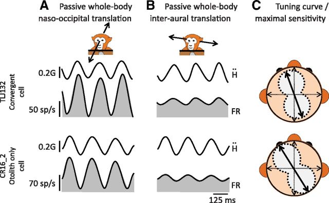
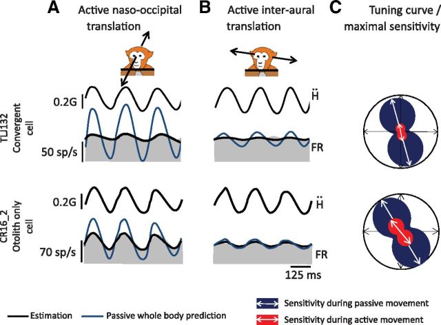
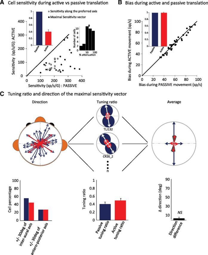
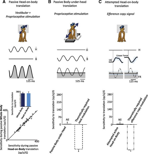
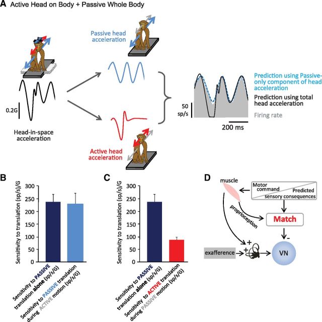
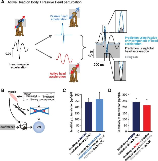
The ability to keep track of where we are going as we navigate through our environment requires knowledge of our ongoing location and orientation. In response to passively applied motion, the otolith organs of the vestibular system encode changes in the velocity and direction of linear self-motion (i.e., heading). When self-motion is voluntarily generated, proprioceptive and motor efference copy information is also available to contribute to the brain's internal representation of current heading direction and speed. However to date, how the brain integrates these extra-vestibular cues with otolith signals during active linear self-motion remains unknown. Here, to address this question, we compared the responses of macaque vestibular neurons during active and passive translations. Single-unit recordings were made from a subgroup of neurons at the first central stage of sensory processing in the vestibular pathways involved in postural control and the computation of self-motion perception. Neurons responded far less robustly to otolith stimulation during self-generated than passive head translations. Yet, the mechanism underlying the marked cancellation of otolith signals did not affect other characteristics of neuronal responses (i.e., baseline firing rate, tuning ratio, orientation of maximal sensitivity vector). Transiently applied perturbations during active motion further established that an otolith cancellation signal was only gated in conditions where proprioceptive sensory feedback matched the motor-based expectation. Together our results have important implications for understanding the brain's ability to ensure accurate postural and motor control, as well as perceptual stability, during active self-motion.

Keywords: corollary discharge; efference copy; head motion; neuron; otolith; proprioception.


Figures

Figure 1.
Figure 1.
Activity of two example neurons (unit TLJ132: convergent cell and unit CR16_2: otolith only cell) during passive whole-body naso-occipital translation (A) and interaural translation (B). A head movement based model (Eq. 1; solid black trace) is superimposed on the firing rate traces. C, The spatial sensitivity of the neuron: tuning curve (dashed line around the gray area) and maximal sensitivity vector (thick arrow). Ḧ, Head acceleration; FR, firing rate.
Figure 2.
Figure 2.
Activity of the same example neurons presented in Figure 1 (unit TLJ132: convergent cell and unit CR16_2: otolith only cell) during self-generated naso-occipital (A) and interaural (B) head translation. Superimposed on the firing rate traces are response predictions based on each neuron's sensitivity to passive translation (blue trace) and the best fits of this model to the actual data (Eq. 1; solid black trace). C, Comparison of the tuning curves computed during self-generated head motion (red area) and those computed during passive head motion (replotted from Fig. 1; blue area). Abbreviations as in Figure 1.
Figure 3.
Figure 3.
Responses to active versus passive translation: population summary. A, Neuronal sensitivities to passive whole-body versus active head-on-body translations. Filled circles represent the neurons for which we were able to compute the maximal sensitivity vector (n = 18), whereas open circle represent the sensitivity of the cells tested along their preferred major axis (naso-occipital or interaural axis). Note that all data points fall below the unity line, demonstrating a relative reduction in sensitivity to active translation. Left, Inset, Mean neuronal sensitivities to self-generated head motion (red) normalized to whole-body sensitivity (blue) further illustrate the attenuation in response. Right, Inset, Distribution histogram showing each neuron's percentage attenuation. B, Comparison of the resting bias estimated for neuronal responses revealed no significant difference for the same two conditions. Inset, Mean resting bias estimated during self-generated head motion (red) normalized to resting bias estimated during whole-body translation (blue). C, Comparison of maximal vector orientation and tuning ratio for passive whole-body and active head-on-body translations. Left, The spatial distribution of the maximal sensitivity vector for passive (blue) and the active motion (red) across the neuronal population. Left, Bottom, The mean repartition of the maximal vector orientation around the interaural axis and the naso-occipital axis. The tuning ratio (ratio between the maximal and the minimal sensitivity) for two example cells is presented in the middle. Population average of the tuning ratio does not differ for passive (blue) and self-generated (red) head movement stimulation conditions (middle, bottom). To further compare passive and active motion, maximal sensitivity vectors for active motion were normalized in magnitude and orientation relative to those computed during passive motion (right). The population average is represented on the right panel. Neither the tuning ratio nor the orientation of the maximal sensitivity vector differed for passive (blue) and self-generated (red) head-movement stimulation conditions. Population average of the difference in preferred orientation is presented below (right, bottom).
Figure 4.
Figure 4.
Neurons are not responsive to either the passive stimulation of proprioceptors or the generated efference copy signals in the absence of corresponding head motion. A, An example neuron's response to combined passive, vestibular and proprioceptive stimulation (head-on-body translation; cell TLJ132; Figs. 1–3). Superimposed on the neuronal firing rate (FR) is the best estimate of the neuronal response (thick black trace). Bottom, Scatter plot showing each neuron's sensitivity to passive whole-body-translation versus passive head-on-body translation. Inset, Bar graph compares population averages for sensitivities to passive whole-body-translation (dark blue) and passive head-on-body translation (light blue). No difference in sensitivity was observed in these two conditions. B, The same example neuron's response to neck proprioceptive stimulation: the body was sinusoidally translated under the stationary head. Superimposed on the neuronal FR is the best estimate of the neuronal response (thick black trace) and a prediction based on the neck modulation that would be theoretically required to account for the attenuation in response observed during active head-on-body motion (dashed line). Bottom, Bar graph summarizes population averaged neuronal sensitivities as well as the average of this latter theoretical prediction. C, The same neuron's response when motor commands are generated in the absence of corresponding head motion (attempted head-on-body translation). The linear force produced by the monkey's neck is shown by the blue traces. Superimposed on the neuronal FR is the best estimate of the neuronal response (thick black trace) and a prediction based on a motor efference copy-driven modulation that would be theoretically required to account for the attenuation in response observed during active head-on-body motion (dashed black line). All neurons were unresponsive to the production of motor efference copy signals in this condition. Bottom, Bar graph summarizes population averaged neuronal sensitivities in this condition, as well as the average of this latter theoretical prediction.
Figure 5.
Figure 5.
A, Neuronal responses to simultaneous passive and active head motion when sensory expectations and feedback correspond. Black traces represent total head-in-space acceleration. Blue traces represent the component of the head motion that was passively applied. Red traces represent the component of the head motion that was actively generated. Two predictions are superimposed on the recorded firing rates: (1) the total vestibular prediction which assumes unattenuated sensitivity to the total head motion (black trace) and (2) the passive-only prediction which assumes unattenuated preferential encoding of the passive component of the head motion (blue dashed line). When active and passive translation occurred concurrently, the neuron selectively encoded the passive component (A, compare black and blue dashed fits). B, C, Summary of population responses during combined stimulation. Neuronal sensitivities to passive whole-body translation were comparable when applied alone or simultaneously during the production of active head-on-body translations (B). In contrast, neuronal responses to the active component of the head motion were consistently attenuated (C). D, A model schematic formalizing the hypothesis that a match between the brain's expectation of the sensory consequences of active translation and actual sensory feedback is required to gate in the otolith cancellation signal.
Figure 6.
Figure 6.
Neuronal responses to simultaneous passive and active head motion when proprioceptive feedback and sensory expectations do not correspond. A, Black traces represent the head-in-space acceleration. Blue traces represent the component of the head motion produced by the passive head perturbation (see Materials and Methods). Red traces represent the component of the head motion that was actively generated. Two predictions are superimposed on the recorded firing rates: (1) the total vestibular prediction, which assumes unattenuated sensitivity to the total head motion (black trace), and (2) the passive-only prediction which assumes unattenuated preferential encoding of the passive component of the head motion (blue dashed line). In this condition, the neuron was not able to discriminate the active and passive component of head motion and instead encoded all vestibular information (right, insets, compare black solid and blue dashed traces). B, A model schematic formalizing this experiment in which a match between the brain's expectation of the sensory consequences of active translation and the actual sensory feedback does not occur during active translation. C, D, Summary of population responses during combined stimulation. Sensitivities to passive motion applied alone or simultaneously with active head translation were comparable (C). Sensitivities to the active component of the motion were not attenuated when produced during simultaneously passive head-on-body perturbations (D).

Similar articles

Cited by

References

    1. Angelaki DE. Generation of two-dimensional spatial and temporal properties through spatiotemporal convergence between one-dimensional neurons. IEEE Trans Biomed Eng. 1993;40:686–692. doi: 10.1109/10.237698. - DOI - PubMed
    1. Angelaki DE, Cullen KE. Vestibular system: the many facets of a multimodal sense. Ann Rev Neurosci. 2008;31:125–150. doi: 10.1146/annurev.neuro.31.060407.125555. - DOI - PubMed
    1. Angelaki DE, Dickman JD. Spatiotemporal processing of linear acceleration: primary afferent and central vestibular neuron responses. J Neurophysiol. 2000;84:2113–2132. - PubMed
    1. Angelaki DE, Gu Y, Deangelis GC. Visual and vestibular cue integration for heading perception in extrastriate visual cortex. J Physiol. 2011;589:825–833. doi: 10.1113/jphysiol.2010.194720. - DOI - PMC - PubMed
    1. Bassett JP, Zugaro MB, Muir GM, Golob EJ, Muller RU, Taube JS. Passive movements of the head do not abolish anticipatory firing properties of head direction cells. J Neurophysiol. 2004;93:1304–1316. doi: 10.1152/jn.00490.2004. - DOI - PubMed

Publication types

LinkOut - more resources