. 2013 Dec 11;33(50):19579-89.
doi: 10.1523/JNEUROSCI.4318-13.2013.

Progranulin deficiency promotes post-ischemic blood-brain barrier disruption

Affiliations

Progranulin deficiency promotes post-ischemic blood-brain barrier disruption

Katherine Jackman et al. J Neurosci. .

Abstract

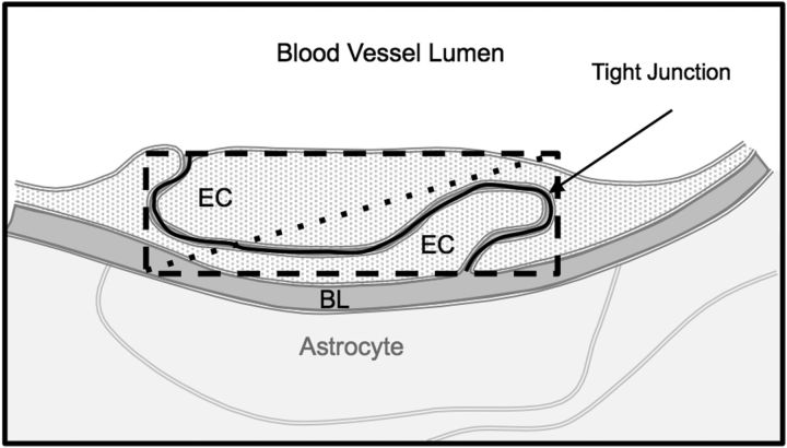
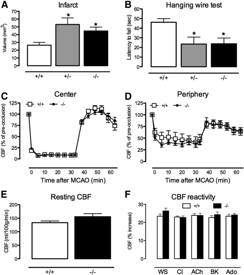
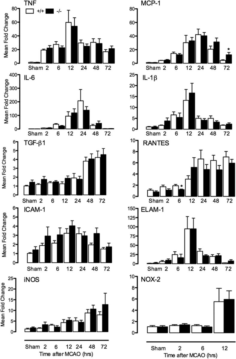
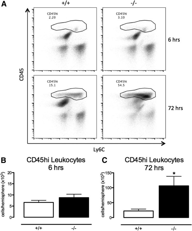
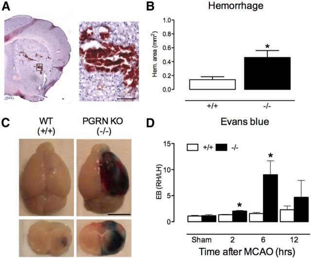
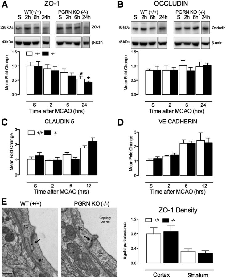
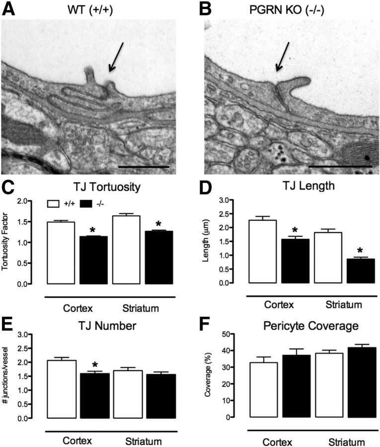
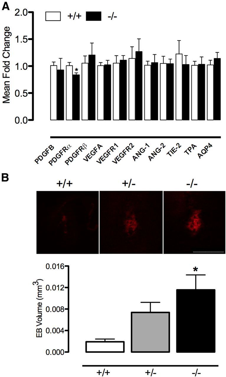
Loss-of-function mutations of progranulin (PGRN) have been linked to frontotemporal dementia, but little is known about the effects of PGRN deficiency on the brain in health and disease. PGRN has been implicated in neurovascular development, inflammation, and Wnt signaling, a pathway involved in the formation of the blood-brain barrier (BBB). Because BBB alterations and inflammation contribute to ischemic brain injury, we examined the role of PGRN in the brain damage produced by ischemia-reperfusion. PGRN(+/-) and PGRN(-/-) mice underwent middle cerebral artery occlusion (MCAO) with monitoring of cerebral blood flow. Infarct volume and motor deficits were assessed 72 h later. Post-ischemic inflammation was examined by expression of inflammatory genes and flow cytometry. BBB structure and permeability were examined by electron microscopy (EM) and Evans blue (EB) extravasation, respectively. MCAO resulted in ~60% larger infarcts in PGRN(+/-) and PGRN(-/-) mice, an effect independent of hemodynamic factors or post-ischemic inflammation. Rather, massive hemorrhages and post-ischemic BBB disruption were observed, unrelated to degradation of tight junction (TJ) proteins or matrix metalloproteinases (MMPs). By EM, TJ were 30-52% shorter, fewer, and less interlocking, suggesting a weaker seal between endothelial cells. Intracerebral injection of platelet-derived growth factor-CC (PDGF-CC), which increases BBB permeability, resulted in a more severe BBB breakdown in PGRN(+/-) and PGRN(-/-) than wild-type mice. We describe a previously unrecognized involvement of PGRN in the expression of key ultrastructural features of the BBB. Such a novel vasoprotective role of PGRN may contribute to brain dysfunction and damage in conditions associated with reduced PGRN function.

Keywords: blood–brain barrier; frontotemporal dementia; neurovascular unit; progranulin; stroke.


Figures

Figure 1.
Figure 1.
TJ tortuosity. Diagram depicting methods used to calculate the tortuosity index presented in Figure 8C. To calculate complexity of the TJ, TJ length (solid black line) is divided by the diagonal (dotted line) of the rectangle (dashed line) that contains the length and height of the complete TJ. EC, Endothelial cell; BL, basal lamina.
Figure 2.
Figure 2.
Ischemic injury is exacerbated in PGRN-deficient mice. A, B, Both PGRN+/− and PGRN-KO (−/−) have larger infarcts and have greater functional impairment than WT (+/+) mice 72 h after MCAO (n = 7–10 per group, *p < 0.05 from WT, ANOVA). C, D, The degree of cerebral ischemia and reperfusion are similar in the center and periphery of the ischemic territory in WT and PGRN-KO (p > 005). E, Resting CBF, measured using ASL-MRI, is similar in naive WT and PGRN-KO (n = 5 per group; p > 0.05, t test). F, Vascular responses to whisker stimulation (WS), the Ca2+ ionophore A23187 (CI), acetylcholine (ACh), bradykinin (BK), and adenosine (Ado) are similar in naive WT and PGRN-KO (−/−) (n = 5 per group; p > 0.05, t test).
Figure 3.
Figure 3.
Post-ischemic inflammatory gene expression. Time course of mRNA expression of genes involved in post-ischemic inflammation in WT (+/+) and PGRN-KO (−/−) (n = 5–8 per group; *p < 0.05 from respective WT, t test).
Figure 4.
Figure 4.
Post-ischemic inflammation. A, Representative density plots of brain mononuclear cells, expressed as percentage of total number of cells, in WT (+/+) and PGRN-KO (−/−) mice 6 and 72 h after MCAO measured with flow cytometry. B, C, Absolute numbers of infiltrating CD45high leukocytes are comparable in the post-ischemic brain of WT and PGRN-KO mice 6 h after MCAO (n = 5 per group; p > 0.05, t test). Leukocyte number is higher in PGRN-KO mice at 72 h, consistent with greater injury at this time (n = 6–8 per group; *p < 0.05, t test).
Figure 5.
Figure 5.
Post-ischemic hemorrhage and BBB disruption are enhanced in PGRN-KO. A, Representative H&E-stained brain section illustrating hemorrhage 6 h after MCAO in PGRN-KO mice. Early tissue injury is also seen as pallor in the dorsal striatum. Scale bar, 100 μm. B, The area of post-ischemic hemorrhage is greater in PGRN-KO (−/−) mice 6 h after MCAO compared with WT (+/+) mice (n = 8 per group; *p < 0.05, t test) C, Representative images of EB extravasation in WT and PGRN-KO mice 6 h after MCAO. Notice the increased BBB permeability in PGRN-KO mice. Scale bar, 5 mm. D, Temporal profile of EB extravasation after MCAO, expressed as ratio between ischemic and non-ischemic hemisphere, showing greater BBB permeability in PGRN-KO (n = 7–10 per group; *p < 0.05 from respective WT, t test).
Figure 6.
Figure 6.
TJ proteins do not differ between WT and PGRN-KO mice after MCAO. A, Protein expression of ZO-1 is similar in WT (+/+) and PGRN-KO (−/−) mice at all time points after MCAO (S, sham). At 24 h, ZO-1 expression is reduced in both WT and PGRN-KO mice (n = 5–8 per group; *p < 0.05 from respective sham, ANOVA). B, Protein expression of occludin is similar in WT and PGRN-KO mice after MCAO (n = 5–8 per group; p > 0.05). C, D, No differences were detected in the mRNA expression of CLDN5 and VE-cadherin. E, Both the spatial distribution at the TJ cleft (representative images) and the ZO-1 immunogold density were similar in WT and PGRN-KO mice (n = 43–65 endothelial cells per group). Scale bar, 200 nm.
Figure 7.
Figure 7.
MMP expression and activity do not differ between WT and PGRN-KO mice after MCAO. A, MMP-9 mRNA expression is similar in WT (+/+) and PGRN-KO (−/−) mice at early time points (2–24 h) but tends to be higher in PGRN-KO mice at 48 and 72 h (n = 5–8 per group; p > 0.05). B, MMP-2 mRNA is induced at 48 and 72 h after MCAO, but expression is similar in WT and PGRN-KO mice (n = 5–8 per group; *p < 0.05 from respective sham, ANOVA). C, Activity of pro-MMP-9 was assessed by gel zymography. Intensity of pro-MMP-9 in samples, expressed relative to pro-MMP-9 standard, is similar in WT and PGRN-KO mice 6 h after MCAO (n = 8 per group; p > 0.05).
Figure 8.
Figure 8.
BBB ultrastructure is altered in PGRN-KO. A, B, Representative micrographs illustrating TJ morphology in WT (+/+) and PGRN-KO (−/−) mice. TJ complexity and length are reduced in PGRN-KO mice (arrows). Scale bar, 500 nm. C, TJ tortuosity (see Fig. 1) is reduced in PGRN-KO mice in neocortex (n = 108 to 129 TJ per group) and striatum (n = 65–109 TJ per group; *p < 0.05, t test). D, TJ are shorter in PGRN-KO in neocortex (n = 108–29 TJ per group) and striatum (n = 59–86 TJ per group; *p < 0.05, t test). E, TJ are fewer in PGRN-KO cortex (n = 42–47 vessels per group), but not striatum (n = 37–48 vessels per group; *p < 0.05, t test). F, Pericyte coverage is similar in WT and PGRN-KO cortex (n = 33–51 vessels per group) and striatum (n = 49–59 vessels per group). For all graphs, data was acquired in n = 3 WT and n = 3 PGRN-KO mice.
Figure 9.
Figure 9.
PGRN deficiency increases BBB permeability to PDGF-CC. A, No major differences were observed in the mRNA expression of factors involved in vascular development and stability, except for PDGFRα (n = 6 per group; *p < 0.05, t test). B, After injection of PDGF-CC, EB leakage/BBB permeability is enhanced in PGRN+/− and KO (−/−) mice compared with WT (+/+) (n = 6–7 per group; *p < 0.05 from WT, ANOVA). Scale bar, 500 μm. AQP4, Aquaporin-4.

References

    1. Abe T, Shimamura M, Jackman K, Kurinami H, Anrather J, Zhou P, Iadecola C. Key role of CD36 in Toll-like receptor 2 signaling in cerebral ischemia. Stroke. 2010;41:898–904. doi: 10.1161/STROKEAHA.109.572552. - DOI - PMC - PubMed
    1. Almeida S, Zhang Z, Coppola G, Mao W, Futai K, Karydas A, Geschwind MD, Tartaglia MC, Gao F, Gianni D, Sena-Esteves M, Geschwind DH, Miller BL, Farese RV, Jr, Gao F-B. Induced pluripotent stem cell models of progranulin-deficient frontotemporal dementia uncover specific reversible neuronal defects. Cell Rep. 2012;2:789–798. doi: 10.1016/j.celrep.2012.09.007. - DOI - PMC - PubMed
    1. Armulik A, Genové G, Mäe M, Nisancioglu MH, Wallgard E, Niaudet C, He L, Norlin J, Lindblom P, Strittmatter K, Johansson BR, Betsholtz C. Pericytes regulate the blood-brain barrier. Nature. 2010;468:557–561. doi: 10.1038/nature09522. - DOI - PubMed
    1. Armulik A, Genové G, Betsholtz C. Pericytes: developmental, physiological, and pathological perspectives, problems, and promises. Dev Cell. 2011;21:193–215. doi: 10.1016/j.devcel.2011.07.001. - DOI - PubMed
    1. Baker M, Mackenzie IR, Pickering-Brown SM, Gass J, Rademakers R, Lindholm C, Snowden J, Adamson J, Sadovnick AD, Rollinson S, Cannon A, Dwosh E, Neary D, Melquist S, Richardson A, Dickson D, Berger Z, Eriksen J, Robinson T, Zehr C, Dickey CA, Crook R, McGowan E, Mann D, Boeve B, Feldman H, Hutton M. Mutations in progranulin cause tau-negative frontotemporal dementia linked to chromosome 17. Nature. 2006;442:916–919. doi: 10.1038/nature05016. - DOI - PubMed

Publication types

MeSH terms

LinkOut - more resources