. 2014 Feb;196(2):455-70.
doi: 10.1534/genetics.113.155218. Epub 2013 Dec 13.

Feedback regulation of SIN by Etd1 and Rho1 in fission yeast

Affiliations

Feedback regulation of SIN by Etd1 and Rho1 in fission yeast

María Alcaide-Gavilán et al. Genetics. 2014 Feb.

Abstract

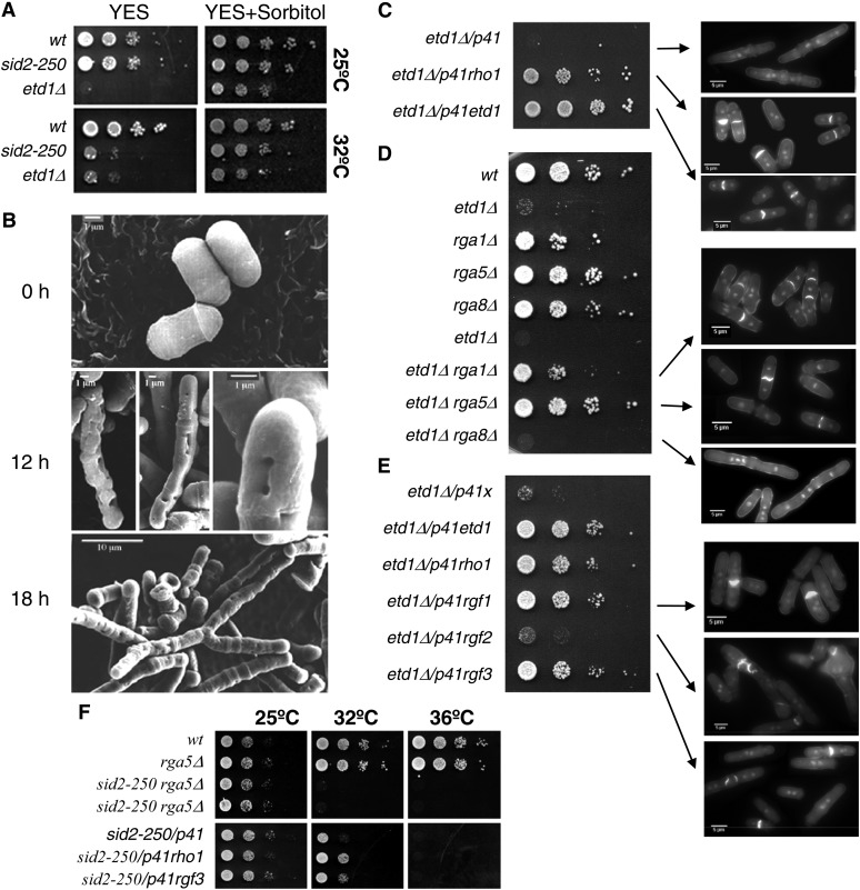
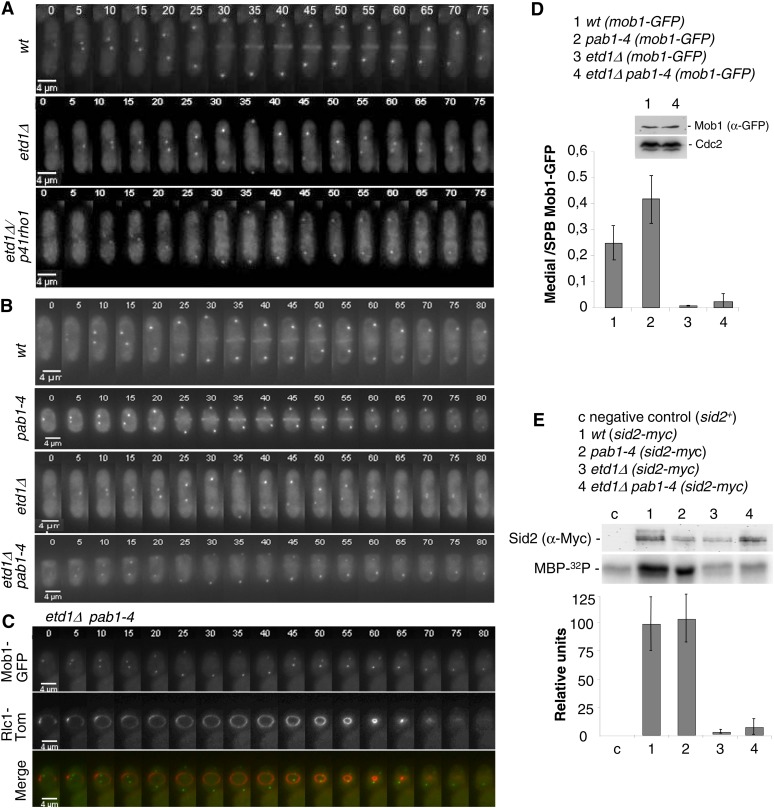
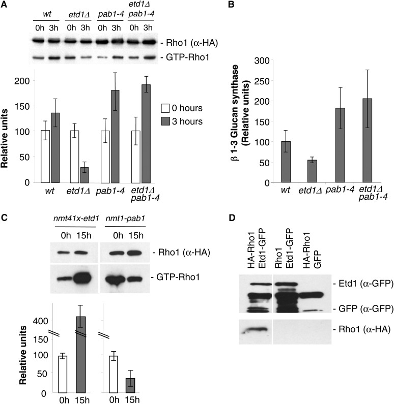
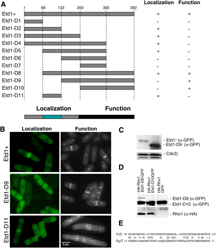
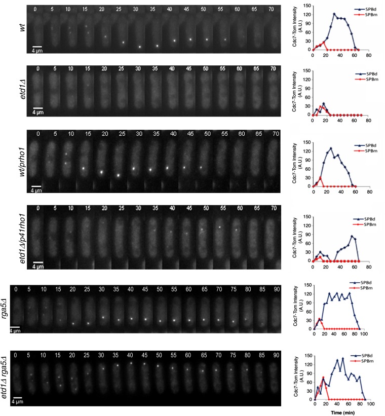
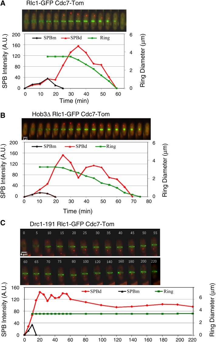
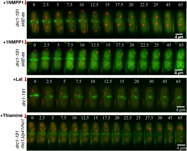
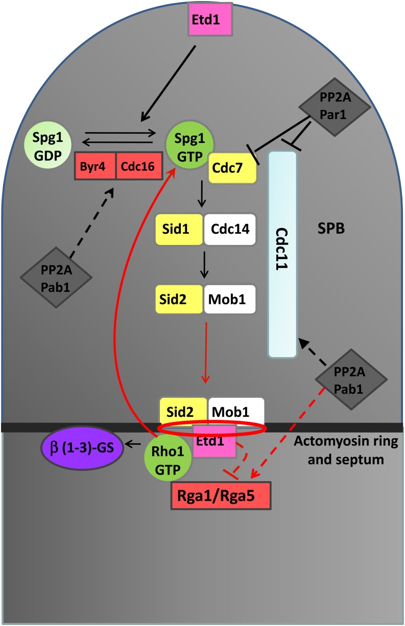
In fission yeast, the septation initiation network (SIN) is thought to promote cytokinesis by downstream activation of Rho1, a conserved GTPase that controls cell growth and division. Here we show that Etd1 and PP2A-Pab1, antagonistic regulators of SIN, are Rho1 regulators. Our genetic and biochemical studies indicate that a C-terminal region of Etd1 may activate Rho1 by directly binding it, whereas an N-terminal domain confers its ability to localize at the growing tips and the division site where Rho1 functions. In opposition to Etd1, our results indicate that PP2A-Pab1 inhibits Rho1. The SIN cascade is upstream-regulated by the Spg1 GTPase. In the absence of Etd1, activity of Spg1 drops down prematurely, thereby inactivating SIN. Interestingly, we find that ectopic activation of Rho1 restores Spg1 activity in Etd1-depleted cells. By using a cytokinesis block strategy, we show that Rho1 is essential to feedback-activate Spg1 during actomyosin ring constriction. Therefore, activation of Spg1 by Rho1, which in turn is regulated by Etd1, uncovers a novel feedback loop mechanism that ensures SIN activity while cytokinesis is progressing.

Keywords: Etd1; PP2A-Pab1; Rho1; SIN; Schizosaccharomyces pombe; Spg1; cytokinesis.


Figures

Figure 1
Figure 1
Etd1-depleted cells are deficient for cell-wall integrity and septation, a phenotype suppressed by high Rho1 activity. (A) Growth assay (serial dilution drop tests on plates) of etd1Δcells in YES media (left panels) and YES with 1.2 M sorbitol (right panels). The wild-type and sid2-250 strains were used as controls. Cells were grown (at 37° for etd1Δ and wt and at 25° for sid2-250), and 10-fold dilutions (starting with 104 cells) were spotted and incubated at 25° or 32°. (B) Electron microscopy images of etd1Δ cells grown at 37° (permissive temperature for etd1Δ after 0-, 12-, and 18-hr incubations at 25° (restrictive temperature for etd1Δ). Etd1-deficient cells show severe defects in cell-wall integrity. (C) Growth assay (serial dilution drop tests on plates) of etd1Δ cells overexpressing Rho1 protein under nmt41x expression in the pREP41x vector (p41rho1, left). Cells were grown at 37°, and 10-fold dilutions (starting with 104 cells) were spotted and incubated at 30° in the absence of thiamine. The pREP41x-driven expression of Etd1 (p41etd1) and the empty pREP41x plasmid (p41) were used as positive and negative controls, respectively. DAPI- and Calcofluor-stained cells of these etd1Δ and etd1Δ/p41rho1 strains are shown (right panels). (D) Growth assay (serial dilution drop tests on plates) (left) and cell phenotype (DAPI and Calcofluor staining) (right panels) of etd1Δ rga1Δ, etd1Δ rga5Δ, and etd1Δ rga8Δ cells. Cells were grown at 37°, and 10-fold dilutions (starting with 104 cells) were spotted and incubated at 30°. Wild-type (wt) and single-deletion strains were used as control. (E) Growth assay (as described in C) and cell phenotype (DAPI and Calcofluor staining) (right panels of etd1Δ cells overexpressing Rgf1, Rgf2, or Rgf3 proteins under nmt41x expression in the pREP41x vector (p41rgf1, p41rgf2, and p41rgf3, respectivelly) (left). p41etd1 and p41rho1 were used as positive controls. The empty p41 plasmid was used as a negative control. (F) Growth assay of sid2-250 cells deleted for rga5 (single sid2-250 and rga5Δ mutant strains were used as controls) (top panels) or overexpressing rgf3 (p41rgf3) (bottom panels). The p41rho1 construct was used as a positive control. The empty p41 plasmid was used as a negative control. Cells were grown at 25°, and 10-fold dilutions (starting with 104 cells) were spotted and incubated at 25°, 32°, or 36° in the absence of thiamine in the case of overexpression assays (bottom panels).
Figure 2
Figure 2
Etd1 is required for Sid2-Mob1 kinase activity and proper localization at the division site. (A) Mob1-GFP was imaged in live wild-type cells (top), etd1Δ mutant cells (middle) and etd1Δ/p41rho1 cells (bottom) by time-lapse microscopy at 5-min intervals at 25°, restrictive conditions for etd1Δ mutants. (B) Mob1-GFP was imaged in live wild-type cells (top), etd1Δ mutant cells (middle), pab1-4 (middle), and etd1Δ pab1-4 cells (bottom) by time-lapse microscopy as described in A. (C) Mob1-GFP and the actomyosin ring marker Rlc1-Tom were co-expressed and imaged in these cells as above. (D) Mob1-GFP fluorescence intensity was quantified in five cells of each strain, and maximum values of medially located Mob1-GFP with respect to SPB-associated Mob1-GFP are represented. Levels of Mob1-GFP in etd1Δ pab1-4 and wild-type cells, determined by Western blot analysis, are shown. The amount of Cdc2 was tested as control. (E) Sid2 kinase activity in wt, etd1Δ, pab1-4, and etd1Δ pab1-4 strains (in a sid2-myc genetic background) was determined in cell extracts by 32P incorporation into MBP-32P. Cells were grown at 37° (permissive temperature for etd1Δ cells) and post-incubated for 3 hr at 25° (restrictive temperature for this strain). Total Sid2 protein (from the sid2-myc construct) was analyzed by Western blot (α-Myc), and a wild-type sid2+ strain was used as negative control. Kinase activity was quantified and represented (mean value was derived from three experiments). Error bars indicate SEM.
Figure 3
Figure 3
Etd1 and PP2A-Pab1 are antagonistic regulators of Rho1 activity. (A) Determination of GTP-bound Rho1 (GTP-Rho1) levels vs. the total amount of Rho1 protein (α-HA) in extracts from wt, etd1Δ, pab1-4, and etd1Δ pab1-4 strains (in a HA-rho1 genetic background) at time 0 (incubated at 37°, the permissive temperature for etd1Δ cells) and at 3 hr post-incubation at the restrictive growth temperature for etd1Δ (25°). Quantification of relative GTP-Rho1 levels is shown (mean value derived from three experiments). Error bars indicate SEM. (B) 1,3-β-glucan synthesis activity was analyzed in cell extracts from the indicated strains and temperature (restrictive for the corresponding conditional mutant) (mean value derived from three experiments). Error bars indicate SEM. (C) Determination of GTP-bound Rho1 (Rho1p-GTP) levels as compared to the total amount of Rho1 protein (α-HA) in extracts from chromosomally tagged HA-rho1 strains overexpressing Etd1 (nmt41x-etd1 in plasmid pREP41x) or Pab1 (nmt1-pab1 in plasmid pREP3x) at 0 and 18 hr after nmt de-repression at 25° (see Materials and Methods for experimental details). Quantification of relative GTP-Rho1 levels is shown (mean value derived from three experiments). Error bars indicate SEM. (D) Physical association of Etd1 and Rho1. Protein extracts prepared from cells expressing Etd1-GFP (rho1+ genetic background), HA-Rho1 (with GFP expression as a control), or both were immunoprecipitated with anti-GFP antibodies; the immunoprecipitates were run on SDS–PAGE gels and probed with anti-GFP and anti-HA antibodies. Western blots of total extracts were also probed with anti-GFP and anti-HA antibodies to check the levels of tagged proteins.
Figure 4
Figure 4
Functional analysis of different Etd1 domains. (A) The indicated PCR-derived fragments of the Etd1-coding region (Etd1-D1 to Etd1-D10) were expressed in the pREP41x-GFP plasmid. Position of amino-acid residues flanking each Etd1 fragment is indicated. For cell localization assays, the GFP-fused constructs were expressed in wild-type cells in the absence of thiamine at 25°. For the functional complementation test, these constructs were expressed in the etd1Δ strain. Plasmids containing etd1Δ cells were grown at 37°, and the ability to rescue growth and division in this strain was assessed in the absence of thiamine at 25°. Etd1-truncated variants were classified as functional (+) or nonfunctional (−) according to their cell localization and complementation properties. The fragment containing the entire Etd1-coding region was used as control. Essential regions required for proper Etd1 cell localization and function are indicated. (B) Cell localization assay and functional complementation test of Etd1+ (control), Etd1-D9, and Etd1-D11 fragments. GFP fluorescence of wild-type cells expressing Etd1-GFP, Etd1-D9-GTP, or Etd1-D11-GTP constructs (left panels). DAPI and Calcofluor staining of etd1Δ cells under the expression of these constructs (right panels). (C) Western blot analysis of Etd1+ and Etd1-D9 (GFP-tagged). Cdc2 protein was used as a control. (D) Physical association of functional truncated variants Etd1-D9 and Etd1-D10 (GFP-tagged) and Rho1 (HA-tagged). Protein extracts prepared from cells expressing HA-Rho1 and Etd1-D9-GFP, Etd1-D10-GFP or GFP (negative control) were immunoprecipitated with anti-GFP antibodies; the immunoprecipitates were run on SDS–PAGE gels and probed with anti-GFP and anti-HA antibodies. Western blots of total extracts were also probed with anti-GFP and anti-HA antibodies to check the levels of tagged proteins. (E) Sequence similarity between Etd1 and Rgf2 in a region that largely overlaps with the Etd1 localization domain.
Figure 5
Figure 5
Effects of high Rho1 activity on Spg1 activation in Etd1-depleted cells. Cdc7-Tom was imaged by time-lapse microscopy at 5-min intervals in living wild-type (wt), etd1Δ, wt/p41rho1 (nmt41x-driven expression of rho1 in plasmid pREP41x), etd1Δ/p41rho1, rga5Δ, and etd1Δ rga5Δ cells under restrictive conditions for the etd1Δ mutation (25°). Fluorescence intensity was quantified (arbitrary units) and represented for each SPB (SPBd and dSPBm).
Figure 6
Figure 6
Kinetics of Spg1 activation and actomyosin ring contraction. Cdc7-Tom (Spg1 activity marker) and Rlc1-GFP (medial ring marker) were imaged by time-lapse microscopy at 5-min intervals in living (A) wild-type cells, (B) hob3Δ mutants (slows down medial ring constriction), and (C) drc1-191 mutants (blocks medial ring constriction) at 36° (restrictive temperature for drc1-191). Fluorescence intensity of Cdc7-Tom (arbitrary units) at each SPB (SPBd and SPBm) and medial ring diameter (μm) according to Rlc1-GFP signal are represented.
Figure 7
Figure 7
Involvement of Sid2, Etd1, and Rho1 in signaling Spg1 activation in drc1-191-blocked cells. Cdc7-Tom (Spg1 activity marker) and Rlc1-GFP (medial ring marker) were imaged by time-lapse microscopy at 2.5-min intervals in living drc1-191 sid2-as, drc1-191, and drc1-191 rho1D/p41rho1 mutants at 36°. Sid2-as inactivation (+10 μM 1NMPP1) before and after asymmetric localization of active Spg1, actomyosin ring disassembly (+100 µM latrunculin B), and repression of nmt41x-driven expression of rho1 in the p41rho1 plasmid (+ Thiamine) are indicated (Materials and Methods).
Figure 8
Figure 8
Schematic representation of mechanisms suggested for Spg1 and Rho1 coordination. The model includes the Cdc16 Spg1-specific GAP; Rga1 and Rga5 Rho1 GAPs; Etd1 and PP2A complexes regulating SIN; and the proposed feedback loop mechanism operating during cytokinesis. In red are the new pathways and regulatory mechanisms proposed in this study. Dashed lines represent hypothetical regulatory mechanisms.

Similar articles

Cited by

References

    1. Arellano M., Duran A., Perez P., 1996. Rho 1 GTPase activates the (1–3)beta-D-glucan synthase and is involved in Schizosaccharomyces pombe morphogenesis. EMBO J. 15: 4584–4591. - PMC - PubMed
    1. Arellano M., Duran A., Perez P., 1997. Localisation of the Schizosaccharomyces pombe rho1p GTPase and its involvement in the organisation of the actin cytoskeleton. J. Cell Sci. 110: 2547–2555. - PubMed
    1. Bardin A. J., Visintin R., Amon A., 2000. A mechanism for coupling exit from mitosis to partitioning of the nucleus. Cell 102: 21–31. - PubMed
    1. Calonge T. M., Arellano M., Coll P. M., Perez P., 2003. Rga5p is a specific Rho1p GTPase-activating protein that regulates cell integrity in Schizosaccharomyces pombe. Mol. Microbiol. 47: 507–518. - PubMed
    1. Cerutti L., Simanis V., 1999. Asymmetry of the spindle pole bodies and spg1p GAP segregation during mitosis in fission yeast. J. Cell Sci. 112: 2313–2321. - PubMed

Publication types

MeSH terms

LinkOut - more resources