. 2014 Jan 31;114(3):469-79.
doi: 10.1161/CIRCRESAHA.114.302414. Epub 2013 Dec 11.

Cooperative interaction of trp melastatin channel transient receptor potential (TRPM2) with its splice variant TRPM2 short variant is essential for endothelial cell apoptosis

Affiliations

Cooperative interaction of trp melastatin channel transient receptor potential (TRPM2) with its splice variant TRPM2 short variant is essential for endothelial cell apoptosis

Claudie M Hecquet et al. Circ Res. .

Abstract

Rationale: Oxidants generated by activated endothelial cells are known to induce apoptosis, a pathogenic feature of vascular injury and inflammation from multiple pathogeneses. The melastatin-family transient receptor potential 2 (TRPM2) channel is an oxidant-sensitive Ca2+ permeable channel implicated in mediating apoptosis; however, the mechanisms of gating of the supranormal Ca2+ influx required for initiating of apoptosis are not understood.

Objective: Here, we addressed the role of TRPM2 and its interaction with the short splice variant TRPM2 short variant (TRPM2-S) in mediating the Ca2+ entry burst required for induction of endothelial cell apoptosis.

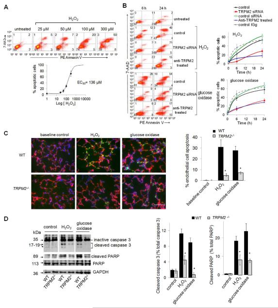
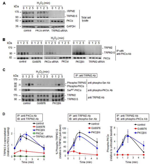
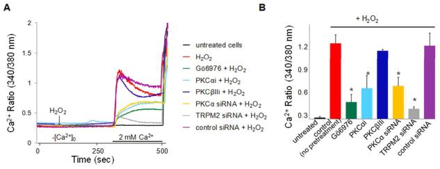
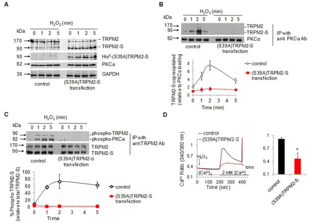
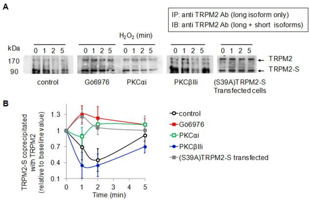
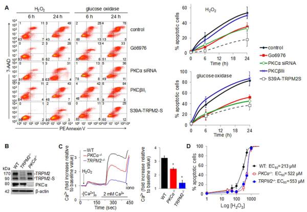
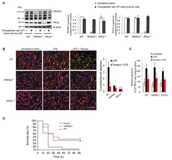
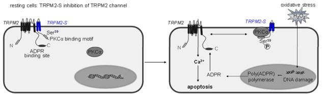
Methods and results: We observed that TRPM2-S was basally associated with TRPM2 in the endothelial plasmalemma, and this interaction functioned to suppress TRPM2-dependent Ca2+ gating constitutively. Reactive oxygen species production in endothelial cells or directly applying reactive oxygen species induced protein kinase C-α activation and phosphorylation of TRPM2 at Ser 39. This in turn stimulated a large entry of Ca2+ and activated the apoptosis pathway. A similar TRPM2-dependent endothelial apoptosis mechanism was seen in intact vessels. The protein kinase C-α-activated phosphoswitch opened the TRPM2 channel to allow large Ca2+ influx by releasing TRPM2-S inhibition of TRPM2, which in turn activated caspase-3 and cleaved the caspase substrate poly(ADP-ribose) polymerase.

Conclusions: Here, we describe a fundamental mechanism by which activation of the trp superfamily TRPM2 channel induces apoptosis of endothelial cells. The signaling mechanism involves reactive oxygen species-induced protein kinase C-α activation resulting in phosphorylation of TRPM2-S that allows enhanced TRPM2-mediated gating of Ca2+ and activation of the apoptosis program. Strategies aimed at preventing the uncoupling of TRPM2-S from TRPM2 and subsequent Ca2+ gating during oxidative stress may mitigate endothelial apoptosis and its consequences in mediating vascular injury and inflammation.

Keywords: apoptosis; capillary permeability; endothelium; inflammation.


Figures

Figure 1
Figure 1. TRPM2 is required for H2O2-mediated apoptosis of endothelial cells
(A-B) Confluent endothelial cell monolayers (HPAECs) challenged with H2O2 or glucose oxidase/glucose were labeled with PE Annexin V-FITC and 7-AAD and analyzed by flow cytometry. (A) Concentration-response curve for H2O2-induced apoptosis after 24-h period of H2O2 exposure. Mean apoptotic values (± SEM) obtained by flow cytometry (top panel) are plotted as % apoptotic cells vs. [H2O2] (EC50 = 136 μmol/L ; bottom panel) (n=3). (B) Left, representative flow cytometry histograms 6 or 24 h after exposure to 300 μmol/L H2O2 or glucose oxidase/glucose (90 min), with or without prior TRPM2 silencing or inhibition with blocking TRPM2 antibody. Right panel, mean percent apoptotic cells at 0, 6, or 24 h after H2O2 treatment (± SEM, n=3). Baseline values were not significantly altered by TRPM2 silencing or TRPM2 blocking Ab. (C-D) Apoptosis in lungs of TRPM2−/− and wild-type (WT) mice measured 3 h after perfusion with solution containing H2O2 (300 μmol/L) or glucose oxidase/glucose (75 min). (C) Right, immunofluorescent staining of frozen lung sections using VE-cadherin (red) and cleaved-PARP (green) antibodies and DAPI (blue)(n=3). Scale bar: 50 μm. Left, quantification of apoptotic endothelial cells (±SEM; n=6), * p ≤ 0.007 vs. WT lungs (t-test). (D) Western immunoblots (Right) and quantification of inactive or cleaved caspase-3 and active or cleaved PARP in lung homogenates (Left). GAPDH was used as loading control. (±SEM; n=3), * p ≤ 0.002, ** p ≤ 0.009 vs. WT perfused-lungs.
Figure 2
Figure 2. PKCα binding and phosphorylation of TRPM2-S
HPAECs were transduced with siRNA to suppress the expression of TRPM2 or PKCα or pretreated with PKC inhibitors (100 nmol/L Gö6976, 1 μmol/L PKCαi or 1 μmol/L PKCβIIi). Cells were challenged with 300 μmol/L H2O2 for the indicated times at 37°C. (A) Western blots from HPAEC lysates identifying expression of TRPM2 isoforms and PKCα; GAPDH was used as loading control. Endothelial cell expression of TRPM2-S (90 kDa) and two TRPM2 splice variants (140 and 171 kDa) were suppressed by TRPM2 silencing and PKCα protein expression (82 kDa) was blocked by PKCα silencing. (B) H2O2 induced association of PKCα and TRPM2-S. PKCα was immunoprecipitated from cell lysates. Following SDS-PAGE and electrophoretic transfer, co-immunoprecipitated TRPM2 was detected using an antibody recognizing both TRPM2 and TRPM2-S. TRPM2-S associated with PKCα immediately after H2O2 exposure whereas inhibition of PKCα activation prevented the association. (C) H2O2 induced PKCα-dependent phosphorylation of 90kDa TRPM2-S splice variant. TRPM2 was immunoprecipitated from same cell lysates using an Ab that recognized either TRPM2 isoform. Top panel: Blots showing phosphorylation of TRPM2 using monoclonal anti-phospho-Ser antibody. Membrane was reblotted with anti-phospho-PKCα (Ser 657) Ab to check the amount of phosphorylated PKCα in the complex (middle panel). Immunoprecipitation of TRPM2 was verified using an anti-TRPM2 Ab (lower panel). (D) Mean densitometric values (± SEM; n=3-4) obtained in B-C showing that PKCα inhibition prevented H2O2-induced association of PKCα with TRPM2-S and phosphorylation of TRPM2-S
Figure 3
Figure 3. PKCα and TRPM2 cooperation mediates Ca2+ entry in endothelial cells required for signaling apoptosis
(A) Ca2+ repletion transients generated by “Ca2+-add-back” in the presence of H2O2. Cultured HPAECs were loaded with Fura-2 Ca2+ dye, washed, and transferred to Ca2+-free medium. In control cells, H2O2 (100 μM) elicited a marked Ca2+ transient on Ca repletion (red trace). Ca2+ transients were blocked by TRPM2 silencing (grey trace) and reduced by Gö6976 (100 nmol/L; green trace), PKCαi (1 μmol/L; cyan trace), or after PKCα silencing (yellow trace); PKCβIIi (1 μmol/L, navy trace) and control siRNA (purple trace) had no effect. Ordinate gives [Ca2+]i as 340:380 nm ratio. (B) Summary of mean ratiometric data (± SEM) for the peak intracellular [Ca2+]i obtained in (E) (n = 3 to 5). *P ≤ 0.0002 vs. control (t-test).
Figure 4
Figure 4. PKCα binds TRPM2-S at Ser 39 and activates apoptosis-inducing Ca2+ entry signal in endothelial cells
The sole predicted PKCα phosphorylation site near the TRPM2-S N-terminus at Ser 39 was mutated by Ala substitution (resulting in phosphodefective mutant). HPAEC monolayers transduced with mutant TRPM2-S (tagged on its carboxy-terminal end with poly-His residues) were grown to confluence for Western blot analysis (A through C) or intracellular Ca2+ measurements using fura-2 (D). (A-C) Cells were exposed to 300 μM H2O2 for the indicated times. (A) Western blots for TRPM2, PKCα, and GAPDH expression in cells transduced with phosphodefective construct. Transfected protein was detected with an anti-His Ab confirming the expression of mutant TRPM2-S construct. (B) PKCα was immunoprecipitated from cell lysates with an antibody and co-immunoprecipitated TRPM2 protein was detected using an Ab recognizing both forms of TRPM2. Graph in B shows mean densitometric values (± SEM; n=3-4). Mutation of Ser 39 with Ala in TRPM2-S prevented TRPM2-S association with PKCα. (C) TRPM2 was immunoprecipitated from the same lysates and phosphorylated TRPM2 was detected using anti-phospho-Ser Ab. Graph in C shows mean densitometric values (± SEM; n=3-4). Ala substitution at Ser 39 abrogated H2O2-induced phosphorylation of TRPM2-S confirming the importance of the PKCα phosphorylation site on TRPM2-S at Ser 39. (D) Ca2+ mobilization assay was carried out using the “Ca2+ add-back” protocol. Transduction of phosphodefective TRPM2-S mutant suppressed H2O2-induced Ca2+ entry. * p = 0.0001 compared with control (t-test). (n = 3 per bar); error bars, ± SEM.
Figure 5
Figure 5. PKCα phosphorylation of TRPM2-S mediates TRPM2-S dissociation from TRPM2 resulting apoptosis-inducing Ca2+ entry signal
HPAEC monolayers were pretreated with PKCα and PKCβII (control) inhibitors 45 min prior to experiments. (A) TRPM2 was immunoprecipitated from cell lysates with an anti-TRPM2 antibody recognizing the region present only on the long isoform following H2O2 (300 μM) exposure for indicated times. Co-immunoprecipitated short isoform was then detected using an Ab that recognizes both TRPM2 and TRPM2-S. At 0 time, TRPM2 in plasma membrane was associated with its short isoform, and H2O2 application induced rapid dissociation of TRPM2-S from TRPM2. H2O2-mediated dissociation was suppressed by PKCα inhibition and did not occur when Ser 39 of TRPM2-S was substituted by Ala. (B) Western blots were quantified by densitomitry. Co-immunoprecipitated TRPM2-S was quantified as ratio to TRPM2 and plotted relative to zero time value (mean ± SEM; n = 3)..
Figure 6
Figure 6. H2O2-induced endothelial cell apoptosis resulting from TRPM2-mediated Ca2+ influx
HPAECs (A) and mouse lung endothelial cells (B-D) challenged with H2O2 were labeled with PE-Annexin/7-AAD. (A) Flow-cytometry histograms (Left) and summary plots of apoptosis (Right) 0, 6, and 24 h after challenge with H2O2 (300 μmol/L) or glucose oxidase/glucose to generate 320 nmol/L H2O2/min for 90 min, as a function of PKCα inhibition or silencing (± SEM, n=5). (B-D) Mouse endothelial cells were isolated from lungs of TRPM2−/−, PKCα−/−, and WT mice. (B) Western blot verifying absence of PKCα expression in PKCα−/− cells and of TRPM2 expression in TRPM2−/− cells. (C) Left, Ca2+ mobilization assay using “Ca2+ add-back” protocol with the Fluor-3 Ca2+ indicator. Right, Mean ratiometric values (± SEM) for steady-state [Ca2+]i (n=6). *p ≤ 0.0001 vs. WT cells (t-test). (D) Dose-response curve for H2O2-induced apoptosis in mouse endothelial cells detected by flow cytometry. Deletion of PKCα or TRPM2 caused ~2.5-fold rightward shift in the dose-response curve.
Figure 7
Figure 7. PKCα interaction with TRPM2 in mice is required lung endothelial cell apoptosis in response to LPS and contributes to mortality
WT, TRPM2−/− and PKCα−/− mice were transplanted with bone marrow cells isolated from WT mice 8 weeks prior to Western blotting (A), apoptosis (B-C) and survival (D) studies. (A) Representative Western blots verifying expression of TRPM2, PKCα, and β-actin in bone marrow of transplanted mice (molecular masses of 171, 82, and 45 kDa, respectively). Left, protein was quantified by densitometry. TRPM2 and PKCα densities were normalized to β-actin and plotted as percentage of untreated control (mean ± SEM for n=3). (B-C) Endothelial apoptosis (B) and oxidant production (C) were determined in lungs of mice 4h after intraperitoneal injection of lipopolysaccharide (LPS, 40 mg/kg) and in lungs of mice treated with the oxidant scavenger Tempol (100 mg/kg, IP) 30 min before injection of LPS. (B) Right, immunofluorescent staining of frozen lung sections using VE-cadherin (red) antibody, TUNEL (green), and DAPI (blue)(n=3). Scale bar: 50 μm. Left, quantification of apoptotic endothelial cells (±SEM; n=3), * p ≤ 0.001 and # p ≤ 0.005 vs. LPS-treated WT lungs. TRPM2 and PKCα deletion significantly reduced LPS-induced endothelial apoptosis. (C) Lungs were homogenized and assayed for H2O2 accumulation using the horseradish peroxide-linked Amplex Red assay. H2O2 was determined spectrophotometrically from its absorbance at 570 nM and corrected for total protein (± SEM, n=3). * p ≤ 0.005 vs. LPS-treated control lungs (D) Deletion of either TRPM2 or PKCα reduced LPS-induced lethality in mice. LPS (30 mg/kg) was injected intraperitoneally and survival was assessed every 12 h during the experiment (WT, n=16; TRPM2−/−, n= 16; and PKCα−/−, n=12). Statistical analysis was performed using the log-rank test. *p ≤ 0.03 and #p ≤ 0.05 vs. WT cells.
Figure 8
Figure 8. PKCα-induced phosphorylation of TRPM2-S mediates oxidant-induced TRPM2 channel opening and supra-normal Ca2+ entry required for activation of the apoptosis program
In resting cells, TRPM2-S associates with TRPM2 to restrict Ca2+ entry (the channel is in a closed conformation state). Oxidative stress induces PKCα activation resulting in binding and phosphorylation of TRPM2-S on Ser 39. TRPM2-S uncoupling from TRPM2 in turn opens the channel and Ca2+ entry resulting in activation of executioner caspases and endothelial apoptosis. In this model the generation of the endogenous TRPM2 agonist, ADPR may also contribute to opening TRPM2 channel.

References

    1. Aarts MM, Tymianski M. TRPMs and neuronal cell death. Pflugers Arch; 2005;451:243–249. - PubMed
    1. Nazıroğlu M. TRPM2 cation channels, oxidative stress and neurological diseases: Where are we now? Neurochem Res. 2011;36:355–366. - PubMed
    1. Nagamine K, Kudoh J, Minoshima S, Kawasaki K, Asakawa S, Ito F, Shimizu N. Molecular cloning of a novel putative Ca2+ channel protein (TRPC7) highly expressed in brain. Genomics. 1998;54:124–131. - PubMed
    1. Kraft R, Grimm C, Grosse K, Hoffmann A, Sauerbruch S, Kettenmann H, Schultz G, Harteneck C. Hydrogen peroxide and ADP-ribose induce TRPM2-mediated calcium influx and cation currents in microglia. Am J Physiol Cell Physiol. 2004;286:C129–C137. - PubMed
    1. Hecquet CM, Ahmmed GU, Vogel SM, Malik AB. Role of TRPM2 channel in mediating H2O2-induced Ca2+ entry and endothelial hyperpermeability. Circ Res. 2008;102:347–355. - PubMed

Publication types