. 2014 Jun;13(6):715-26.
doi: 10.1128/EC.00273-13. Epub 2013 Dec 13.

Role of the Apt1 protein in polysaccharide secretion by Cryptococcus neoformans

Affiliations

Role of the Apt1 protein in polysaccharide secretion by Cryptococcus neoformans

Juliana Rizzo et al. Eukaryot Cell. 2014 Jun.

Abstract

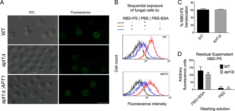
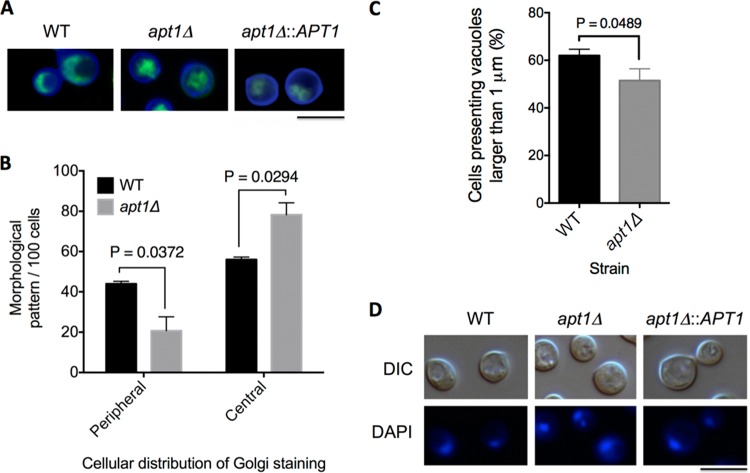
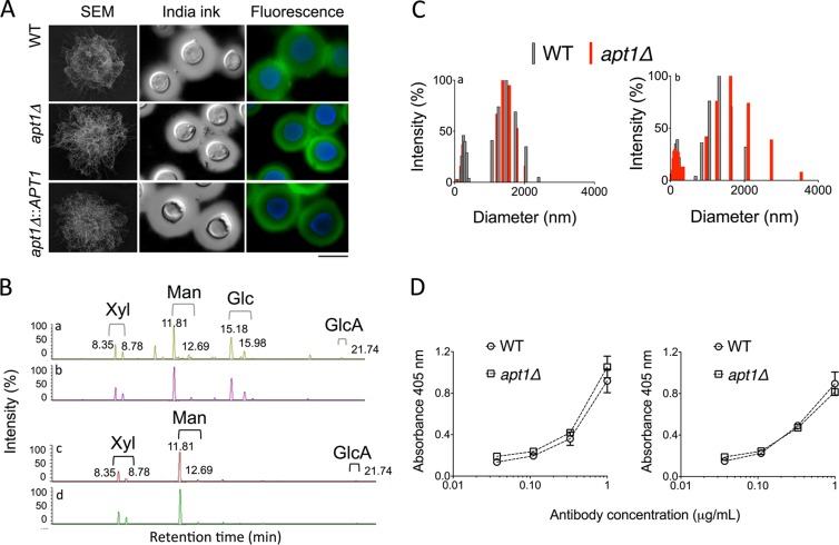
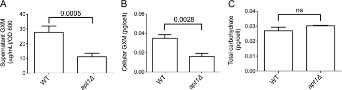
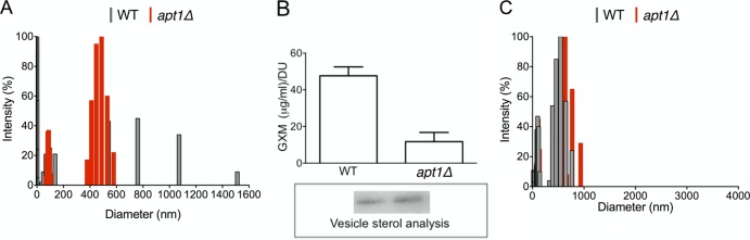
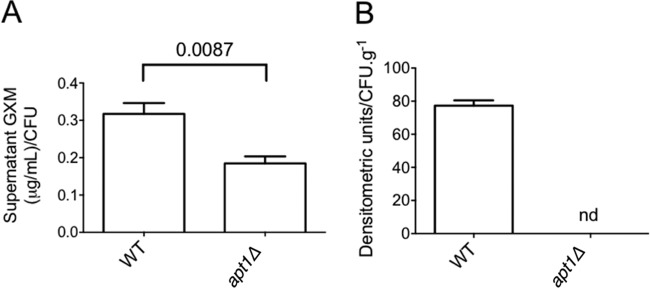
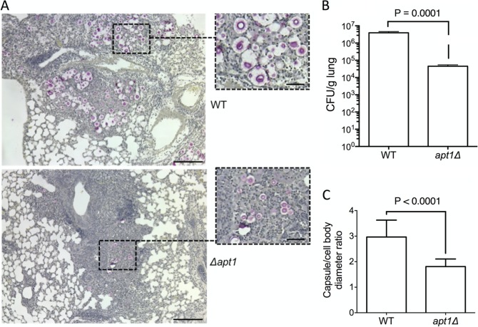
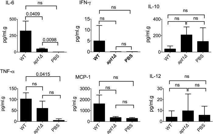
Flippases are key regulators of membrane asymmetry and secretory mechanisms. Vesicular polysaccharide secretion is essential for the pathogenic mechanisms of Cryptococcus neoformans. On the basis of the observations that flippases are required for polysaccharide secretion in plants and the putative Apt1 flippase is required for cryptococcal virulence, we analyzed the role of this enzyme in polysaccharide release by C. neoformans, using a previously characterized apt1Δ mutant. Mutant and wild-type (WT) cells shared important phenotypic characteristics, including capsule morphology and dimensions, glucuronoxylomannan (GXM) composition, molecular size, and serological properties. The apt1Δ mutant, however, produced extracellular vesicles (EVs) with a lower GXM content and different size distribution in comparison with those of WT cells. Our data also suggested a defective intracellular GXM synthesis in mutant cells, in addition to changes in the architecture of the Golgi apparatus. These findings were correlated with diminished GXM production during in vitro growth, macrophage infection, and lung colonization. This phenotype was associated with decreased survival of the mutant in the lungs of infected mice, reduced induction of interleukin-6 (IL-6) cytokine levels, and inefficacy in colonization of the brain. Taken together, our results indicate that the lack of APT1 caused defects in both GXM synthesis and vesicular export to the extracellular milieu by C. neoformans via processes that are apparently related to the pathogenic mechanisms used by this fungus during animal infection.


Figures

FIG 1
FIG 1
APT1 deletion does not affect the global flippase activity of C. neoformans. (A) Determination of phosphatidylserine (PS) exposure after treatment of fungal cells with FITC-annexin V reveals similar profiles of partial staining in wild-type (WT), mutant (apt1Δ), and complemented (apt1Δ::APT1) strains. Scale bar, 10 μm. (B) Analysis of the uptake of NBD-PS, a fluorescent analog of PS, by WT and mutant cells reveals that both strains were similarly efficient in incorporating the phospholipid derivative (red histograms) in comparison to unstained cells (black histograms). Treatment of C. neoformans cells with PBS-BSA for sequestration of NBD-PS molecules distributed into the external phospholipid layer of the plasma membrane resulted in cells with similar levels of fluorescence (blue histograms). (C and D) Accordingly, the levels of PS translocation were similar in WT and apt1Δ cells (C), as were the residual amounts of NBD-PS in the supernatants of cells that were washed with PBS alone or with PBS-BSA (D). P values resulting from the statistical comparison between WT and apt1Δ cells were higher than 0.5 in all cases.
FIG 2
FIG 2
Involvement of APT1 in morphological aspects of the Golgi apparatus in C. neoformans. (A) The Golgi apparatus of wild-type (WT), mutant (apt1Δ), and complemented (apt1Δ::APT1) cells was stained with C6-NBD-ceramide (green fluorescence), and the cell wall was stained with Uvitex 2B (blue fluorescence). (B) Quantitative analysis of the morphological profiles that predominated in WT and apt1Δ cells. (C) Quantification of intracellular vacuoles exceeding 1 μm in diameter in WT and mutant cells. (D) Analysis of nuclear morphology in WT, mutant, and complemented strains. C. neoformans cells are shown under differential interferential contrast (DIC) and fluorescence modes. Scale bar, 5 μm.
FIG 3
FIG 3
Morphological, structural, and serological analyses of capsular components in WT and Δapt1 cells of C. neoformans. (A) Morphological aspects of the capsule in WT, apt1Δ mutant, and complemented cells were visualized by scanning electron microscopy (SEM), India ink counterstaining, and fluorescence microscopy (green fluorescence, GXM; blue fluorescence, cell wall chitin). (B) GXM was isolated from C. neoformans WT (a) or mutant (b) cells or culture supernatants (c, WT cells; d, apt1Δ mutant) and analyzed by GC-MS. The chromatographic separation of monosaccharide components revealed no differences between fractions from WT and mutant cells. (C) Determination of molecular dimensions of cellular (a) or extracellular (b) polysaccharide fractions obtained from WT and apt1Δ cells reveals polysaccharide distributions in similar size ranges. (D) Serological tests with MAb 18B7 reveal that cellular (left) and extracellular (right) GXM fractions from WT and apt1Δ cells are similarly recognized by the antibody.
FIG 4
FIG 4
Lack of Apt1 results in attenuated GXM synthesis. (A) Quantification of supernatant GXM in cultures of WT and apt1Δ cells reveals a significantly decreased concentration of the polysaccharide in cultures of the mutant in comparison with the parental strain. (B) Similar results were obtained when cellular extracts were analyzed. (C) The defect in polysaccharide synthesis manifested by the mutant is apparently specific for GXM, since the total carbohydrate contents in both cells are similar. ns, not statistically significant (P > 0.05).
FIG 5
FIG 5
Lack of Apt1 affects C. neoformans EVs. (A) Diameter distribution of cryptococcal EVs. (B) Quantitative determination of vesicular GXM after sterol analysis (boxed area) of EVs produced by WT and mutant strains. DU, densitometry units used for normalization of GXM content to sterol concentration. The GXM concentration was significantly higher (P < 0.0001) in vesicles produced by WT cells. (C) Size determination of GXM fibers extracted from vesicles produced by WT and apt1Δ cells.
FIG 6
FIG 6
Apt1 is required for GXM secretion during infection of host cells. (A) Quantification of GXM in macrophage cultures by ELISA after infection with C. neoformans reveals a significantly decreased concentration of the polysaccharide when the phagocytes interact with apt1Δ mutant cells in comparison with the parental strain. (B) Soluble GXM is abundantly detected by dot blotting in lung macerates when mice are infected with WT C. neoformans cells. In systems where animals are infected with the apt1Δ mutant, GXM was not detected (nd).
FIG 7
FIG 7
Apt1 affects lung colonization, capsule formation, and host response during murine infection by C. neoformans. (A and B) The histopathology of mouse lungs after infection with WT or apt1Δ mutant cells (A) suggests a lower fungal burden when the Apt1-lacking cells are used for in vivo experimentation, which was confirmed by CFU determination (B). (C) Microscopic determination of capsule size (C) confirmed the supposition, based on visual analysis of higher-magnification fields, of reduced capsule formation in the mutant. Scale bars correspond to 200 μm (large panels) and 50 μm (insets). Data are representative of two experiments with similar results.
FIG 8
FIG 8
Cytokine (IL-6, IL-10 IL-12, IFN-γ, and TNF-α) and chemokine (MCP-1) determination in the lungs of mice infected with WT or apt1Δ mutant cells versus mice receiving PBS as controls. Statistical comparisons between the values obtained from the lungs of mice infected with WT or apt1Δ mutant cells revealed that only IL-6 was differentially induced in the two systems. ns, not significant (P > 0.05).

Similar articles

Cited by

References

    1. Ding Y, Wang J, Wang J, Stierhof YD, Robinson DG, Jiang L. 2012. Unconventional protein secretion. Trends Plant Sci. 17:606–615. 10.1016/j.tplants.2012.06.004 - DOI - PubMed
    1. Schekman RW. 1994. Regulation of membrane traffic in the secretory pathway. Harvey Lect. 90:41–57 - PubMed
    1. Lyman SK, Schekman R. 1996. Polypeptide translocation machinery of the yeast endoplasmic reticulum. Experientia 52:1042–1049. 10.1007/BF01952100 - DOI - PubMed
    1. Nickel W. 2010. Pathways of unconventional protein secretion. Curr. Opin. Biotechnol. 21:621–626. 10.1016/j.copbio.2010.06.004 - DOI - PubMed
    1. Rodrigues ML, Nakayasu ES, Almeida IC, Nimrichter L. 2014. The impact of proteomics on the understanding of functions and biogenesis of fungal extracellular vesicles. J. Proteomics 97:177–186. 10.1016/j.jprot.2013.04.001 - DOI - PMC - PubMed

Publication types

MeSH terms