. 2014 Jan 15;192(2):714-21.
doi: 10.4049/jimmunol.1302384. Epub 2013 Dec 11.

TNFR-associated factor 6 and TGF-β-activated kinase 1 control signals for a senescence response by an endosomal NK cell receptor

Affiliations

TNFR-associated factor 6 and TGF-β-activated kinase 1 control signals for a senescence response by an endosomal NK cell receptor

Sumati Rajagopalan et al. J Immunol. .

Abstract

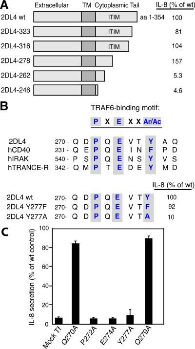
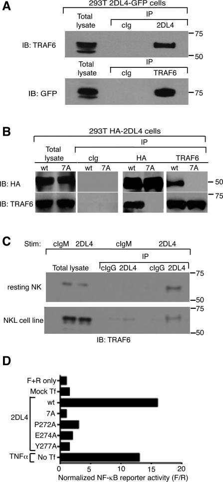
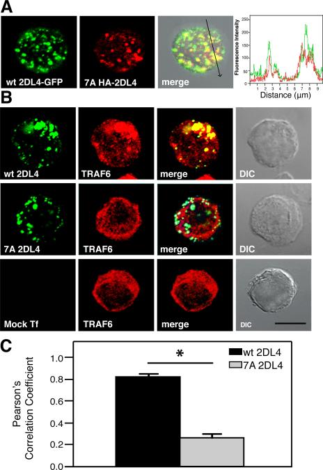
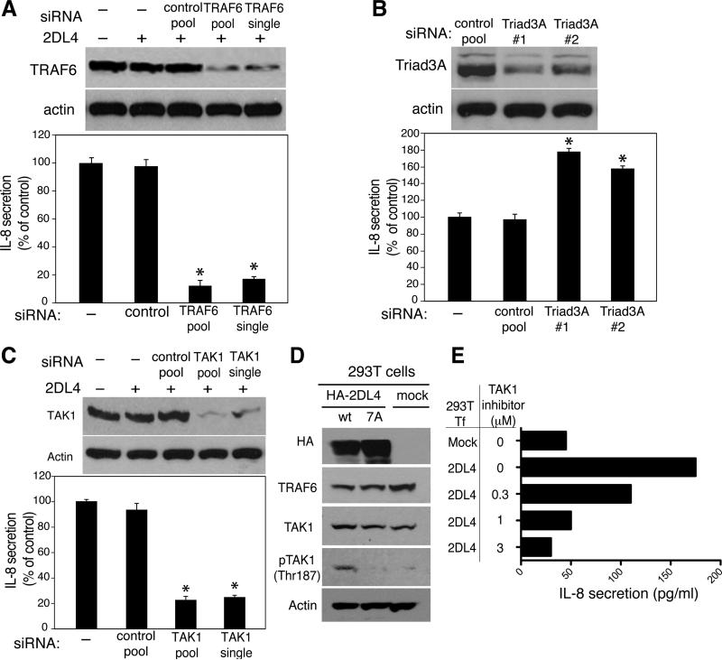
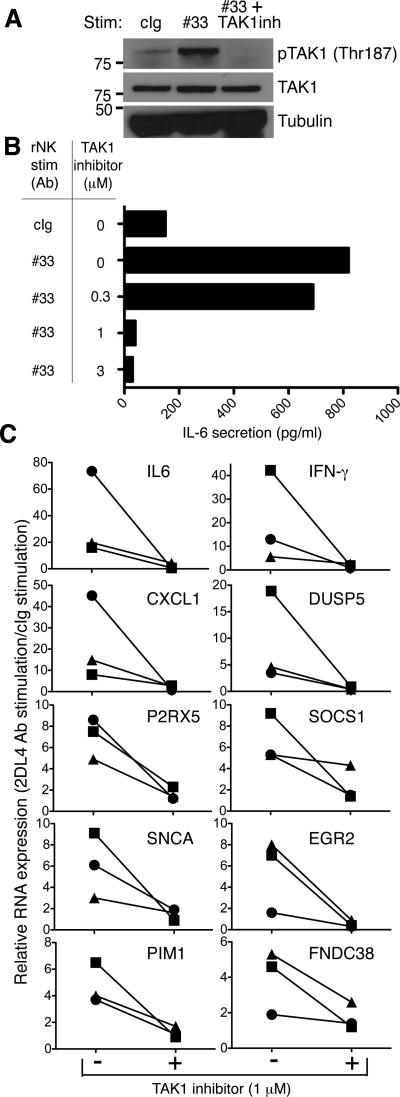
The endosomal innate receptor CD158d (killer cell Ig-like receptor 2DL4) induces cellular senescence in human NK cells in response to soluble ligand (HLA-G or agonist Ab). These senescent NK cells display a senescence-associated secretory phenotype, and their secretome promotes vascular remodeling and angiogenesis. To understand how CD158d initiates signaling for a senescence response, we mapped the region in its cytoplasmic tail that controls signaling. We identified a conserved TNFR-associated factor 6 (TRAF6) binding motif, which was required for CD158d-induced NF-κB activation and IL-8 secretion, TRAF6 association with CD158d, and TRAF6 recruitment to CD158d(+) endosomes in transfected cells. The adaptor TRAF6 is known to couple proximal signals from receptors such as endosomal TLRs and CD40 through the kinase TGF-β-activated kinase 1 (TAK1) for NF-κB-dependent proinflammatory responses. Small interfering RNA-mediated silencing of TRAF6 and TAK1, and inhibition of TAK1 blocked CD158d-dependent IL-8 secretion. Stimulation of primary, resting NK cells with soluble Ab to CD158d induced TRAF6 association with CD158d, induced TAK1 phosphorylation, and inhibition of TAK1 blocked the CD158d-dependent reprogramming of NK cells that produces the senescence-associated secretory phenotype signature. Our results reveal that a prototypic TLR and TNFR signaling pathway is used by a killer cell Ig-like receptor that promotes secretion of proinflammatory and proangiogenic mediators as part of a unique senescence phenotype in NK cells.


Figures

FIGURE 1
FIGURE 1. Mutagenesis of the 2DL4 cytoplasmic domain identifies critical residues for signaling
A, The 2DL4 cytoplasmic tail truncations, as indicated, were transfected into HEK293T cells. After 48 h, IL-8 secretion was measured by ELISA. IL-8 secreted after transfection of wt 2DL4 was 430 pg/ml. The data shown is representative of 2 to 4 experiments for each truncation. B, The TRAF6-binding motif P-X-E-X-X-Ar/Ac, (Ar, aromatic; Ac, acidic) in the 2DL4 cytoplasmic tail is aligned with similar motifs in human CD40, IRAK, and TRANCE-R. The aromatic Y277 in the 2DL4 cytoplasmic tail was mutated to either aromatic F or aliphatic A. IL-8 secretion was measured 48 h after transfection of HEK293T cells with 2DL4 and the Y277F or Y277A mutants. IL-8 secreted after transfection of wt 2DL4 was 1750 pg/ml. The data shown is representative of 2 experiments. C, Plasmids encoding point mutations of the indicated 2DL4 cytoplasmic tail residues were transfected into HEK293T cells. After 48 h, IL-8 secretion was measured by ELISA. Mock Tf: Mock-transfected cells. Results represent the mean ± SEM of triplicate samples from one representative experiment out of 3 experiments. IL-8 secreted after transfection of wt 2DL4 ranged from 888 to 1110 pg/ml.
FIGURE 2
FIGURE 2. A canonical TRAF6-binding motif in the 2DL4 cytoplasmic tail is required for NF-κB activation
A, TRAF6 associates with 2DL4. Cell lysates from HEK293T–2DL4-GFP cells were immunoprecipitated (IP) with control antibody (cIg), anti-2DL4 antibody, or anti-TRAF6 antibody, as indicated, and analyzed by immunoblotting (IB) with antibodies against TRAF6 and GFP. B, Mutagenesis of the TRAF6-binding region in the cytoplasmic tail of 2DL4 abrogates binding. Wild-type (wt), HA-tagged 2DL4 (HA-2DL4) and the 7A mutant (amino acids 271-277 replaced by alanine) were transfected in HEK293T cells. After 48 h, cell lysates were immunoprecipitated with control antibody (cIg), or antibodies against HA or TRAF6 followed by immunoblotting using anti-HA and anti-TRAF6 antibodies. C, TRAF6 association with 2DL4 in resting NK cells. Resting NK cells (top panel) or rested NKL cells (bottom panel) were stimulated with IgM Abs to 2DL4 and control IgM (cIgM) as indicated for 16 h and lysed. Lysates were immunoprecipitated as indicated with control Ab (cIgG) or anti-2DL4 IgG Ab and analyzed by immunoblotting with antibodies against TRAF6. D, NF-κB reporter activity in HEK293T cells transfected with either wt 2DL4 or the indicated 2DL4 mutants. Mock Tf: mock transfection. F+R: Firefly and Renilla luciferase constructs only. Stimulation of untransfected cells with TNF-α (10 ng/ml) during the final 5 hours of culture served as a positive control. These data are representative of 3 independent experiments.
FIGURE 3
FIGURE 3. TRAF6 colocalizes with endosomal 2DL4
A, The endocytosed 7A 2DL4 mutant colocalizes with wt 2DL4-gfp in HEK293T cells. Intact cells were incubated with anti-HA Alexa Fluor 594 at 37°C for 2 h, fixed and analyzed by confocal microscopy. The arrow in the third panel indicates the axis along which the fluorescence intensities shown in the fourth panel were traced. B, An intact TRAF6-binding domain is required for co-localization with endosomal 2DL4. HEK293T cells were mock-transfected (bottom panel) or transiently transfected with plasmids encoding either wt 2DL4 (top panel) or 7A 2DL4 (middle panel). Intact cells were incubated with anti-HA Alexa Fluor 488, fixed, permeabilized, stained with anti-TRAF6 antibody plus secondary antibody conjugated with Alexa Fluor 564, and analyzed by confocal microscopy. wt and 7A 2DL4 are shown in green, and endogenous TRAF6 is shown in red. Scale bar: 20 μM C, Pearson's Correlation Coefficient for co-localization of endogenous TRAF6 with wt or 7A 2DL4 in the transfected HEK293T cells. Results are presented as mean ± SEM of 12 cells transfected with WT 2DL4 and 10 cells transfected with 7A 2DL4 (*P<0.0001; Student's t-test).
FIGURE 4
FIGURE 4. TRAF6 and TAK1 silencing impairs 2DL4-mediated signaling
A, siRNA-mediated silencing of endogenous TRAF6 in HEK293T cells impairs 2DL4-mediated IL-8 secretion. Total cell lysates of HEK293T cells transfected with the indicated siRNAs and HA-2DL4 were analyzed for TRAF6 expression by immunoblotting. Actin was used as a loading control. IL-8 secretion by these cells was measured by ELISA. Values are mean ± SEM of two replicates per experimental group. The data shown are representative of at least eight independent experiments (*P<0.0006, compared with 2DL4-induced IL-8 secretion by cells transfected with negative control siRNA; Student's t-test). B, Triad3A silencing enhances IL-8 production by 2DL4 in HEK293T cells. Silencing of endogenous Triad3A in HEK293T cells was as described for TRAF6 and monitored by immunoblotting. IL-8 secretion by HEK293T cells was measured by ELISA 72 h post-transfection of Triad3A siRNA. Values are mean ± SEM of triplicate samples from two independent experiments. (*P<0.0001, compared with control siRNA; Student's t-test). C, Silencing of TAK1 in HEK293T cells by siRNA was monitored after 72 h by immunoblotting. IL-8 secretion was measured by ELISA after 72 h. Values are mean ± SEM of duplicate samples from two independent experiments. (*P=0.003, compared with control siRNA; Student's t-test). D, Lysates of HEK293T cells transfected with plasmids encoding wt HA-2DL4 or 7A HA-2DL4 for 48 h, were analyzed by immunoblotting, using the indicated antibodies. Actin was used as a loading control. E, TAK1 inhibitor (5Z-7-Oxozeanol) blocks 2DL4-mediated IL-8 secretion in HEK293T cells. 24 h after transfection with HA-2DL4, medium in the cultures was replaced with fresh medium containing 1 μM TAK1 inhibitor. 48 h later, supernatants were tested for IL-8 secretion by ELISA.
FIGURE 5
FIGURE 5. TAK1 inhibition in primary NK cells blocks the induction of SASP
A, Freshly isolated primary NK cells were treated with either control IgG (cIg) or agonist Ab to 2DL4 (#33) for 16 h. TAK1 inhibitor 5Z-7-Oxozeanol (as indicated) was added 30 min before stimulation of cells by Ab #33. Total cell lysates were blotted for phospho-TAK1, TAK1 and tubulin. Data is representative of that obtained from 3 independent donors. B, IL-6 secretion of supernatants from cells treated as described in A was measured by ELISA. C, qRT-PCR analysis of genes induced during the senescence response mediated by 2DL4. Fold increase in gene expression (2DL4 stimulation/cIg stimulation) in the presence or absence of TAK1 inhibitor in resting NK cells of 3 donors, after stimulation for 16 h. Each symbol (circle, square, triangle) represents data from the same donor. Different scales are used according to strength of gene induction.

Similar articles

Cited by

References

    1. Parham P, Moffett A. Variable NK cell receptors and their MHC class I ligands in immunity, reproduction and human evolution. Nat Rev Immunol. 2013;13:133–144. - PMC - PubMed
    1. Long EO, Sik Kim H, Liu D, Peterson ME, Rajagopalan S. Controlling natural killer cell responses: integration of signals for activation and inhibition. Annu Rev Immunol. 2013;31:227–258. - PMC - PubMed
    1. Rajagopalan S, Long EO. KIR2DL4 (CD158d): An activation receptor for HLA-G. Front Immunol. 2012;3:258. - PMC - PubMed
    1. Rajagopalan S, Bryceson YT, Kuppusamy SP, Geraghty DE, van der Meer A, Joosten I, Long EO. Activation of NK cells by an endocytosed receptor for soluble HLA-G. PLoS Biol. 2006;4:e9. - PMC - PubMed
    1. Rajagopalan S, Moyle MW, Joosten I, Long EO. DNA-PKcs controls an endosomal signaling pathway for a proinflammatory response by natural killer cells. Science Signaling. 2010;3:ra14. - PMC - PubMed

Publication types

MeSH terms