. 2014 Jan 15;192(2):589-602.
doi: 10.4049/jimmunol.1202802. Epub 2013 Dec 11.

IL-32 promotes angiogenesis

Affiliations

IL-32 promotes angiogenesis

Claudia A Nold-Petry et al. J Immunol. .

Abstract

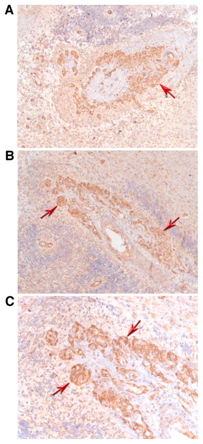
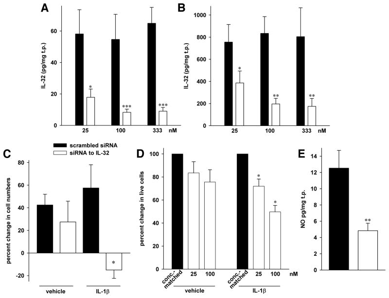
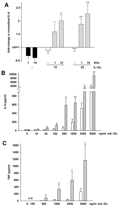
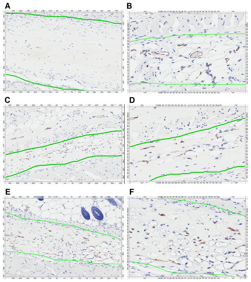
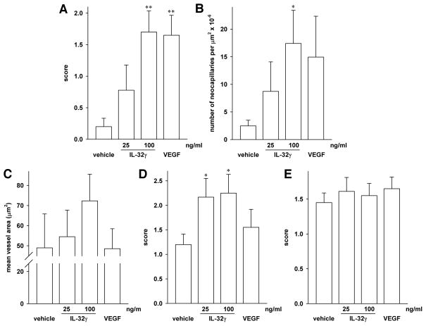
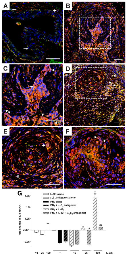
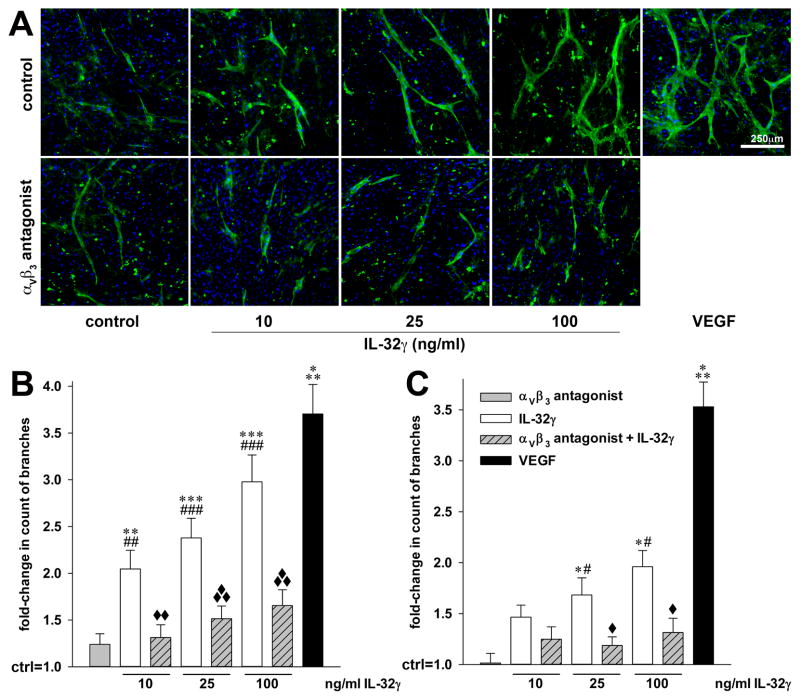
IL-32 is a multifaceted cytokine with a role in infections, autoimmune diseases, and cancer, and it exerts diverse functions, including aggravation of inflammation and inhibition of virus propagation. We previously identified IL-32 as a critical regulator of endothelial cell (EC) functions, and we now reveal that IL-32 also possesses angiogenic properties. The hyperproliferative ECs of human pulmonary arterial hypertension and glioblastoma multiforme exhibited a markedly increased abundance of IL-32, and, significantly, the cytokine colocalized with integrin αVβ3. Vascular endothelial growth factor (VEGF) receptor blockade, which resulted in EC hyperproliferation, increased IL-32 three-fold. Small interfering RNA-mediated silencing of IL-32 negated the 58% proliferation of ECs that occurred within 24 h in scrambled-transfected controls. Reduction of IL-32 neither affected apoptosis (insignificant changes in Bak-1, Bcl-2, Bcl-xL, lactate dehydrogenase, annexin V, and propidium iodide) nor VEGF or TGF-β levels, but siIL-32-transfected adult and neonatal ECs produced up to 61% less NO, IL-8, and matrix metalloproteinase-9, and up to 3-fold more activin A and endostatin. In coculture-based angiogenesis assays, IL-32γ dose-dependently increased tube formation up to 3-fold; an αVβ3 inhibitor prevented this activity and reduced IL-32γ-induced IL-8 by 85%. In matrigel plugs loaded with IL-32γ, VEGF, or vehicle and injected into live mice, we observed the anticipated VEGF-induced increase in neocapillarization (8-fold versus vehicle), but unexpectedly, IL-32γ was equally angiogenic. A second signal such as IFN-γ was required to render cells responsive to exogenous IL-32γ; importantly, this was confirmed using a completely synthetic preparation of IL-32γ. In summary, we add angiogenic properties that are mediated by integrin αVβ3 but VEGF-independent to the portfolio of IL-32, implicating a role for this versatile cytokine in pulmonary arterial hypertension and neoplastic diseases.


Figures

FIGURE 1
FIGURE 1
IL-32 in PAH. (A–D) Triple-label immunofluorescence images of human lung vessels. IL-32 is stained in red, vWF in green, and nuclei in blue (DAPI). Scale bar: 50 μm. (A) Staining of a normal pulmonary arteriole (arrow) from a patient not suffering from PAH. Image is representative for 3 similar results. (B–D) Specimen from one out of a total of 3 similar patients with idiopathic PAH. (B) An affected, but not obliterated arteriole with activated EC is depicted. Dotted lines indicate areas that are enlarged in insets on right. The arrowheads point to EC expression of IL-32. (C, D) A plexiform lesion is shown, in which the lumen of the blood vessel is obliterated by hyperproliferative EC. Dotted line indicates area that is enlarged in (D), and yet more detail is provided in insets on right. The arrowheads point to the hyperproliferative EC that stain positive for IL-32 and vWF. (E–H) Classical immunohistochemistry of IL-32 in lung specimen of PAH patients. After incubation with the primary antibody to IL-32, slides were stained with diaminobenzidine and hematoxylin. Diseased, hyperproliferative pulmonary blood vessels with a markedly thickened endothelium are shown in overview in panel E (100x magnification) and in detail in panels F–H (200x). The innermost endothelial layer contains a considerable amount of IL-32 protein (red arrows). Staining is also seen in alveolar epithelial cells (golden arrows). Inages are representative for n = 3 patients. (I) HUVEC were plated and grown for 24h. Thereafter, the medium was changed to stimulation medium (2% FCS, see Methods) and either semaxanib (10 μM) or vehicle was added. Cells were harvested after the indicated periods of time and lysates were assayed for IL-32 protein and total protein. IL-32 abundance was normalized to total protein, fold-increases in normalized IL-32 abundance were calculated (IL-32 in semaxanib-treated cells divided by IL-32 in vehicle-treated cells), and IL-32 abundance in vehicle-treated cells was set at 1. Each line indicates fold-change in semaxanib-treated cells over vehicle-treated cells in one time course experiment. n = 4; *, p < 0.05 for semaxanib vs vehicle.
FIGURE 2
FIGURE 2
IL-32 in glioblastoma multiforme (GBM). Immunohistochemistry with an antibody against IL-32 in sections of brains affected by GBM was performed and is depicted at an original magnification of 100x (A, B) or x 200 (C). Strong staining for IL-32 protein is observed in areas affected by the tumor (red arrows). Images are representative for those obtained from a total of three GBM patients.
FIGURE 3
FIGURE 3
Proliferation and NO abundance after silencing of IL-32 in HUVEC. All statistical comparisons are *, p < 0.05; **, p < 0.01; and ***, p < 0.001 for siIL-32 vs scrambled siRNA. (A, B) These panels are reproduced from (4), since the results in this Figure were obtained from the same lysates and supernatants as those in Figs. 4 and 5 in (4). Transfection of HUVEC with either the indicated concentrations of siIL-32 or scrambled siRNA was followed by stimulation with 10 ng/ml IL-1β (B) or vehicle (A) for 20h. Mean ± SEM of IL-32 abundance in cell lysates normalized to total protein (t.p.) is depicted; n = 10. (C) HUVEC were gently detached from the plates and viable cells were counted by hand using the trypan blue exclusion method. The number of cells that was seeded after transfection was set as baseline. Mean percent change in siIL-32-transfected cells compared to scr-transfected cells ± SEM is shown; n = 4. (D) After transfection with the indicated concentration of siIL-32 or the same concentration of scrambled siRNA, cells were plated, IL-1β (10 ng/ml) or vehicle was added, and the cultures were incubated for 3d. Thereafter, MTS assays were performed. The graph shows means of percent changes ± SEM in the number of live cells, comparing 25 or 100 nM siIL-32 to the appropriate concentration of scrambled siRNA, which is set at 100%; n = 6. (E) Supernatants from HUVEC transfected with 100 nM of siIL-32 or scrambled siRNA were collected after 48h and assayed for total nitrate/nitrite concentration. Mean concentration of total NO normalized to total protein (t.p.) ± SEM is depicted; n = 6.
FIGURE 4
FIGURE 4
IFNγ sensitizes cells to exogenous IL-32γ. (A) After the medium was changed from growth- to stimulation medium, HUVEC were treated with the indicated concentrations of IFNγ (in ng/ml) or vehicle for 24h. Thereafter, recombinant IL-32γ or vehicle was added at the concentrations indicated (ng/ml). IL-6 was measured in the culture supernatants after another 24h. The graph shows means of fold-changes in normalized IL-6 protein abundance comparing vehicle (control, set as 1) with stimulated conditions ± SEM; n = 7; *, p < 0.05 and **, p < 0.01 for IFNγ alone vs IL-32γ plus IFNγ. (B, C) PBMC were incubated with 10 ng/ml of IFNγ or vehicle for 24h, followed by addition of synthetic (s)IL-32γ at the indicated final concentrations. Supernatants were harvested 24h later and assayed for IL-6 (B) and TNF (C). Absolute concentrations of both cytokines ± SEM are depicted, n = 4 healthy donors. Grey bar (very left), medium alone; black bar (second from left), IFNγ alone; open bars, sIL-32γ alone; striped bars, sIL-32γ plus IFNγ. *, p < 0.05 and **, p < 0.01 for sIL-32γ alone vs sIL-32γ plus IFNγ. n.d., not detected.
FIGURE 5
FIGURE 5
IL-32γ induces angiogenesis in vivo. Growth factor-reduced high concentration matrigel was mixed with vehicle (A), 25 ng/ml VEGF (B), or recombinant IL-32γ at 25 ng/ml (C, D) or at 100 ng/ml (E, F); n = 10 plugs for each condition. Two 200 μl aliquots of matrigel with non-identical contents were then injected on the right and left sides of the abdominal wall of male ICR mice. The plugs were harvested 14d later, followed by cutting and staining for CD31 (chromagens were diaminobenzidine and hematoxylin). The coworkers involved in the analysis of the matrigel experiments were blinded to the reagent with which each individual plug was loaded. Blinding was lifted only after the completion of all aspects of the analysis. Representative plugs loaded with vehicle (A, 10x magnification), VEGF at 25 ng/ml (B, 20x magnification), or IL-32γ at 25 ng/ml (C, D at 10x and 20x magnification, respectively) or 100 ng/ml (E, F at 10x and 20x magnification, respectively) are depicted. The green line highlights the matrigel plugs, differentiating them from the surrounding tissue, which was left in situ.
FIGURE 6
FIGURE 6
Quantitative analysis of neocapillarization, lipoidosis, and influx of inflammatory cells into the matrigel plugs. After cutting and CD31- as well as H&E staining, the slides of the matrigel plugs (n = 10 per group) were assessed for the formation of capillaries (A, B) and for the surface area of these capillaries (C), as well as for the presence of adipocytes (D) and white blood cells (E). (A) Neocapillarization was determined by hand-counting and scored semi-quantitatively with 0 representing no increase in neocapillarization, 1 minimal increase, 2 mild increase, 3 moderate increase, and 4 marked increase in neocapillarization. Mean score ± SEM is shown; **, p < 0.01 for IL-32γ or VEGF vs vehicle. (B) The same slides were assessed by automated analysis using Aperio’s microvessel algorithm. The graph depicts means of absolute numbers of microvessels per μm2 × 10−6 ± SEM; *, p < 0.05 for IL-32γ vs vehicle. (C) Computer-based analysis of the surface area of the capillaries in the plugs. Means of the surface area of the microvessels on each slide ± SEM are shown. (D, E) The same semi-quantitative scoring system as in (A) was used to categorize the degree of lipoidosis (D) and inflammation (E). Mean score ± SEM is shown; *, p < 0.05 for IL-32γ vs vehicle.
FIGURE 7
FIGURE 7
Colocalization of integrin αVβ3 with IL-32 and functional relevance of the interaction. (A–F) Representative optical sections acquired by confocal microscopy of double immunofluorescence staining for IL-32 (red) and αVβ3 (green). Nuclei are counterstained with DAPI (blue). (A) In lung tissue from a representative control patient (without pulmonary vascular disease) single αVβ3+ cells are seen in the endothelial layer or perivascular region of a pulmonary artery, indicated by double-headed arrow. One αVβ3+/IL-32+ cell (single-headed arrow) was found in the perivascular tissue (cytoplasmic staining). (B–F) In lung tissue from a representative patient, αVβ3 colocalized with IL-32 in the plexiform lesions. This colocalization occurred in the nucleus, as indicated by arrowheads, as well as in the cytoplasm and the cell membrane, pointed at by arrows. Cells positive for IL-32 alone are indicated with open arrows. (C and E) depict the regions enclosed by the dotted line in (B and D, respectively) at higher magnification. Original magnification was 400x. Scale bar: 50 μm. n = 3 controls and 3 PAH patients. (G) HUVEC were pretreated with IFNγ (10 ng/ml) or vehicle for 24h, followed by addition of recombinant IL-32γ at the indicated concentrations (ng/ml) and/or the αVβ3 inhibitor cyclo [Arg-Gly-Asp-D-Phe-Val] at 10 μM. Six hours thereafter, cells were lysed and subjected to real-time PCR analysis. Fold-changes in abundance of IL-8 mRNA normalized to 18S over control (which is set at 1) ± SEM are depicted. n = 3; *, p < 0.05 and ***, p < 0.001 for IFNγ alone vs IL-32γ plus IFNγ; #, p < 0.05 and ##, p < 0.01 for IL-32γ plus IFNγ vs IL-32γ plus IFNγ plus cyclo [Arg-Gly-Asp-D-Phe-Val].
FIGURE 8
FIGURE 8
IL-32γ-induced in vitro EC tube formation requires functional αVβ3. Human dermal fibroblasts were grown to confluence for 3d, followed by careful addition of HUVEC or HAoEC. Four hours later, treatment with the indicated concentrations of recombinant IL-32γ and/or 10 μM of the αVβ3 inhibitor cyclo [Arg-Gly-Asp-D-Phe-Val] was commenced. Treatment with recombinant human VEGF-165 (40 ng/ml) was used as internal assay control. On day 7 of co-culture, newly formed EC tubes were stained with sambuccus nigra lectin-FITC (green), nuclei were labeled with DAPI (blue), and the cells were then examined microscopically. Five fields of view per condition were randomly chosen and photographed. (A) One represesentative image of five independently performed HAoEC experiments is shown. (B, C) The number of visible branches in the co-cultures containing HAoEC (B) or HUVEC (C) was counted using ImageJ. Graphs illustrate the fold-changes in the number of branches of the newly formed EC tubes over vehicle-stimulated co-cultures (which are set as 1). *, p < 0.05, **, p < 0.01, and ***, p < 0.001 for IL-32γ or VEGF vs control; #, p < 0.05, ##, p < 0.01, and ###, p < 0.001 for IL-32γ vs the αVβ3 inhibitor; and ◆, p < 0.05, ◆◆, p < 0.01, and ◆◆◆, p < 0.001 for IL-32γ vs IL-32γ plus the αVβ3 inhibitor.

Similar articles

Cited by

References

    1. Kim SH, Han SY, Azam T, Yoon DY, Dinarello CA. Interleukin-32: a cytokine and inducer of TNFalpha. Immunity. 2005;22:131–142. - PubMed
    1. Goda C, Kanaji T, Kanaji S, Tanaka G, Arima K, Ohno S, Izuhara K. Involvement of IL-32 in activation-induced cell death in T cells. Int Immunol. 2006;18:233–240. - PubMed
    1. Imaeda H, Andoh A, Aomatsu T, Osaki R, Bamba S, Inatomi O, Shimizu T, Fujiyama Y. A new isoform of interleukin-32 suppresses IL-8 mrna expression in the intestinal epithelial cell line ht-29. Mol Med Report. 2011;4:483–487. - PubMed
    1. Nold-Petry CA, Nold MF, Zepp JA, Kim SH, Voelkel NF, Dinarello CA. IL-32-dependent effects of IL-1{beta} on endothelial cell functions. Proc Natl Acad Sci U S A 2009 - PMC - PubMed
    1. Heinhuis B, Netea MG, van den Berg WB, Dinarello CA, Joosten LA. Interleukin-32: A predominantly intracellular proinflammatory mediator that controls cell activation and cell death. Cytokine 2012 - PubMed

Publication types

MeSH terms