. 2013 Dec 11;5(215):215ra173.
doi: 10.1126/scitranslmed.3007092.

Dietary therapy mitigates persistent wake deficits caused by mild traumatic brain injury

Affiliations

Dietary therapy mitigates persistent wake deficits caused by mild traumatic brain injury

Miranda M Lim et al. Sci Transl Med. .

Abstract

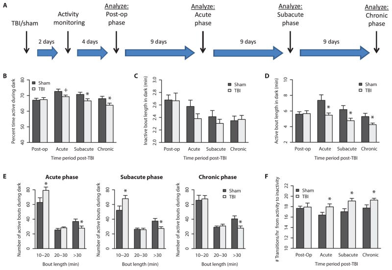
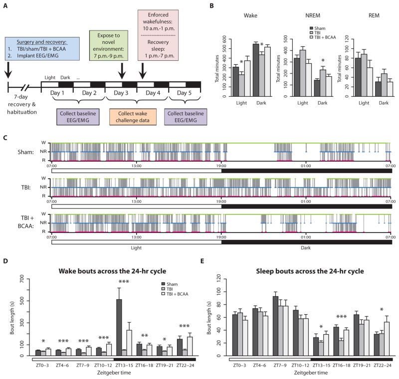
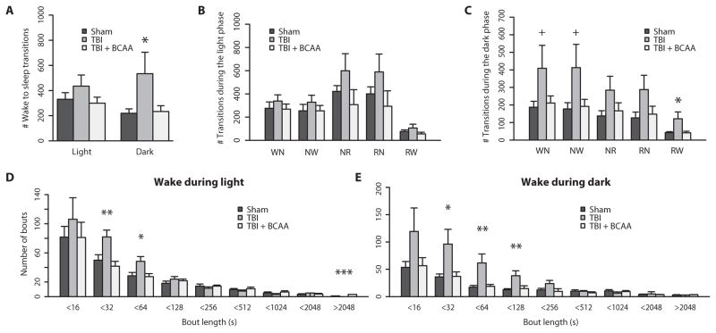
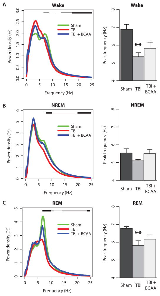
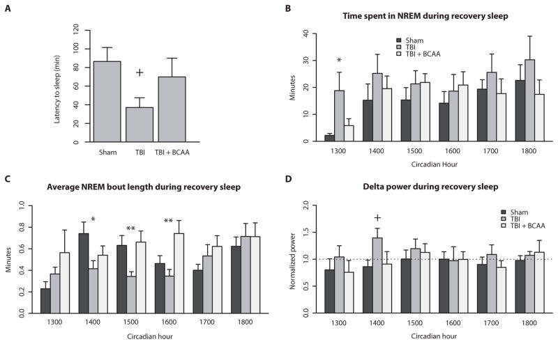
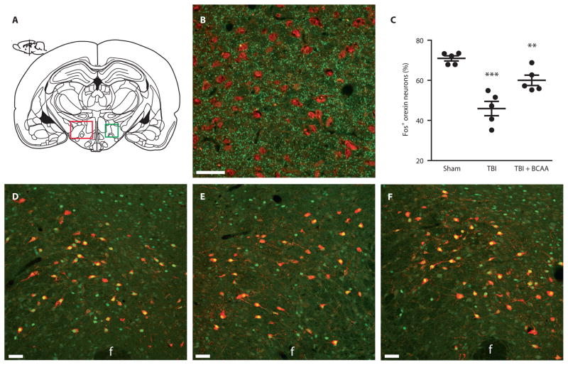
Sleep disorders are highly prevalent in patients with traumatic brain injury (TBI) and can significantly impair cognitive rehabilitation. No proven therapies exist to mitigate the neurocognitive consequences of TBI. We show that mild brain injury in mice causes a persistent inability to maintain wakefulness and decreases orexin neuron activation during wakefulness. We gave mice a dietary supplement of branched-chain amino acids (BCAAs), precursors for de novo glutamate synthesis in the brain. BCAA therapy reinstated activation of orexin neurons and improved wake deficits in mice with mild brain injury. Our data suggest that dietary BCAA intervention, acting in part through orexin, can ameliorate injury-induced sleep disturbances and may facilitate cognitive rehabilitation after brain injury.


Conflict of interest statement

Competing interests: A.S.C. and the Children’s Hospital of Philadelphia hold a provisional patent for the use of BCAAs as a therapeutic intervention for traumatic brain injury: U.S. Provisional Patent Application Nos. 61/883,526 and 61/812,352, filed under the title “Compositions and methods for the treatment of brain injury.”

Figures

Fig. 1
Fig. 1. Mild TBI decreased overall activity and fragmented activity patterns during dark phase
(A) Activity monitoring timeline. Activity patterns were binned into post-op, acute, subacute, and chronic time points after injury (n = 18) or sham (n = 12) surgery. (B) The percent time spent active during the dark phase was decreased across acute, subacute, and chronic periods after TBI. (C) The average length of each inactivity bout did not significantly differ between groups. (D) However, the average length of each activity bout was significantly decreased after TBI across the three time points. (E) Active bouts longer than 30 min were particularly affected after TBI across all three phases. (F) The number of transitions from active to inactive bouts was significantly increased after TBI across the three time points. +P < 0.1, *P < 0.05, Student’s two-tailed independent t tests. See table S1 for detailed statistics.
Fig. 2
Fig. 2. Baseline EEG recording over 24 hours showed alterations in wake/sleep patterns, which were ameliorated by BCAA treatment
(A) Experimental timeline for EEG/EMG recording. (B) TBI mice (n = 6) spent less time awake and more time in NREM sleep compared to sham mice (n = 7) and TBI + BCAA mice (n = 6). (C) Representative hypnograms from sham, TBI, and TBI + BCAA mice. Wake, green; NREM, blue; and REM, red. Note the absence of the long wake period (green) at 7:00 p.m. (lights off) in the TBI mouse, which was restored in the TBI + BCAA mouse. (D) Distribution of wake bout length over the circadian cycle showed that TBI significantly shortened wake bouts throughout the light and dark phases, and the normal diurnal fluctuation in wake bout length was abolished. (E) Distribution of sleep (NREM + REM) bout length over the circadian cycle showed significantly shorter sleep bouts in TBI mice during the dark phase. ZT0–3 = 7:00 a.m. to 10:00 a.m., ZT13–15 = 7:00 p.m. to 10:00 p.m., and so forth. *P < 0.05, **P < 0.01, ***P < 0.001, one-way analysis of variance (ANOVA) with Dunnett’s post hoc test. See table S2 for detailed statistics.
Fig. 3
Fig. 3. TBI causes behavioral state instability, which is restored by BCAA therapy
(A) TBI significantly increased the total number of wake to sleep (NREM + REM) transitions during the dark or active phase. (B) Transitions subcategorized as Wake to NREM (WN), NREM to Wake (NW), NREM to REM (NR), REM to NREM (RN), and REM to Wake (RW) during the light phase. (C) Transition subcategories during the dark phase. TBI mice had more transitions to and from Wake (WN, NW, RW), and this was ameliorated by BCAA therapy. Also, group differences were more robust in the dark phase compared to the light phase. (D and E) TBI mice had significantly more short wake bouts compared to sham mice, and BCAA therapy restored the distribution of long wake bouts. Group differences in wake bout lengths were more robust in the dark phase compared to the light phase. +P < 0.1, *P < 0.05, one-way ANOVA with Dunnett’s post hoc test. See table S3 for detailed statistics.
Fig. 4
Fig. 4. TBI alters baseline power spectra and theta peak frequency
(A) Wake power spectra. Statistical group differences denoted as a black bar above the power spectral curves from 7 to 24 Hz. Note the loss of the theta peak (5 to 8 Hz) after TBI. (B) NREM power spectra showing a similar flattening of the theta peak after TBI. Group differences are denoted as a black bar above the power spectral curves from 7 to 24 Hz. (C) REM power spectra showing the decreased theta peak after TBI. Group differences are denoted as a black bar above the power spectral curves ranging from 7 to 24 Hz. Green, sham; red, TBI; blue, TBI + BCAA. Thin black bar, P < 0.10; medium black bar, P < 0.05; thick black bar, P < 0.01. **P < 0.01, one-way ANOVA with Dunnett’s post hoc test. See table S4 for detailed statistics.
Fig. 5
Fig. 5. TBI mice exhibit a higher sleep pressure during situations challenging wakefulness
(A) After being placed in a novel environment, TBI mice showed a shorter latency to the first sleep episode, which was reinstated with BCAA therapy. (B) After a relatively short 3-hour period of sleep deprivation, TBI mice spent more time in NREM sleep during the first hour of the recovery period. BCAA therapy decreased NREM sleep back to sham levels. (C) Despite greater NREM sleep time, average NREM bout length was significantly shorter in TBI compared to sham mice, indicating sleep fragmentation. BCAA therapy lengthened the average NREM bout particularly in hours 3 and 4 of recovery sleep. (D) Delta power, a proxy for sleep pressure, was increased in TBI mice during hour 2 of recovery sleep compared to sham and TBI + BCAA mice. +P < 0.10, *P < 0.05, **P < 0.01, one-way ANOVA with Dunnett’s post hoc test. See table S7 for detailed statistics.
Fig. 6
Fig. 6. Orexin neuron activation after 3 hours of enforced wakefulness was decreased after TBI, and was restored with BCAA therapy
(A) Schematic coronal section of a mouse brain showing the lateral hypothalamus (LH, red box), the region where orexin neurons reside and where orexin cells were counted. The green box represents the area depicted in photomicrographs in (D) to (F). (B) Photomicrograph of the lateral hypothalamus showing presynaptic glutamate vesicles (VGLUT1 immuno-labeling, green) in close proximity to orexin cell bodies (orexin-A immuno-labeling, red) at ×40 magnification. (C) Quantification of orexin neurons expressing c-Fos, a marker of neural activation, after the 3-hour wake challenge. There was decreased orexin activation after TBI compared to sham mice, which was restored with BCAA therapy (F = 22.47, P < 0.0001; TBI versus sham P < 0.001, TBI versus TBI + BCAA P < 0.01; one-way ANOVA followed by Dunnett’s post hoc test; n = 5 per group). (D to F) Representative photomicrographs of the lateral hypothalamus from sham, TBI, and TBI + BCAA mice showing orexin (red) colocalization with c-Fos (green) at ×20 magnification. F, fornix. Scale bars, 50 μm. **P < 0.01, ***P < 0.001.

References

    1. Castriotta RJ, Murthy JN. Sleep disorders in patients with traumatic brain injury: A review. CNS Drugs. 2011;25:175–185. - PubMed
    1. Castriotta RJ, Wilde MC, Lai JM, Atanasov S, Masel BE, Kuna ST. Prevalence and consequences of sleep disorders in traumatic brain injury. J Clin Sleep Med. 2007;3:349–356. - PMC - PubMed
    1. Baumann CR, Werth E, Stocker R, Ludwig S, Bassetti CL. Sleep-wake disturbances 6 months after traumatic brain injury: A prospective study. Brain. 2007;130:1873–1883. - PubMed
    1. Kempf J, Werth E, Kaiser PR, Bassetti CL, Baumann CR. Sleep-wake disturbances 3 years after traumatic brain injury. J Neurol Neurosurg Psychiatry. 2010;81:1402–1405. - PubMed
    1. Makley MJ, English JB, Drubach DA, Kreuz AJ, Celnik PA, Tarwater PM. Prevalence of sleep disturbance in closed head injury patients in a rehabilitation unit. Neurorehabil Neural Repair. 2008;22:341–347. - PubMed

Publication types