. 2014 Jan 24;289(4):2250-9.
doi: 10.1074/jbc.M113.523654. Epub 2013 Dec 11.

A role for peroxisome proliferator-activated receptor γ coactivator 1 (PGC-1) in the regulation of cardiac mitochondrial phospholipid biosynthesis

Affiliations

A role for peroxisome proliferator-activated receptor γ coactivator 1 (PGC-1) in the regulation of cardiac mitochondrial phospholipid biosynthesis

Ling Lai et al. J Biol Chem. .

Abstract

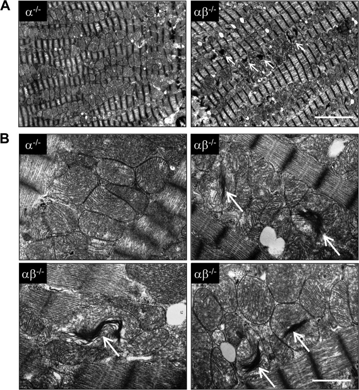
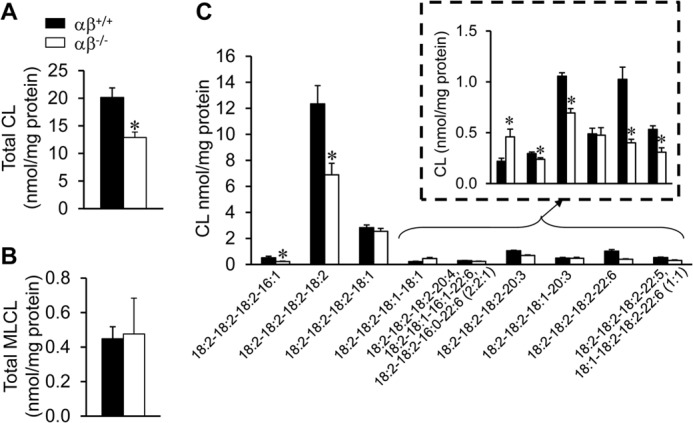
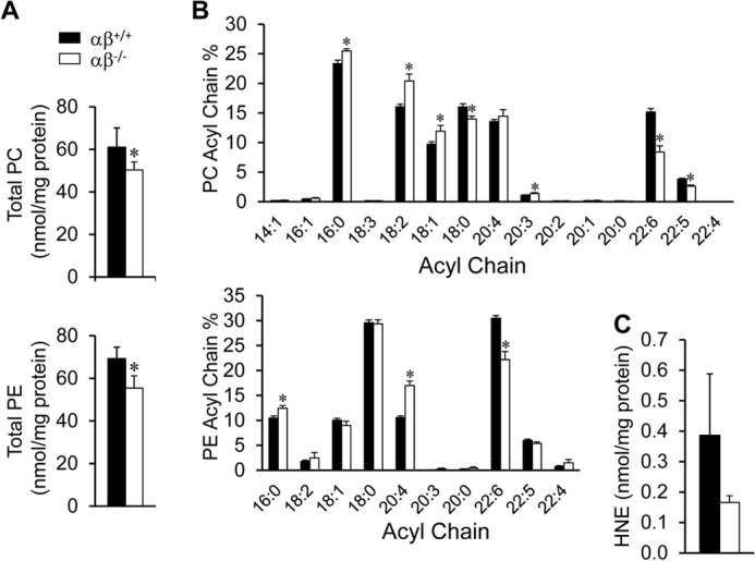
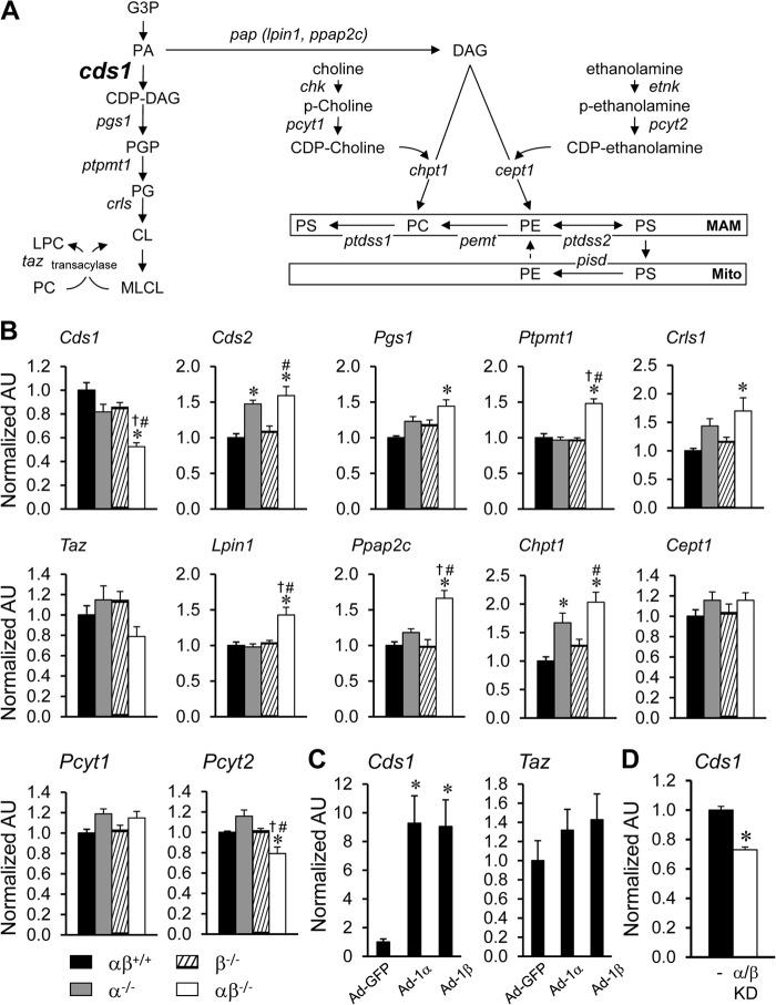
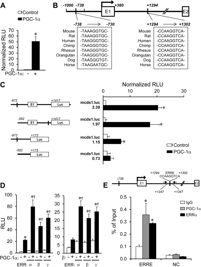
The energy demands of the adult mammalian heart are met largely by ATP generated via oxidation of fatty acids in a high capacity mitochondrial system. Peroxisome proliferator-activated receptor γ coactivator 1 (PGC-1)-α and -β serve as inducible transcriptional coregulators of genes involved in mitochondrial biogenesis and metabolism. Whether PGC-1 plays a role in the regulation of mitochondrial structure is unknown. In this study, mice with combined deficiency of PGC-1α and PGC-1β (PGC-1αβ(-/-)) in adult heart were analyzed. PGC-1αβ(-/-) hearts exhibited a distinctive mitochondrial cristae-stacking abnormality suggestive of a phospholipid abnormality as has been described in humans with genetic defects in cardiolipin (CL) synthesis (Barth syndrome). A subset of molecular species, containing n-3 polyunsaturated species in the CL, phosphatidylcholine, and phosphatidylethanolamine profiles, was reduced in PGC-1αβ-deficient hearts. Gene expression profiling of PGC-1αβ(-/-) hearts revealed reduced expression of the gene encoding CDP-diacylglycerol synthase 1 (Cds1), an enzyme that catalyzes the proximal step in CL biosynthesis. Cds1 gene promoter-reporter cotransfection experiments and chromatin immunoprecipitation studies demonstrated that PGC-1α coregulates estrogen-related receptors to activate the transcription of the Cds1 gene. We conclude that the PGC-1/estrogen-related receptor axis coordinately regulates metabolic and membrane structural programs relevant to the maintenance of high capacity mitochondrial function in heart.

Keywords: Cardiac Metabolism; Cardiolipin; Gene Regulation; Heart; Lipidomics; Mitochondria; Phospholipids; Polyunsaturated Fatty Acids; Transcriptional Coregulators.


Figures

FIGURE 1.
FIGURE 1.
Mitochondrial ultrastructural abnormalities in hearts of adult PGC-1αβ−/− mice. Representative electron micrographs of sections taken from the left ventricular papillary muscle from 16-week-old PGC-1α−/−−/−) and PGC-1αβ−/− (αβ−/−) mice at two different magnifications. Arrows indicate the structurally abnormal mitochondria with “stacked” cristae (white arrows). Scale bar for each magnification is shown at the bottom right corner: A, 1 μm; B, 5 μm.
FIGURE 2.
FIGURE 2.
Cardiolipin deficiency in adult PGC-1αβ−/− hearts. Total CL (A) and MLCL (B) levels in 12-week-old wild-type (αβ+/+) and PGC-1αβ−/− (αβ−/−) hearts. C, quantitative analysis of representative CL molecular species determined in hearts of two genotypes as denoted. Bars represent mean ± S.D. *, p < 0.05 compared with αβ+/+.
FIGURE 3.
FIGURE 3.
Abnormal PC and PE profiles in PGC-1αβ−/− hearts. A, total PC and PE analyzed from 12-week-old wild-type (αβ+/+) and PGC-1αβ−/− (αβ−/−) hearts. B, percentage of the fatty acyl chain groups in PC and PE. C, HNE content. Bars represent mean ± S.D. *, p < 0.05 compared with αβ+/+.
FIGURE 4.
FIGURE 4.
Altered expression of phospholipid biosynthesis pathway genes in adult PGC-1αβ−/− mice. A, schematic representation of de novo PL biosynthesis and remodeling pathways in mammalian cells. B, results of qRT-PCR analysis of RNA extracted from hearts of 12-week-old wild-type (αβ+/+), PGC-1α−/−−/−), PGC-1β−/−−/−), and PGC-1αβ−/− (αβ−/−) mice for the following: CDP-DAG synthase (Cds); phosphatidylglycerophosphate synthase 1 (Pgs1); protein-tyrosine phosphatase, mitochondrial 1 (Ptpmt1); cardiolipin synthase (Crls); tafazzin (Taz); lipin 1 (Lpin1); phosphatidic acid phosphatase type 2C (Chptc); choline phosphotransferase 1 (Chpt1); choline/ethanolaminephosphotransferase 1 (Cept1); phosphate cytidylyltransferase 1; choline (Pcyt1); and phosphate cytidylyltransferase 2, ethanolamine (Pcyt2). Bars represent mean ± S.E. *, p < 0.05 compared with αβ+/+; †, p < 0.05 compared with α−/−; and #, p < 0.05 compared with β−/−. C, quantitative analysis of RNA extracted from cultured NRCMs infected with Ad-PGC-1α (Ad-1α) and Ad-PGC-1β (Ad-1β) adenovirus. Bars represent mean ± S.E. *, p < 0.05 compared with NRCMs infected with Ad-GFP control. D, qRT-PCR analysis of RNA extracted from cultured NRCMs infected with Ad-shPGC-1α and Ad-shPGC-1β (PGC-1α/β knockdown (KD)) adenovirus. Bars represent mean ± S.E. *, p < 0.05 compared with NRCMs infected with Ad-control (−). Additional abbreviations used are as follows: MAM, mitochondrion-associated endoplasmic reticulum membrane; Mito, mitochondria; G3P, glycerol 3-phosphate; PA, phosphatidic acid, CDP-DAG, CDP-diacylglycerol; PGP, phosphatidylglycerolphosphate; PG, phosphatidylglycerol; CL, cardiolipin; MLCL, monolysocardiolipin; LPC, lysophosphatidylcholine; PAP, phosphatidic acid phosphatase; DAG, diacylglycerol; Chk, choline kinase; p-choline, phosphorylcholine; Etnk, ethanolamine kinase; p-ethanolamine, phosphorylethanolamine; PS, phosphatidylserine; PC, phosphatidylcholine; PE, phosphatidylethanolamine; Ptdss, phosphatidylserine synthase; Pemt, phosphatidylethanolamine N-methyltransferase; Pisd, phosphatidylserine decarboxylase; AU, arbitrary units.
FIGURE 5.
FIGURE 5.
Transcriptional activation of the Cds1 gene by PGC-1/ERR. A, transient transfections were performed in C2C12 myoblasts (differentiated into myotubes) using a mouse Cds1 promoter reporter construct, mcds1.luc.2.39, containing a putative ERR-binding site at −738 and +1294, as denoted in B, cotransfected with pcDNA3.1-PGC-1α construct (+) compared with vector backbone alone (−). The bars represent mean ± S.E. promoter-reporter activity shown as relative light units (RLU) normalized to the condition transfected with vector backbone alone. C, results of transient transfection performed with mouse Cds1 reporter mcds1.luc.2.39 and truncation mutants of mcds1.luc.1.97, 1.15, or 0.73 with the pcDNA3.1-PGC-1α construct in C2C12 myoblasts (differentiated into myotubes). D, C2C12 myoblasts were transfected with mouse Cds1 reporter mcds1.luc.2.39 in the presence of pcDNA3.1-PGC-1α or pCATCH-PGC-1β and expression vector of ERRα, ERRβ, or ERRγ. *, p < 0.05 compared with pcDNA3.1 control; †, p < 0.05 compared with PGC-1α or PGC-1β alone. E, quantification of ChIP assays performed with chromatin isolated from C2C12 myoblasts (differentiated into myotubes) infected with Ad-PGC-1α or control virus using anti-PGC-1α, anti-ERRα or IgG (negative control). Schematics above the graphs indicate the relative positions of primers used for amplification (black arrows), and the position of the downstream conserved ERRE. Upstream region, −4799/−4709 primer set that does not contain an ERR-responsive region was used as an intergenic negative control (NC). Bars represent the % of input of ChIP ± S.E. *, p < 0.05 compared with IgG control.

Similar articles

Cited by

References

    1. Zambrano F., Fleischer S., Fleischer B. (1975) Lipid composition of the Golgi apparatus of rat kidney and liver in comparison with other subcellular organelles. Biochim. Biophys. Acta 380, 357–369 - PubMed
    1. Bissler J. J., Tsoras M., Göring H. H., Hug P., Chuck G., Tombragel E., McGraw C., Schlotman J., Ralston M. A., Hug G. (2002) Infantile dilated X-linked cardiomyopathy, G4.5 mutations, altered lipids, and ultrastructural malformations of mitochondria in heart, liver, and skeletal muscle. Lab. Invest. 82, 335–344 - PubMed
    1. Hallman M. (1971) Changes in mitochondrial respiratory chain proteins during perinatal development. Evidence of the importance of environmental oxygen tension. Biochim. Biophys. Acta 253, 360–372 - PubMed
    1. Smolich J. J., Walker A. M., Campbell G. R., Adamson T. M. (1989) Left and right ventricular myocardial morphometry in fetal, neonatal, and adult sheep. Am. J. Physiol. 257, H1–H9 - PubMed
    1. Marin-Garcia J., Ananthakrishnan R., Goldenthal M. J. (2000) Heart mitochondrial DNA and enzyme changes during early human development. Mol. Cell. Biochem. 210, 47–52 - PubMed

Publication types

MeSH terms

Substances

LinkOut - more resources