. 2014 Jan 31;289(5):2736-44.
doi: 10.1074/jbc.M113.508200. Epub 2013 Dec 11.

Tumor necrosis factor and transforming growth factor β regulate clock genes by controlling the expression of the cold inducible RNA-binding protein (CIRBP)

Affiliations

Tumor necrosis factor and transforming growth factor β regulate clock genes by controlling the expression of the cold inducible RNA-binding protein (CIRBP)

Martin Lopez et al. J Biol Chem. .

Abstract

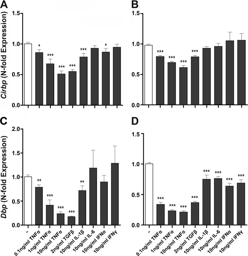
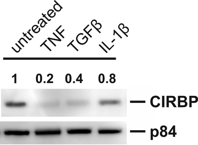
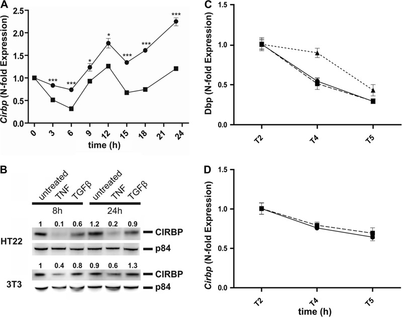
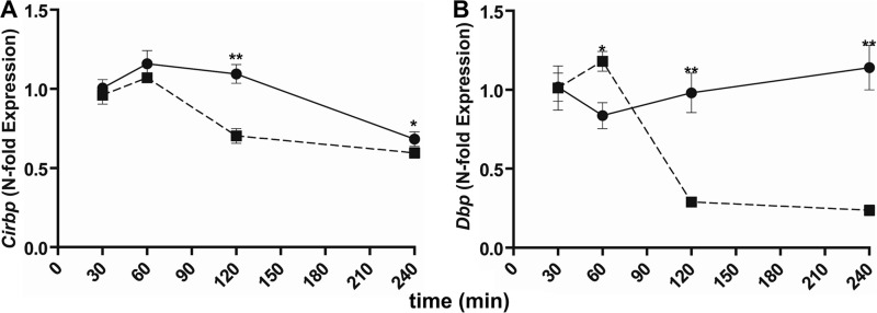
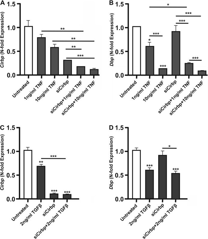
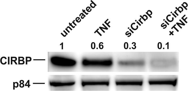
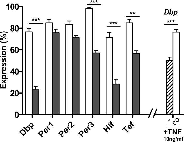
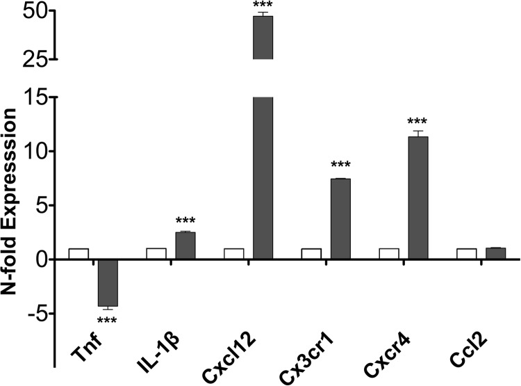
The circadian clock drives the rhythmic expression of a broad array of genes that orchestrate metabolism, sleep wake behavior, and the immune response. Clock genes are transcriptional regulators engaged in the generation of circadian rhythms. The cold inducible RNA-binding protein (CIRBP) guarantees high amplitude expression of clock. The cytokines TNF and TGFβ impair the expression of clock genes, namely the period genes and the proline- and acidic amino acid-rich basic leucine zipper (PAR-bZip) clock-controlled genes. Here, we show that TNF and TGFβ impair the expression of Cirbp in fibroblasts and neuronal cells. IL-1β, IL-6, IFNα, and IFNγ do not exert such effects. Depletion of Cirbp is found to increase the susceptibility of cells to the TNF-mediated inhibition of high amplitude expression of clock genes and modulates the TNF-induced cytokine response. Our findings reveal a new mechanism of cytokine-regulated expression of clock genes.

Keywords: Circadian Rhythms; Cytokine; Gene Expression; Innate Immunity; Metabolism; Sickness Behavior; Sleep.


Figures

FIGURE 1.
FIGURE 1.
TNF and TGFβ inhibit the expression of Cirbp (A and B) and of Dbp (C and D) mRNA in NIH-3T3 (A and C) and HT22 cells (B and D) exposed for 4 h to cytokines (gray bars) (untreated control, white bars). Data of RT-qPCR assays of Cirbp and Dbp expression show the mean ± S.E. (error bars) of triplicates from one representative experiment of three; independent t test, *, p < 0.05; **, p < 0.01; ***, p < 0.001.
FIGURE 2.
FIGURE 2.
Effects on the expression of Cirbp (A) and Dbp (B) after the co-treatment of NIH-3T3 cells with TNF and TGFβ. *, p <0. 05; **, p <0.01; ***, p <0.001. Error bars, S.E.
FIGURE 3.
FIGURE 3.
Western blot analysis of CIRBP in NIH-3T3 cells treated for 4 h with TNF (10 ng/ml), TGFβ (2 ng/ml), and IL1-β (10 ng/ml). As a loading control p84 was used. Numbers indicate the densitometric ratio comparing the bands with the untreated control.
FIGURE 4.
FIGURE 4.
Time kinetic experiments and mRNA stability. A, time course experiments of Cirbp mRNA expression in MEFs cells treated with TNF (10 ng/ml) (black squares) or media control (black circles). In TNF-treated cultures a significant reduction of the circadian expression of Cirbp mRNA is seen at all time points analyzed. B, Western blot of TNF- (10 ng/ml) and TGFβ- (2 ng/ml) treated HT22 and NIH-3T3 cells, the cultures being exposed to the cytokines for 8 h and 24 h. p84 was used as a loading control. C and D, Dbp (C) and Cirbp (D) mRNA stability of untreated MEFs (black circles) and TNF- (10 ng/ml) treated cells (black squares). Data of Cirbp−/− MEFs (untreated) are shown as black triangles. After 1 h of serum deprivation, at T2 ActD (5 μg/ml) was added and the WT MEFs analyzed at the indicated time points with qPCR. Error bars, S.E.
FIGURE 5.
FIGURE 5.
Time-dependent expression of Cirbp (A) and Dbp (B) mRNA in TNF-treated NIH-3T3 cells. Cultures were treated with TNF (10 ng/ml) (broken line) or PBS control (solid line). RNA was harvested at various time points after treatment. One representative experiment of three is shown; values are mean of triplicate experiments ± S.E. (error bars); independent t test, *, p < 0.05; **, p < 0.01.
FIGURE 6.
FIGURE 6.
The expression of Cirbp and Dbp mRNA was tested in Cirbp-depleted cells that were treated with TNF (A and B) and TGFβ (C and D). The data show the effect of cytokines and of siRNA against Cirbp and Dbp that were added to the cultures alone or in combination. Error bars, S.E. *, p < 0.05; **, p <0.01; ***, p <0.001.
FIGURE 7.
FIGURE 7.
Control Western blot to show the effective repression of CIRBP by down-regulating Cirbp with siRNA. p84 was used as a loading control; the numbers show the ratio from the protein of interest to the loading control.
FIGURE 8.
FIGURE 8.
MEFs from WT mice (open bars) were compared with Cirbp−/− MEFs (gray bars) for their responsiveness to TNF. Data show the TNF-induced expression of the indicated genes as percentage from their expression in untreated MEFs. In a separate experiment the extent of TNF-induced inhibition of Dbp was assessed in Cirbp−/− MEFs which were complemented after transfection with pCMV6::Cirbp (CO) and in mock transfected Cirbp−/− MEFs (−). Error bars, S.E. **, p <0.01; ***, p <0.001.
FIGURE 9.
FIGURE 9.
Cirbp overexpression in MEF cells treated with TNF (A and B) and TGFβ (C and D). WT MEFs (−) and Cirbp-overexpressing MEFs (+) were treated with cytokines (gray bars) or medium control (open bars); the expression of Cirbp (A) and of Dbp (B) was assayed. Error bars, S.E. *, p <0.05; **, p <0.01; ***, p <0.001.
FIGURE 10.
FIGURE 10.
The effect of TNF on the expression of cytokines and cytokine receptors was tested on Cirbp-depleted cells (gray bars) and WT MEFs (open bars). The expression of cytokine and cytokine receptor genes was set as 1-fold expression. Error bars, S.E. ***, p <0.001.

Similar articles

Cited by

References

    1. Eckel-Mahan K., Sassone-Corsi P. (2013) Metabolism and the circadian clock converge. Physiol. Rev. 93, 107–135 - PMC - PubMed
    1. Franken P., Dijk D. J. (2009) Circadian clock genes and sleep homeostasis. Eur. J. Neurosci. 29, 1820–1829 - PubMed
    1. Dibner C., Schibler U., Albrecht U. (2010) The mammalian circadian timing system: organization and coordination of central and peripheral clocks. Annu. Rev. Physiol. 72, 517–549 - PubMed
    1. Ko C. H., Takahashi J. S. (2006) Molecular components of the mammalian circadian clock. Hum. Mol. Genet. 15, R271–R277 - PubMed
    1. Keller M., Mazuch J., Abraham U., Eom G. D., Herzog E. D., Volk H. D., Kramer A., Maier B. (2009) A circadian clock in macrophages controls inflammatory immune responses. Proc. Natl. Acad. Sci. U.S.A. 106, 21407–21412 - PMC - PubMed

Publication types

MeSH terms

Substances

LinkOut - more resources