. 2014 Jan 31;289(5):2658-74.
doi: 10.1074/jbc.M113.528505. Epub 2013 Dec 11.

Amino acids activate mammalian target of rapamycin (mTOR) complex 1 without changing Rag GTPase guanyl nucleotide charging

Affiliations

Amino acids activate mammalian target of rapamycin (mTOR) complex 1 without changing Rag GTPase guanyl nucleotide charging

Noriko Oshiro et al. J Biol Chem. .

Abstract

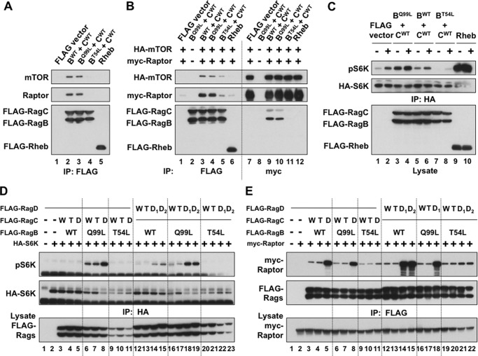
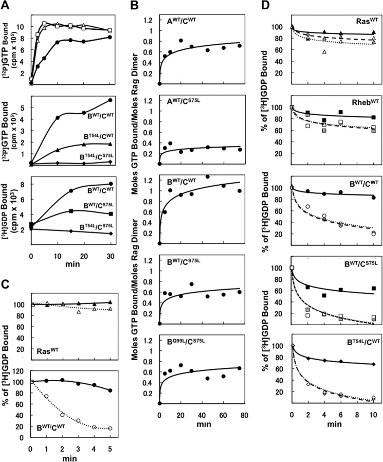
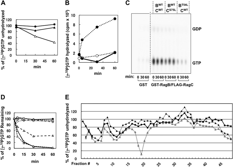
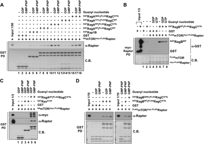
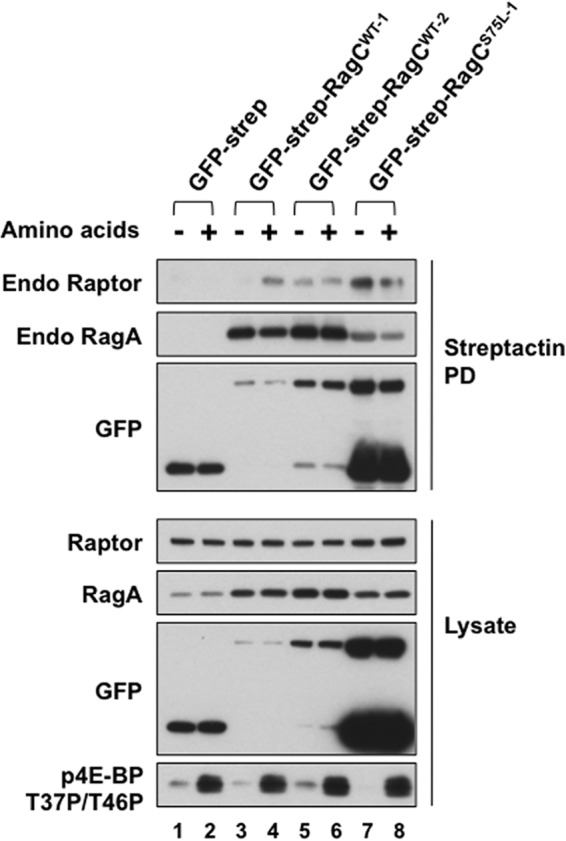
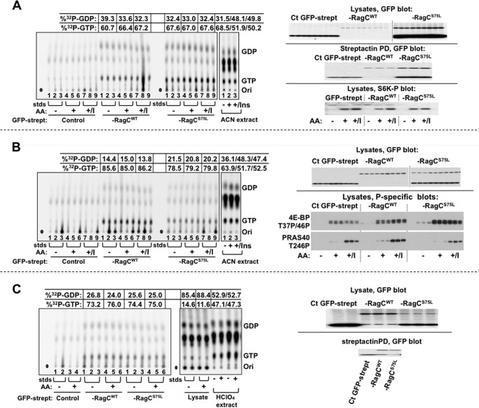
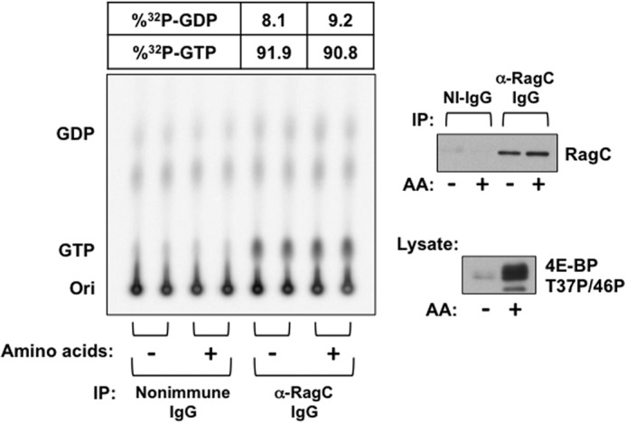
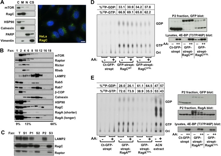
Activation of mammalian target of rapamycin complex 1 (mTORC1) by amino acids is mediated in part by the Rag GTPases, which bind the raptor subunit of mTORC1 in an amino acid-stimulated manner and promote mTORC1 interaction with Rheb-GTP, the immediate activator. Here we examine whether the ability of amino acids to regulate mTORC1 binding to Rag and mTORC1 activation is due to the regulation of Rag guanyl nucleotide charging. Rag heterodimers in vitro exhibit a very rapid, spontaneous exchange of guanyl nucleotides and an inability to hydrolyze GTP. Mutation of the Rag P-loop corresponding to Ras(Ser-17) abolishes guanyl nucleotide binding. Such a mutation in RagA or RagB inhibits, whereas in RagC or RagD it enhances, Rag heterodimer binding to mTORC1. The binding of wild-type and mutant Rag heterodimers to mTORC1 in vitro parallels that seen with transient expression, but binding to mTORC1 in vitro is entirely independent of Rag guanyl nucleotide charging. HeLa cells stably overexpressing wild-type or P-loop mutant RagC exhibit unaltered amino acid regulation of mTORC1. Despite amino acid-independent raptor binding to Rag, mTORC1 is inhibited by amino acid withdrawal as in parental cells. Rag heterodimers extracted from (32)P-labeled whole cells, or just from the pool associated with the lysosomal membrane, exhibit constitutive [(32)P]GTP charging that is unaltered by amino acid withdrawal. Thus, amino acids promote mTORC1 activation without altering Rag GTP charging. Raptor binding to Rag, although necessary, is not sufficient for mTORC1 activation. Additional amino acid-dependent steps couple Rag-mTORC1 to Rheb-GTP.

Keywords: Amino Acids; Cell Signaling; GTPase; Insulin; Lysosomes; Rag GTPase; Raptor; mTOR Complex (mTORC).


Figures

FIGURE 1.
FIGURE 1.
mTOR complex 1 regulation by and binding to transiently expressed wild-type and mutant Rag heterodimers. A and B, binding of endogenous (A) and recombinant (B) mTORC1 by various RagB/C dimers or Rheb in amino acid-replete cells. The FLAG-tagged RagB and RagC variants indicated or wild-type Rheb were expressed in HEK293T cells together with or without HA-mTOR and myc-Raptor. Anti-FLAG or anti-myc immunoprecipitates (IP) were immunoblotted as indicated. C, regulation of S6K phosphorylation by coexpressed RagB/C dimers or Rheb with or without amino acid withdrawal. 48 h after cotransfection of HA-S6K with the FLAG-RagB/C dimers indicated or with FLAG-Rheb to HEK293T cells, the medium was replaced by fresh DMEM for 1 h and then by DPBS. After 50 min, the medium of plates AA- was replaced with fresh DPBS, whereas that of plates AA+ was replaced with DMEM, with harvest 10 min thereafter. The extracts and anti-HA immunoprecipitates were immunoblotted for the proteins indicated and for the phosphorylation of HA-S6K. D, regulation of S6K phosphorylation by coexpressed RagB/C and RagB/D dimers in amino acid-deprived cells. 48 h after cotransfection of HA-S6K with the FLAG-RagB/C or FLAG-RagB/D dimers indicated, the HEK293T cells were incubated in fresh DMEM for 1 h and deprived of amino acids by DPBS treatment for 1 h. The extracts and the anti-HA immunoprecipitates were immunoblotted for the proteins indicated and for the phosphorylation of HA-S6K. The RagC identified as D was S75L, and that identified as T was Q120L. The RagD identified as D1 was S76L, and that identified as D2 was S77L, whereas that labeled T was Q121L. E, the binding of recombinant Raptor to coexpressed RagB/C and RagB/D dimers in amino acid-deprived cells. 48 h after cotransfection of myc-Raptor with the FLAG-RagB/C or FLAG-RagB/D dimers indicated, the HEK293T cells were treated as in D, and the extracts and anti-FLAG immunoprecipitates were immunoblotted as indicated. The labeling of RagC and RagD is as in D.
FIGURE 2.
FIGURE 2.
Guanyl nucleotide binding to and dissociation from Rag heterodimers. A, time course of guanyl nucleotide tracer binding to variant RagB/C heterodimers. Equal Coomassie Blue-stained amounts of GST-Ha-Ras (▵, top panel), GST-Rheb (□, top panel), GST-RagB/FLAG-RagC heterodimers (BWT/CWT, ●; BT54L/CWT, ▴; BT54L/CS75L, ♦; BWT/CS75, ■) were incubated with [γ32P]GTP (50 nm, top and center panels) or [3H]GDP (68 μm, bottom panel) at 30 °C. B, stoichiometry of guanyl nucleotide binding to variant RagA or B/C heterodimers. Various GST-RagA or B/FLAG-RagC heterodimers were incubated with 0.1 mm32P]GTP at 25 °C. The ordinate indicates the moles GTP bound per dimer. C, wild-type RagB/C heterodimers exhibit rapid spontaneous exchange of bound [3H]GDP. Equal Coomassie Blue-stained amounts of GST-Ha-Ras (top panel) and wild-type GST-RagB/FLAG-RagC heterodimer (bottom panel) were preincubated with [3H]GDP (68 μm) to a steady state at 30 °C. At t = 0, nonradioactive ADP (5 mm, ▴ and ●) or GDP (5 mm, ▵ and ○) was added, and the protein-bound [3H]GDP was measured at intervals thereafter. D, [3H]GDP bound to wild-type RagB or RagC within heterodimers is displaced by GTP and GDP with similar efficacy. GST-Ha-Ras (first panel), GST-Rheb (second panel), and various GST-RagB/FLAG-RagC heterodimers were incubated with 0.5 μm [3H]GDP to a steady state at 25 °C. At t = 0, water (▴, ■, ●, and ♦), nonradioactive GTP (5 μm, gray symbols), or GDP (5 μm, ▵, □, ○, and ♢) were added, and the protein-bound [3H]GDP was measured at intervals thereafter.
FIGURE 3.
FIGURE 3.
Rag heterodimers lack detectable GTPase activity in vitro. A, the RagB/C wild-type heterodimer does not hydrolyze [γ-32P]GTP in vitro. GST-Ha-RasWT (▵), GST-Ha-RasG12V (▴), and GST-RagBWT/FLAG-RagCWT (●) were charged with [γ-32P]GTP and examined for intrinsic GTPase activity. The amount of unhydrolysed [γ-32P]GTP on proteins was measured at intervals thereafter by nitrocellulose filter binding assay. B, a recombinant catalytic fragment of NF1 stimulates GTP hydrolysis by Ha-RasWT but not Ha-RasG12V or RagBWT/CWT. GST-Ha-RasWT (●, dashed line), GST-Ha-RasG12V (○, dashed line), or GST-RagBWT/FLAG-RagCWT (●, solid line) were charged with [γ-32P]GTP, and a catalytic fragment of NF1 was added. The release of 32Pi at 30 °C was measured at intervals thereafter by the charcoal method. C, neither RagB wild-type or RagC wild-type within a heterodimer hydrolyze [α-32P]GTP detectably in vitro. GST and various GST-RagB/FLAG-RagC heterodimers were charged with [α-32P]GTP, and at intervals thereafter, nucleotides were extracted and separated on TLC on PEI cellulose. D, extracts from HEK293T cells do not promote the ability of RagB/C to hydrolyze [γ-32P]GTP. HEK293T cells were lysed in the absence of detergent (solid line) or with CHAPS (0.3%, dotted line), TritonX-100 (1%, short dashes), or RIPA buffer (Pierce) (long dashes). GST-Ha-RasWT (triangles) and GST-RagBWT/FLAG-RagCWT (circles) charged with [γ-32P]GTP were mixed with each of these extracts at 30 °C, and the amount of protein-bound [γ-32P]GTP was measured by filtration through nitrocellulose filters. E, MonoQ anion exchange chromatography of a HEK293T cell extract contains GAP activity toward Ha-Ras wild-type, not RagB/C wild-type. HEK293T cells were extracted by freezing and thawing and separated by anion exchange chromatography. Each fraction was incubated at 30 °C with GST-Ha-RasWT (▵, dashed lines), GST-Ha-RasG12V (▴, dashed lines), and GST-RagBWT/FLAG-RagCWT, each charged with [γ-32P]GTP. After 30 min, protein-bound [γ-32P]GTP was measured as in C.
FIGURE 4.
FIGURE 4.
The binding of mTOR complex1 to various RagA or B/C heterodimers in vitro. A, variant RagA/C heterodimers bind mTOR complex1 specifically and in a guanyl nucleotide-independent manner. Equal amounts of GST tagged proteins indicated were immobilized on GSH-Sepharose; loaded with 0.2 mm GDP, GMP-PNP, or water; and incubated with purified HA-mTOR/myc-FLAG-Raptor. The bound Raptor was analyzed by immunoblotting, and GST-proteins were detected by Coomassie Blue staining (C.B.). PD, pull-down. B, immobilized mTOR complex1 binds GST-RagBWT/FLAG-RagCS75L in a guanyl nucleotide-independent manner. Coexpressed FLAG-mTOR/myc-FLAG-Raptor was isolated using Sepharose-bound anti-myc and mixed with GST or GST-RagBWT/FLAG-RagCS75L charged with 0.2 mm GDP, GTP, or water. After washing, the Sepharose-bound proteins were analyzed by anti-GST immunoblot analysis and Coomassie Blue staining. C, GST-RagA/RagCS75L, but not GST-HA-RasG12V, binds mTOR complex 1 in vitro. GST, GST-RagA/RagCS75L, and GST-HA- RasG12V immobilized on GSH-Sepharose were charged with 0.2 mm GDP or GMP-PNP and incubated with recombinant HA-mTOR/myc-FLAG-Raptor. After incubation and washing, the retained proteins were analyzed by immunoblot analysis for myc (top panel) and Raptor (center panel) and by Coomassie Blue staining. D, the constitutively active GST-RagBQ99L/FLAG-RagCS75L heterodimer and the minimally active GST-RagBWT/FLAG-RagCS75L heterodimer bind mTOR complex1 to a similar extent in vitro. GST or the GST-Rag heterodimers immobilized on GSH-Sepharose were charged with 0.2 mm GDP, GMP-PNP, or water and incubated with recombinant FLAG-mTOR/myc-FLAG-Raptor. After incubation and washing, the retained proteins were immunoblotted with anti-myc and stained with Coomassie Blue.
FIGURE 5.
FIGURE 5.
The amino acid dependence of mTOR complex 1 binding to recombinant Rag heterodimers in HeLa cells stably expressing GFP-streptag-RagC variants at differing abundances. HeLa cells stably expressing GFP-streptag, GFP-streptag-RagCWT, or GFP-streptag-RagCS75L were selected by cell sorting for GFP abundance, and the expression of the full-length recombinant protein was estimated by GFP immunoblot analysis of lysates. The cells were incubated in fresh DMEM for 1 h and then deprived of amino acids by incubation in DPBS for 1.5 h. The medium of plates AA- was replaced with fresh DPBS, whereas that of plates AA+ was replaced with DMEM, with harvest 10 min thereafter. Strep-Tactin pull-downs (PD) and the lysates were analyzed by immunoblotting for endogenous (Endo) Raptor and RagA, for GFP, and for 4E-BP(T37P/T46P).
FIGURE 6.
FIGURE 6.
The effect of amino acid withdrawal and insulin on the [32P]guanyl nucleotide content of stably expressed RagCWT and RagCS75L heterodimeric complexes in 32Pi-labeled HeLa cells. A, B, and C, replicate plates of HeLa cells stably expressing GFP-streptag, GFP-streptag-RagCWT, or GFP-streptag-RagCS75L were incubated in Pi-free DMEM containing 32Pi (0.2 mCi/ml). After 4 h, the cells were rinsed and incubated in either homemade Pi-free medium (AA+) or Pi-free medium lacking amino acids (AA-), each containing 32Pi (0.2 mCi/ml), for another 2 h. In A and B, insulin (1.0 μm) was added to some of the cells in DMEM (AA+/I) 30 min before harvest. The nucleotides bound to Strep-Tactin (strept) pull-downs were extracted and separated by TLC on PEI cellulose. One set of 32P -labeled HeLa cells expressing FP-streptag, treated as described above for AA-, AA+, and AA+/I, were rinsed, extracted directly into acetonitrile, and then the solubilized total nucleotides were separated by TLC. The 32P comigrating with GDP and GTP was quantitated by phosphorimaging. After subtraction of the averaged values found in the GFP-streptag lanes, the percentage of [32P]GTP was calculated as [[32P]GTP / (1.5 × [32P]GDP + [32P]GTP)], and the averaged values are shown. In A, the top and center panels on the right show GFP immunoblot analyses of the cell lysates and representative Strep-Tactin pull-downs (corresponding to lanes 1, 4, and 7 of each set). The bottom panel shows an immunoblot analysis of S6K(T389P) corresponding to lanes 1, 4, and 7 of each set. In B, the top panel on the right shows a GFP immunoblot analysis of the lysates, whereas the center and bottom panels show lysate immunoblot analyses for 4E-BP(T37P/T46P) and PRAS40(S246P), respectively. In C, one set of 32P-labeled HeLa cells expressing GFP-streptag, treated as in Fig. 6, were rinsed, extracted directly into HClO4 (0.3 m, 0 °C, HClO4 extract). The HClO4 supernatants were neutralized with KHCO3, and the [32P]guanyl nucleotides were quantified as above. In addition, the nucleotides in the lysate after pull-down of the Strep-Tactin beads were also analyzed. Immunoblot analyses of the lysates and representative Strep-Tactin pull-downs are shown in the right panel. Ori, origin; std, guanyl nucleotide standards; Ins, insulin; ACN, acetonitrile; Ct, C-terminal.
FIGURE 7.
FIGURE 7.
Effect of amino acid withdrawal on [32P]guanyl nucleotide content of RagC-containing heterodimers endogenous to 32Pi-labeled HEK293T cells. Replicate plates of HEK293T cells were incubated in Pi-free DMEM containing 32Pi (0.2 mCi/ml). After 4 h, the cells were rinsed and incubated in either homemade Pi-free medium (AA+) or Pi-free medium lacking amino acids (AA-), each containing 32Pi (0.2 mCi/ml). Cells were extracted 2 h later, and immunoprecipitation (IP) was performed using nonimmune (NI) rabbit IgG or anti-RagC IgG. Nucleotides were extracted from the washed immunoprecipitates and separated by TLC on PEI cellulose. An immunoblot analysis of the extract for 4E-BP(T37P/T46P) and of the immunoprecipitates for RagC are shown in the right panels. Ori, origin.
FIGURE 8.
FIGURE 8.
The effect of amino acid withdrawal on [32P]guanyl nucleotide content of recombinant Rag heterodimeric complexes associated with lysosomal membranes in 32Pi-labeled HEK293T cells stably expressing recombinant RagA or RagC variants. A, subcellular distribution of endogenous RagC in HEK293E cells. A commercial kit was employed to extract selected subcellular fractions from HEK293E cells. Markers included HSP90 (cytosol, C), calnexin (endoplasmic reticulum and membranes, M), poly-ADP ribose polymerase (PARP) (nucleus, N), and vimentin (intermediate filaments and cytoskeleton, CS). The image shows the immunocytochemical localization of endogenous RagC in HeLa cells. Scale bar = 10 μm. RagC, green; DAPI, blue. B, sucrose density gradient fractionation of HEK293T cells. HEK293T cells were sheared open and fractionated upon a 10-ml 13–60% sucrose density gradient at 100,000 × g for 4.5 h. 500-μl fractions were collected, and 50 μl of each even-numbered fraction (Fxn), plus the top fraction (1), was subjected to SDS-PAGE and analyzed by immunoblotting as indicated. Two exposures are shown for RagA. C, isolation of lysosomes from HeLa cells by differential centrifugation. T, cell homogenate; S1, supernatant from centrifugation of T at 1000 × g for10 min; P1, pellet from centrifugation of T at 1000 × g for10 min; S2, supernatant from centrifugation of S1 at 16,100 × g for 30 min; P2, the pellet from the latter centrifugation; S3, the supernatant after centrifugation of S2 at 105 × g for 30 min. Each fraction was brought to the volume of the original homogenate, and an equal volume was subjected to SDS-PAGE and analyzed by immunoblotting as indicated. D, the effect of amino acid withdrawal on the [32P]guanyl nucleotide content of stably expressed RagCWT and RagCS75L heterodimeric complexes associated with a LAMP2-containing fraction of 32Pi-labeled HEK293T cells. Replicate plates containing the GFP-streptag-RagC variants indicated were incubated with 32P in Pi-free DMEM for 4 h, rinsed, and incubated in either homemade Pi-free medium (AA+) or Pi-free medium lacking amino acids (AA-), each containing 32Pi as in Fig. 6. The cells were extracted two h later, the fraction corresponding to P2 (C) was isolated, and the [32P]guanyl nucleotide content of Strep-Tactin-isolated Rag complexes in P2 was determined as in Fig. 6. Immunoblot analyses of GFP in the P2 fraction and 4E-BP(T37P/T46P) in the lysate are shown in the right panel. E, the effect of amino acid withdrawal on the [32P]guanyl nucleotide content of stably expressed RagAWT and RagAT21L heterodimeric complexes associated with a LAMP2-containing fraction of 32Pi-labeled HEK293T cells. The cells and extracts were processed as described in D. Immunoblots of GFP and RagA in the P2 fraction and 4E-BPT37P/T46P) in the lysates are shown in the right panel. Ori, origin; Ct, C-terminal.

References

    1. Hara K., Maruki Y., Long X., Yoshino K., Oshiro N., Hidayat S., Tokunaga C., Avruch J., Yonezawa K. (2002) Raptor, a binding partner of target of rapamycin (TOR), mediates TOR action. Cell 110, 177–189 - PubMed
    1. Kim D. H., Sarbassov D. D., Ali S. M., King J. E., Latek R. R., Erdjument-Bromage H., Tempst P., Sabatini D. M. (2002) mTOR interacts with raptor to form a nutrient-sensitive complex that signals to the cell growth machinery. Cell 110, 163–175 - PubMed
    1. Loewith R., Jacinto E., Wullschleger S., Lorberg A., Crespo J. L., Bonenfant D., Oppliger W., Jenoe P., Hall M. N. (2002) Two TOR complexes, only one of which is rapamycin sensitive, have distinct roles in cell growth control. Mol. Cell 10, 457–468 - PubMed
    1. Laplante M., Sabatini D. M. (2012) mTOR signaling in growth control and disease. Cell 149, 274–293 - PMC - PubMed
    1. Long X., Lin Y., Ortiz-Vega S., Yonezawa K., Avruch J. (2005) Rheb binds and regulates the mTOR kinase. Curr. Biol. 15, 702–713 - PubMed

Publication types

MeSH terms