Review
. 2014 May;98(5):579-85.
doi: 10.1136/bjophthalmol-2013-304256. Epub 2013 Dec 12.

Techniques and outcomes of minimally invasive trabecular ablation and bypass surgery

Affiliations
Review

Techniques and outcomes of minimally invasive trabecular ablation and bypass surgery

Kevin Kaplowitz et al. Br J Ophthalmol. 2014 May.

Abstract

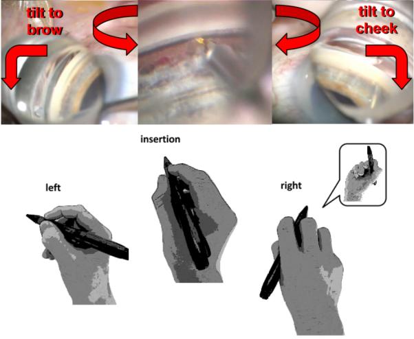
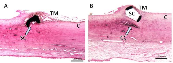
Minimally invasive glaucoma surgeries (MIGS) can improve the conventional, pressure-dependent outflow by bypassing or ablating the trabecular meshwork (TM), or creating alternative drainage routes into the suprachoroidal or subconjunctival space. They have a highly favourable risk profile compared to penetrating surgeries, and lower intraocular pressure with variable efficacy that may depend on the extent of outflow segments accessed. Since they are highly standardised procedures that use clear corneal incisions, they can elegantly be combined with cataract and refractive procedures to improve vision in the same session. There is a growing need for surgeons to become proficient in MIGS to address the increasing prevalence of glaucoma and cataracts in a well-informed, aging population. Techniques of visualisation and instrumentation in an anatomically highly confined space with semitransparent tissues are fundamentally different from other anterior segment surgeries, and present even experienced surgeons with a substantial learning curve. Here, we provide practical tips, and review techniques and outcomes of TM bypass and ablation MIGS.

Keywords: Angle; Glaucoma; Treatment Lasers; Treatment Surgery.


Figures

Figure 1
Figure 1
Procedures enhancing conventional outflow by degree of angle access. Left half: trabecular meshwork bypass stents (counter clockwise): a) iStent Inject, b) iStent G1 and c) Hydrus. Right half: trabecular meshwork ablation devices (clockwise): d) laser assisted endoscopic goniopuncture with single, circular entry and e) trabectome with plasma ablation tip that can remove TM up to 180° (blue half-circle arrows). The naturally segmented and discontinuous Schlemm’s canal limits the extent of access to drainage segments that can be achieved with bypass stents (Background canalography image courtesy of The Glaucoma Imaging Group, UPMC Eye Center, University of Pittsburgh School of Medicine).
Figure 2
Figure 2
Engaging the TM with the iStent G1 and trabectome. A more pointed angle (left inset) allows for easier entry into Schlemm’s canal (blue) compared to a parallel orientation (right inset).
Figure 3
Figure 3
Trabectome. A) Schematic of trabectome ablating TM, opening a view to the white inner wall of Schlemm’s canal (courtesy of Neomedix Corp. , Tustin, CA) and B) scanning electron microscopy of Schlemm’s canal following successful TM ablation with a collector channel seen clearly at far right (courtesy of Douglas H. Johnson for Neomedix).
Figure 4
Figure 4
Atraumatic entry into Schlemm’s canal can be most easily achieved by angling the tip 45° upward, directly anterior to the scleral spur, to avoid collapse of the canal during engagement. The tip is then moved up, in and along Schlemm’s canal (dotted line).
Figure 5
Figure 5
Gonioscopic view and hand position in angle surgery (trabectome shown). After engagement and SC entry at the nasal TM slightly off to the left, ablation is continued for 90° while the lens is rotated in the same direction and the eye (right eye shown) is tilted towards the brow. The same is repeated for the opposite direction. Gonioscopic prism power is increased by lifting the lens off the cornea to float in saline at the final clock hours.
Figure 6
Figure 6
Challenges encountered by microstents. (A) Section through microstent in Schlemm's canal (SC) shows that the lumen can become partially occluded. B) Scaffold device that distends SC effectively but may compress adjacent collector channels (CC; courtesy of Ivantis Inc., Irvine, CA).

Similar articles

Cited by

References

    1. Quigley HA, Broman AT. The number of people with glaucoma worldwide in 2010 and 2020. Br J Ophthalmol. 2006;90(3):262–7. doi: 10.1136/bjo.2005.081224. - PMC - PubMed
    1. Klaver CC, Wolfs RC, Vingerling JR, et al. Age-specific prevalence and causes of blindness and visual impairment in an older population: the Rotterdam Study. Arch Ophthalmol. 1998;116(5):653–8. - PubMed
    1. Gedde SJ, Herndon LW, Brandt JD, et al. Postoperative complications in the Tube Versus Trabeculectomy (TVT) study during five years of follow-up. Am J Ophthalmol. 2012;153(5):804–14. e1 doi: 10.1016/j.ajo.2011.10.024. - PMC - PubMed
    1. Shingleton BJ, Laul A, Nagao K, et al. Effect of phacoemulsification on intraocular pressure in eyes with pseudoexfoliation: single-surgeon series. J Cataract Refract Surg. 2008;34(11):1834–41. doi: 10.1016/j.jcrs.2008.07.025. - PubMed
    1. Huang G, Gonzalez E, Peng PH, et al. Anterior chamber depth, iridocorneal angle width, and intraocular pressure changes after phacoemulsification: narrow vs open iridocorneal angles. Arch Ophthalmol. 2011;129(10):1283–90. doi: 10.1001/archophthalmol.2011.272. - PubMed

Publication types

MeSH terms