. 2014 Feb 7;289(6):3416-31.
doi: 10.1074/jbc.M113.503359. Epub 2013 Dec 13.

Evolutionary and structural analyses of mammalian haloacid dehalogenase-type phosphatases AUM and chronophin provide insight into the basis of their different substrate specificities

Affiliations

Evolutionary and structural analyses of mammalian haloacid dehalogenase-type phosphatases AUM and chronophin provide insight into the basis of their different substrate specificities

Annegrit Seifried et al. J Biol Chem. .

Abstract

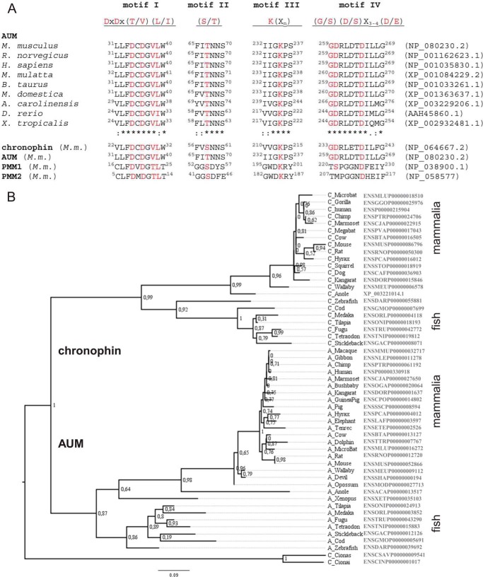
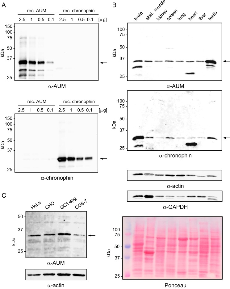
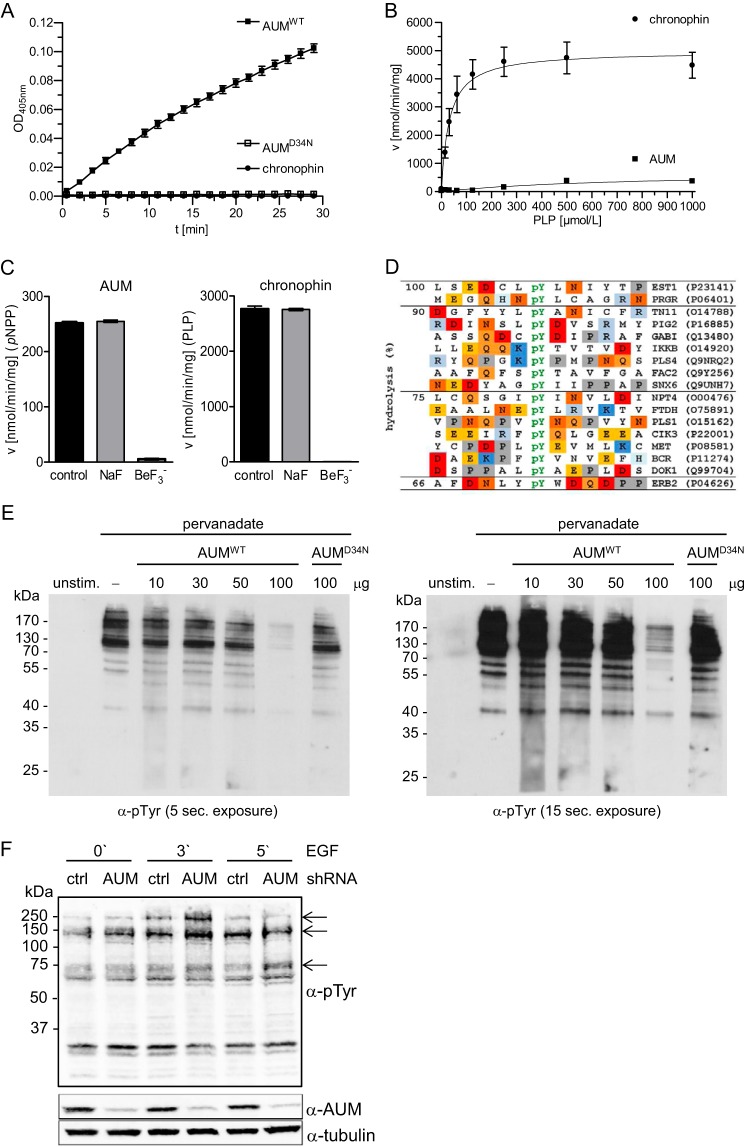
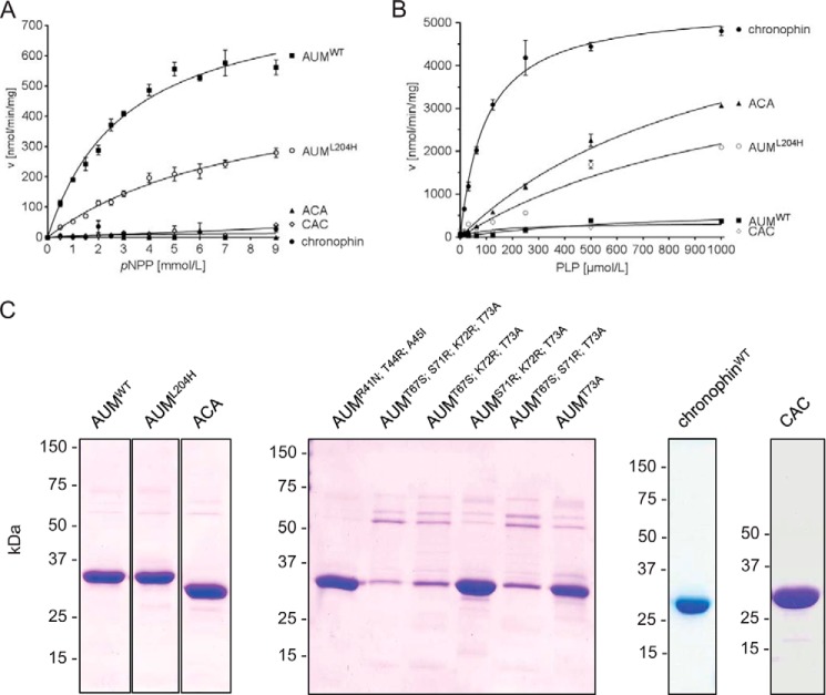
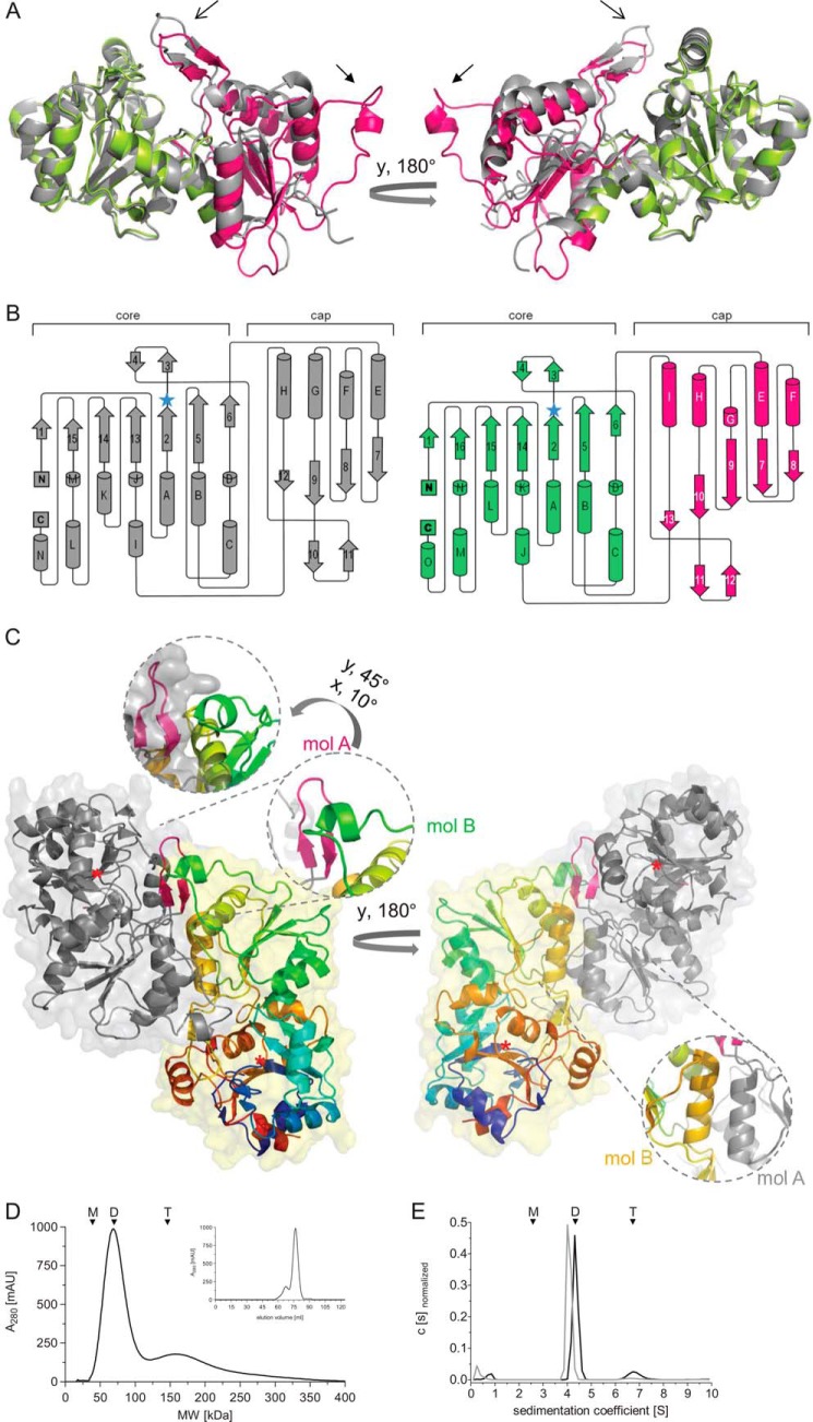
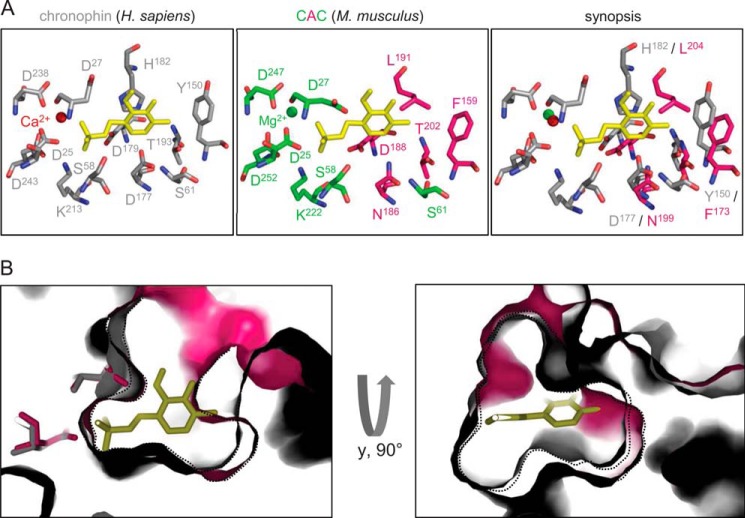
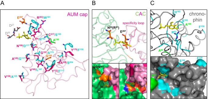
Mammalian haloacid dehalogenase (HAD)-type phosphatases are an emerging family of phosphatases with important functions in physiology and disease, yet little is known about the basis of their substrate specificity. Here, we characterize a previously unexplored HAD family member (gene annotation, phosphoglycolate phosphatase), which we termed AUM, for aspartate-based, ubiquitous, Mg(2+)-dependent phosphatase. AUM is a tyrosine-specific paralog of the serine/threonine-specific protein and pyridoxal 5'-phosphate-directed HAD phosphatase chronophin. Comparative evolutionary and biochemical analyses reveal that a single, differently conserved residue in the cap domain of either AUM or chronophin is crucial for phosphatase specificity. We have solved the x-ray crystal structure of the AUM cap fused to the catalytic core of chronophin to 2.65 Å resolution and present a detailed view of the catalytic clefts of AUM and chronophin that explains their substrate preferences. Our findings identify a small number of cap domain residues that encode the different substrate specificities of AUM and chronophin.

Keywords: Haloacid Dehalogenase Phosphatase; Molecular Evolution; Phosphatase Substrate Specificity; Phosphoglycolate Phosphatase; Pyridoxal Phosphate; Serine/Threonine Protein Phosphatase; Tyrosine-Protein Phosphatase (Tyrosine Phosphatase); X-ray Crystallography.


Figures

FIGURE 1.
FIGURE 1.
Comparison of AUM and chronophin. A, HAD motifs of AUM and chronophin are closely related. Alignment of the HAD motifs of vertebrate AUM orthologs in comparison with chronophin and phosphomannomutase (PMM) 1 and -2. M. m., Mus musculus. The consensus motifs reflect an alignment of 40 human phosphatases. B, phylogenetic tree of vertebrate AUM and chronophin proteins together with urochordate sequences predating the duplication of a common ancestor. Branch support is indicated by the result of the approximate likelihood ratio test (72). Accession numbers are given on the right.
FIGURE 2.
FIGURE 2.
Antibody validation, comparison of AUM and chronophin expression levels in murine tissues, and analysis of AUM expression in cultured cell lines. A, purified, recombinant (rec.) AUM and chronophin (0.1–2.5 μg/lane) were separated by SDS-PAGE, blotted onto nitrocellulose membranes, and probed with rabbit polyclonal α-AUM (upper panel) or with rabbit monoclonal α-chronophin antibodies (lower panel). B, comparison of endogenous AUM and chronophin expression levels in mouse tissues. Mouse tissue lysates (50 μg of protein/lane) were separated by SDS-PAGE, blotted onto nitrocellulose membranes, and analyzed by immunoblotting with α-AUM antibodies. The blot was stripped and reprobed with α-chronophin antibodies. The identity of the faster migrating bands is unknown. Blots were reprobed with α-actin and α-GAPDH antibodies to test for comparable protein loading. Additionally, the Ponceau-stained membrane is shown. C, whole cell lysates (25 μg of protein/lane) of human cervical carcinoma (HeLa), Chinese hamster ovary (CHO), mouse spermatogonial (GC1-spg), and monkey kidney (COS7) cells were separated by SDS-PAGE, blotted onto nitrocellulose membranes, and analyzed by immunoblotting with α-AUM antibodies. A–C, the bands corresponding to the expected AUM or chronophin molecular weights are indicated by arrows.
FIGURE 3.
FIGURE 3.
AUM substrate specificity in vitro and in cells. A, in vitro pNPP phosphatase assays were performed in 96-well microtiter plates in a total assay volume of 100 μl, using recombinantly expressed and purified AUM, AUMD34N, or chronophin (0.8 μg of protein/well) and 3.5 mm pNPP as a substrate. The kinetics of pNP generation were followed spectrophotometrically by measuring the absorbance at 405 nm. B, in vitro PLP phosphatase assays with purified AUM and chronophin were performed in 96-well microtiter plates in a total assay volume of 50 μl, using recombinantly expressed, purified AUM or chronophin (0.16 μg of protein/well), and 0–1 mmol/liter PLP. The reaction was stopped with malachite green, and released phosphate was determined by measuring A620. The enzyme velocity toward increasing PLP concentrations is shown. C, effect of BeF3 on AUM or chronophin activity toward pNPP or PLP. The recombinant purified enzymes (0.8 μg of AUM or 0.16 μg of chronophin/well) were preincubated for 30 min at 37 °C (AUM) or for 10 min at 22 °C (chronophin) in the absence (control) or presence of 1 mm NaF (NaF) or 1 mm NaF + 0.1 mm BeCl2 (BeF3) before phosphatase activity against pNPP (3.5 mm) or PLP (0.5 mm) was measured as described above. Left panel, velocity of AUM-dependent pNPP dephosphorylation ± inhibitors; right panel, velocity of chronophin-dependent PLP dephosphorylation ± inhibitors. A–C, results are mean values ± S.E. of n = 3 independent experiments. D, activity of AUM in phosphopeptide array assays. A set of 720 different phosphopeptides phosphorylated on tyrosine, serine, or threonine residues (final substrate concentration, 10 μm) was incubated with 100 nm purified AUM in a 384-well microtiter plate (final assay volume, 25 μl) for 45 min at 37 °C. The reaction was quenched with malachite green, and released phosphate was determined by measuring A620. Absorbance values were normalized to the peptide substrate yielding the highest reading (100% hydrolysis), and all peptides with absorbance values of ≥66% over the background are listed. In these sequences, acidic residues are highlighted in red, basic residues in blue, and proline residues in gray. Swiss-Prot accession numbers of the corresponding proteins are given on the right. E, determination of AUM and AUMD34N activity toward tyrosine-phosphorylated proteins in overlay assays. HeLa cells were left unstimulated (unstim.) or treated with pervanadate. After SDS-PAGE, cell lysates were blotted onto nitrocellulose, and the membrane was cut and incubated in the absence (−) or presence of the indicated concentrations of AUMWT or AUMD34N. Protein tyrosine phosphorylation was analyzed with 4G10 α-phosphotyrosine antibodies. A short (left panel) and longer exposure (right panel) is shown (n = 3). F, comparison of cellular phosphotyrosine levels in control (ctrl) or AUM-depleted GC1-spg cells. GC1-spg cells expressing control (ctrl) shRNA or AUM shRNA were stimulated with 100 ng/ml EGF for the indicated time points. Cells were lysed, and proteins were separated by SDS-PAGE and transferred onto nitrocellulose membranes for immunoblotting. Cellular tyrosine phosphorylation levels were analyzed with 4G10 α-phosphotyrosine (pTyr) antibodies; AUM depletion was assessed with α-AUM antibodies, and tubulin served as a loading control (n = 3).
FIGURE 4.
FIGURE 4.
Effects of the AUM-L204H substitution and of AUM and chronophin cap domain exchanges on phosphatase specificity. The indicated recombinant proteins were tested for their in vitro phosphatase activity toward pNPP (using 0.8 μg of the respective purified proteins in an assay volume of 100 μl) and PLP (using 0.16 μg of the respective purified proteins in an assay volume of 50 μl). Enzyme velocities against pNPP (A) and PLP (B) are plotted against increasing substrate concentrations. Results are mean values ± S.E. of n = 3 independent experiments. ACA, hybrid protein consisting of AUM core domain fused to the chronophin cap domain; CAC, chronophin core domain fused to the AUM cap domain. C, purity of the employed recombinant proteins. Shown are Coomassie Blue-stained gels (see also Table 2).
FIGURE 5.
FIGURE 5.
Crystal structure of the murine chronophin core/AUM cap hybrid (CAC). A, overall CAC structure (PDB 4BKM) compared with human chronophin in the PLP-bound state (PDB 2P69). Chronophin is in gray; the CAC murine chronophin core is in lime, and the AUM cap is in pink. The catalytic cores of human and murine chronophin are organized in a superimposable manner, whereas the AUM and chronophin caps differ. Open arrows, AUM substrate specificity loop; closed arrows, AUM transverse loop (see C). B, topology diagrams of chronophin and CAC. Left panel, organization of chronophin. Right panel, CAC organization with the core domain in lime and the cap in pink. The β-strands are numbered consecutively, and α-helices are in alphabetical order from the N to the C terminus. Blue star, nucleophilic Asp-25; flap, β-strands 3 and 4; substrate specificity loop, β-strands 10 and 11 (chronophin) or 11 and 12 (CAC). C, overall structure of the CAC homodimer, shown in ribbon representation with transparent surfaces. One protomer (mol A) is represented in gray with the substrate specificity loop shown in magenta. Mol B is in rainbow colors (N terminus in blue). One inset shows the magnified spatial arrangement of the small helix in the transverse loop of the AUM cap (mol B, green) and the specificity loop of the AUM cap (mol A, pink). The 2nd inset shows helix I of mol A (gray) and of mol B (orange) forming the dimer interface. Red star, Asp-25. D, elution profile of purified, untagged murine AUM on a size exclusion chromatography column. The peak elution volume of AUM (theoretical molecular mass, 34.5 kDa) corresponds to a calculated molecular mass of 79.6 kDa. The elution volume of a fraction (19.3%) of AUM corresponds to a calculated molecular mass of 163.6 kDa. E, analytical ultracentrifugation sedimentation velocity experiments of purified AUM and chronophin. AUM exists in equilibrium between dimers (88.3%) and tetramers (11.7%) in solution and has a greater propensity for tetramer formation than chronophin (only 2.2% of chronophin particles sediment as tetramers). D and E, n = 3. Arrowheads indicate the expected/observed position of AUM monomers (M), dimers (D), and tetramers (T).
FIGURE 6.
FIGURE 6.
View of the active sites of chronophin and the CAC chronophin/AUM cap hybrid. A, left panel, view of the chronophin/PLP binding interface (PDB 2P69). PLP is shown in yellow, and the catalytic core residues that build up the binding site for the substrate's phosphate moiety are in gray and consist of HAD motif I (Asp-25 and Asp-27), II (Ser-58 and Ser-61), III (Lys-213), and IV (Asp-238 and Asp243) residues. Middle panel, view into the active site of the CAC hybrid (PDB 4BKM). The catalytic residues of the murine chronophin core domain are shown in lime, and the residues of the AUM cap domain are in pink. Amino acid residues are labeled according to their position in CAC. The PLP molecule is modeled according to the human chronophin structure. Right panel, superposition of the active site residues of human chronophin in the PLP-bound state (gray) and the corresponding residues of the AUM cap domain in the CAC hybrid (pink). Amino acid residues are labeled according to their position in murine chronophin and murine AUM. B, superposition of the CAC (pink) and chronophin (gray) active sites in surface representation, revealing the different spatial arrangements of the substrate binding pockets. Shown is a zoom into the active site underneath the nucleophilic Asp-25 (left panel). A lateral view is presented in the right panel. Black dotted lines outline the borders of the active site sections in CAC or chronophin, respectively.
FIGURE 7.
FIGURE 7.
Localization of differently conserved residues in AUM and chronophin. A, loop diagram representing the overall structure of the AUM cap domain (in pink; Asp-25/27 of the chronophin core in gray, PLP in yellow). Differently conserved AUM or chronophin residues (class II sites) are labeled in pink or cyan, respectively (residue numbering according to murine AUM or chronophin). Chronophin residues are represented according to their spatial orientation in the chronophin cap (which is not shown for clarity) after structural alignment with and superimposition onto the CAC structure. Sites conserved in one subfamily, but not in the other (class I sites), are labeled in orange (AUM) or green (chronophin). B, loop diagram (upper panel) and surface representation (lower panel, two different orientations are shown) of a zoom into the active site of the CAC hybrid (chronophin core domain in lime, AUM cap domain in pink, and PLP in yellow). The position of Glu-207 in the AUM cap and the putative position of AUMArg-41 (corresponding to chronophinAsn-32) in the chronophin core are shown in orange. Both residues are conserved in AUM but not in chronophin. C, loop diagram (upper panel) and surface representation (lower panel) of a magnification of the active center of human chronophin (PDB code 2P69). The differently conserved residue Arg-63 in chronophin (shown in cyan; stick representation in the upper panel) is located at the active site entrance and corresponds to Lys-72 of AUM (see also Table 2). The differently conserved chronophin residues Ser-195, Phe-156, and Gly-117 (cyan) and residues Arg-74 and Ala-198 that are only conserved in chronophin (green) are located on the chronophin surface. Ala-92 is only conserved in chronophin; its function is currently unknown. (Note: Ala-198 is not visible in the lower panel.)

References

    1. Aravind L., Koonin E. V. (1998) The HD domain defines a new superfamily of metal-dependent phosphohydrolases. Trends Biochem. Sci. 23, 469–472 - PubMed
    1. Burroughs A. M., Allen K. N., Dunaway-Mariano D., Aravind L. (2006) Evolutionary genomics of the HAD superfamily: understanding the structural adaptations and catalytic diversity in a superfamily of phosphoesterases and allied enzymes. J. Mol. Biol. 361, 1003–1034 - PubMed
    1. Allen K. N., Dunaway-Mariano D. (2009) Markers of fitness in a successful enzyme superfamily. Curr. Opin. Struct. Biol. 19, 658–665 - PMC - PubMed
    1. Almo S. C., Bonanno J. B., Sauder J. M., Emtage S., Dilorenzo T. P., Malashkevich V., Wasserman S. R., Swaminathan S., Eswaramoorthy S., Agarwal R., Kumaran D., Madegowda M., Ragumani S., Patskovsky Y., Alvarado J., Ramagopal U. A., Faber-Barata J., Chance M. R., Sali A., Fiser A., Zhang Z. Y., Lawrence D. S., Burley S. K. (2007) Structural genomics of protein phosphatases. J. Struct. Funct. Genomics 8, 121–140 - PMC - PubMed
    1. Possemato R., Marks K. M., Shaul Y. D., Pacold M. E., Kim D., Birsoy K., Sethumadhavan S., Woo H. K., Jang H. G., Jha A. K., Chen W. W., Barrett F. G., Stransky N., Tsun Z. Y., Cowley G. S., Barretina J., Kalaany N. Y., Hsu P. P., Ottina K., Chan A. M., Yuan B., Garraway L. A., Root D. E., Mino-Kenudson M., Brachtel E. F., Driggers E. M., Sabatini D. M. (2011) Functional genomics reveal that the serine synthesis pathway is essential in breast cancer. Nature 476, 346–350 - PMC - PubMed

Publication types

Substances

Associated data