. 2014 Jan 31;289(5):3040-54.
doi: 10.1074/jbc.M113.505842. Epub 2013 Dec 14.

Control of helicase loading in the coupled DNA replication and recombination systems of bacteriophage T4

Affiliations

Control of helicase loading in the coupled DNA replication and recombination systems of bacteriophage T4

Amy M Branagan et al. J Biol Chem. .

Abstract

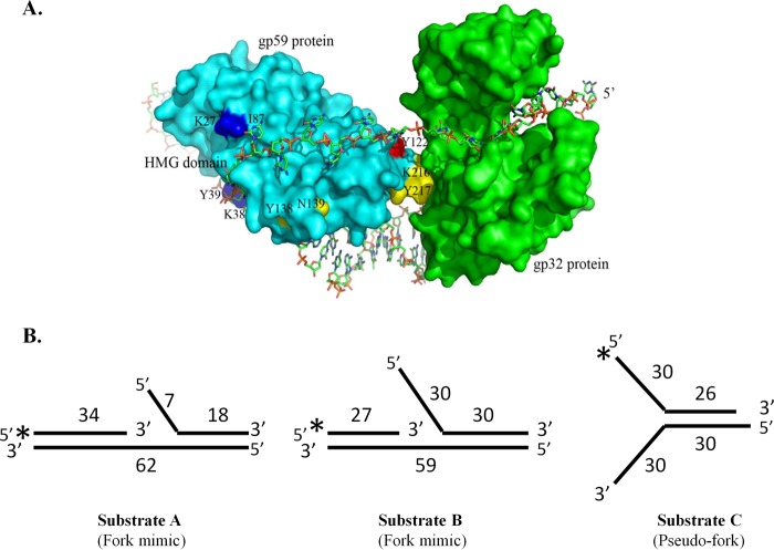
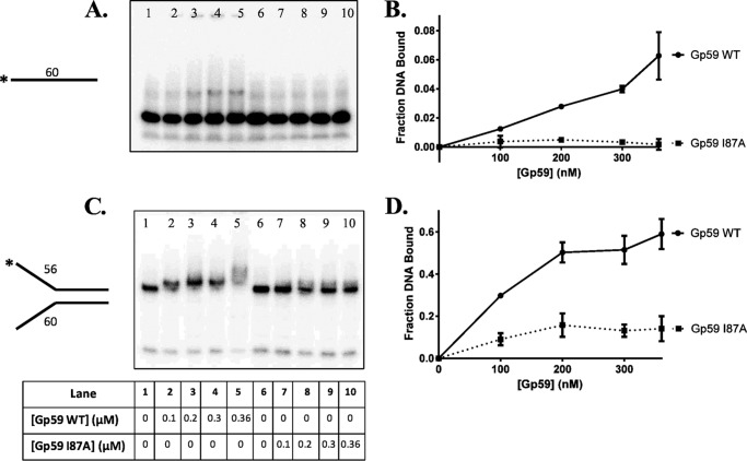
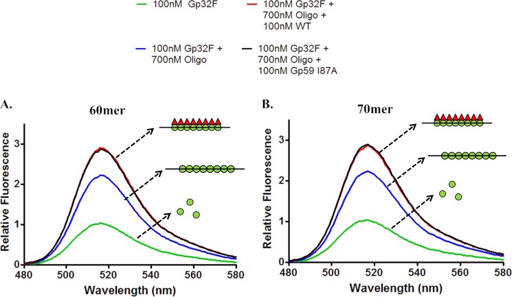
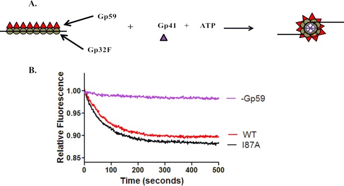
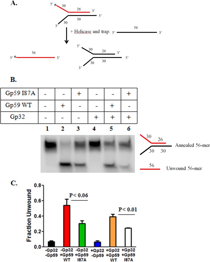
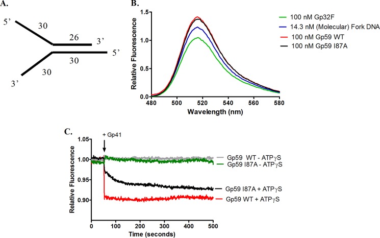
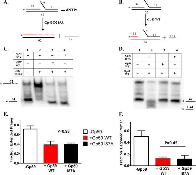
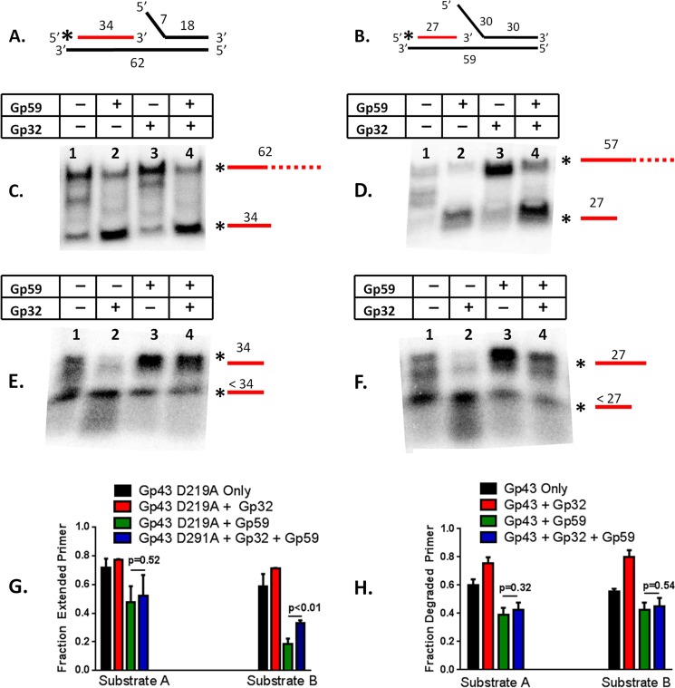
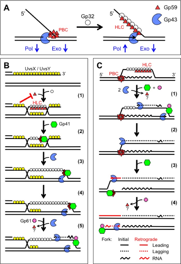
The Gp59 protein of bacteriophage T4 promotes DNA replication by loading the replicative helicase, Gp41, onto replication forks and recombination intermediates. Gp59 also blocks DNA synthesis by Gp43 polymerase until Gp41 is loaded, ensuring that synthesis is tightly coupled to unwinding. The distinct polymerase blocking and helicase loading activities of Gp59 likely involve different binding interactions with DNA and protein partners. Here, we investigate how interactions of Gp59 with DNA and Gp32, the T4 single-stranded DNA (ssDNA)-binding protein, are related to these activities. A previously characterized mutant, Gp59-I87A, exhibits markedly reduced affinity for ssDNA and pseudo-fork DNA substrates. We demonstrate that on Gp32-covered ssDNA, the DNA binding defect of Gp59-I87A is not detrimental to helicase loading and translocation. In contrast, on pseudo-fork DNA the I87A mutation is detrimental to helicase loading and unwinding in the presence or absence of Gp32. Other results indicate that Gp32 binding to lagging strand ssDNA relieves the blockage of Gp43 polymerase activity by Gp59, whereas the inhibition of Gp43 exonuclease activity is maintained. Our findings suggest that Gp59-Gp32 and Gp59-DNA interactions perform separate but complementary roles in T4 DNA metabolism; Gp59-Gp32 interactions are needed to load Gp41 onto D-loops, and other nucleoprotein structures containing clusters of Gp32. Gp59-DNA interactions are needed to load Gp41 onto nascent or collapsed replication forks lacking clusters of Gp32 and to coordinate bidirectional replication from T4 origins. The dual functionalities of Gp59 allow it to promote the initiation or re-start of DNA replication from a wide variety of recombination and replication intermediates.

Keywords: Bacteriophage; D-loop; DNA Helicase; DNA Repair; DNA Replication; Homologous Recombination; Mediator; Origin; R-loop; ssDNA-binding Protein.


Figures

FIGURE 1.
FIGURE 1.
A, model of Gp59 (cyan) bound to pseudo-fork DNA and the ssDNA binding domain of Gp32 (green). The positions of point mutations that affect DNA binding are shown in blue. Residue Ile-87, site of the I87A point mutation that attenuates DNA binding, is located in the N-terminal HMG homology domain of Gp59 and close to the 5′ ssDNA arm of pseudo-fork DNA. The 5′ arm corresponds to the lagging strand ssDNA of a replication fork, the site where Gp59 loads Gp41 helicase. This research was originally published in Hinerman et al. (32). B, representation of the fork DNA substrates used for experiments. Substrates were either fork mimics (Substrates A and B) or pseudo-fork (Substrate C) structures. Sequences of the oligonucleotides used in their construction are shown in Table 1. Asterisks denote the positions of 5′-32P labels.
FIGURE 2.
FIGURE 2.
Electrophoretic mobility shift assays for Gp59-DNA interactions; comparisons of I87A to WT. DNA binding assays were performed as described under “Experimental Procedures.” A, binding to 60-mer ssDNA (Oligo 3). The concentration of 60-mer in each lane is 3 nm (molecules). Lanes 1–5 contain 0–360 nm Gp59 WT as indicated. Lanes 6–10 contain 0–360 nm Gp59-I87A as indicated. B, quantified results from panel A. Plot shows the fraction ssDNA bound versus Gp59 concentration for Gp59 WT (solid line) or I87A (dotted line). C, binding to pseudo-fork DNA (Substrate C). The concentration of Substrate C in each lane is 3 nm (molecules). Protein concentrations are the same as in A. D, quantified results from panel C. The plot shows the fraction pseudo-fork DNA bound versus Gp59 concentration for Gp59 WT (solid line) or I87A (dotted line). Error bars in panels B and D represent S.D. from three separate experiments.
FIGURE 3.
FIGURE 3.
Effects of Gp59-I87A mutation on the formation of Gp59-Gp32-ssDNA HLCs. Gp32F fluorescence assays for HLC formation were carried out as described under “Experimental Procedures.” Complexes were formed using either Oligo 4, an ssDNA 60-mer (A) or Oligo 5, an ssDNA 70mer of different sequence (B). All other conditions were identical between the two panels. Green line, fluorescence spectrum of 100 nm Gp32F alone; blue line, fluorescence spectrum of 100 nm Gp32F + 700 nm (nucleotides) ssDNA; red line, fluorescence spectrum of 100 nm Gp32F + 700 nm ssDNA + 100 nm Gp59 WT; black line, fluorescence spectrum of 100 nm Gp32F + 700 nm ssDNA + 100 nm Gp59-I87A. The dotted arrow from each spectrum points to a schematic of the protein complex present; green circles, Gp32F; solid black lines, ssDNA; red triangles, Gp59 (WT or I87A).
FIGURE 4.
FIGURE 4.
Effects of Gp59-I87A mutation on helicase loading activity. Changes in Gp32F fluorescence were used to monitor the loading of Gp41 helicase onto 70mer ssDNA (Oligo 5) as described under “Experimental Procedures.” The components of the helicase loading complex were added in the following order and preincubated: 100 nm Gp32F, 700 nm ssDNA, and 100 nm Gp59 (WT or I87A). Helicase loading reactions were initiated by the simultaneous addition of 100 nm Gp41 and 1 mm ATP. A, reaction schematic for helicase loading assays. Green circles, Gp32F; solid black line, ssDNA; red triangles, Gp59; purple triangles, Gp41monomers (hexamerizes upon assembly onto ssDNA). B, relative fluorescence of the helicase loading complex versus time after Gp41/ATP addition in the presence of Gp59 WT (red), Gp59 I87A (black), or in the absence of Gp59 (purple).
FIGURE 5.
FIGURE 5.
Effects of Gp59-I87A mutation on the stimulation of Gp41 helicase activity in the presence and absence of Gp32. Helicase assays to monitor the unwinding of pseudo-fork DNA (Substrate C) were carried out as described under “Experimental Procedures.” A, reaction scheme. The unwinding of 30 nm (molecules) Substrate C, a DNA pseudo-fork containing labeled 56-mer (Oligo 6 (red)) annealed to unlabeled 60-mer (Oligo 4 (black)), by 300 nm Gp41 helicase in the presence of a cold trap (120 nm unlabeled 56-mer) releases labeled 56-mer product, which is separated from labeled substrate by non-denaturing PAGE and quantified by phosphorimaging. The substrate was preincubated with different combinations of Gp32, Gp59 WT, and Gp59-I87A as indicated in panel B before initiating the reaction by simultaneous addition of Gp41 and trap. B, visualization of results by non-denaturing PAGE/phosphorimaging for reactions containing no Gp59 and no Gp32 (lane 1), 300 nm Gp59 WT (lane 2), 300 nm Gp59-I87A (lane 3), 300 nm Gp32 (lane 4), 300 nm Gp32 + 300 nm Gp59 WT (lane 5), and 300 nm Gp32 + 300 nm Gp59-I87A (lane 6). C, quantification of results. The fraction of labeled 56-mer appearing as product are plotted for reactions in the absence of Gp32 or Gp59 (black) or in the presence of Gp59 WT (red), Gp59 I87A (green), Gp32 (blue), Gp32 + Gp59 WT (orange), or Gp32 + Gp59 I87A (white). Error bars represent S.D. from three separate experiments. p values shown for adjacent columns in the histogram are based on Student's t test.
FIGURE 6.
FIGURE 6.
Effects of Gp59-I87A mutation on HLC formation and helicase loading on pseudo-fork DNA. A, unlabeled pseudo-fork DNA (Substrate C) contained 56-mer (Oligo 6) annealed to 60-mer (Oligo 4). B, Gp32F fluorescence assays for HLC formation on pseudo-fork DNA were carried out as described under “Experimental Procedures.” Shown are fluorescence emission spectra of 100 nm Gp32F (green), 100 nm Gp32F + 14.3 nm (molecules) Substrate C (blue), 100 nm Gp32F + 14.3 nm Substrate C + 100 nm Gp59 WT (red), and 100 nm Gp32F + 14.3 nm Substrate C + 100 nm Gp59-I87A (black). C, changes in Gp32F fluorescence were used to monitor the loading of Gp41 helicase onto pseudo-fork DNA, as described under “Experimental Procedures.” 100 nm Gp32F was preincubated with 14.3 nm Substrate C followed by the addition of 100 nm concentrations of either Gp59 WT or I87A and a stable signal was attained. 100 nm Gp41 was then added either alone or in combination with 500 μm ATPγS, and the change in fluorescence was monitored versus time. Individual traces represent reactions containing Gp59 WT (gray), Gp59 I87A (green), Gp59 WT + ATPγS (red), or Gp59 I87A + ATPγS (black).
FIGURE 7.
FIGURE 7.
Effects of I87A mutation on the polymerase blocking functions of Gp59. Assays for the inhibition of Gp43 DNA synthesis and exonuclease activities were carried out as described under “Experimental Procedures.” Strand displacement DNA synthesis reaction mixtures contained 150 μm each of dATP, dTTP, dGTP, and dCTP. A, reaction scheme for DNA synthesis assays. The replication fork mimic, Substrate A, containing a labeled 34-mer primer strand (Oligo 7), is shown in red. Extension of the primer to a run-off length of 62 bases is measured by denaturing gel electrophoresis and phosphorimaging. B, reaction scheme for exonuclease assays. Substrate A contained the same labeled 34-mer primer strand (Oligo 7), shown in red. Degradation of the primer is measured by denaturing gel electrophoresis and phosphorimaging. C, visualization of DNA synthesis reactions by denaturing PAGE. Lane 1, 50 nm Substrate A with no proteins added; lane 2, 50 nm Substrate A + 250 nm Gp43 D219A; lane 3, 50 nm Substrate A + 1 μm Gp59 WT + 250 nm Gp43-D219A; Lane 4, 50 nm Substrate A + 1 μm Gp59-I87A + 250 nm Gp43-D219A. D, visualization of exonuclease reactions by denaturing PAGE. Lane 1,- 50 nm Substrate A with no proteins added; lane 2, 50 nm Substrate A + 50 nm Gp43 WT; lane 3, 50 nm Substrate A + 1 μm Gp59 WT + 50 nm Gp43 WT; lane 4, 50 nm Substrate A + 1 μm Gp59-I87A + 50 nm Gp43WT. E, quantification of DNA synthesis data from panel C. The histogram shows the fraction of primer that is extended in the presence of Gp43-D219A (white), Gp43-D219A + Gp59 WT (red), or Gp43-D219A + Gp59-I87A (black). F, quantification of exonuclease data from panel D. The histogram shows the fraction of primer that is degraded in the presence of Gp43 WT (white), Gp43 WT + Gp59 WT (red), or Gp43 WT + Gp59-I87A (black). Error bars in panels E and F represent S.D. from three separate experiments. p values shown for adjacent columns in the histogram are based on Student's t test.
FIGURE 8.
FIGURE 8.
Effects of Gp32 on the polymerase blocking functions of Gp59. Assays for the inhibition of Gp43 DNA synthesis and exonuclease activities were carried out as described under “Experimental Procedures.” Strand displacement DNA synthesis reaction mixtures contained 150 μm each of dATP, dTTP, dGTP, and dCTP. A, schematic representation of the replication fork mimic, Substrate A, containing a 7-base ssDNA flap. The 34-mer primer is radiolabeled. B, schematic representation of the replication fork mimic, Substrate B, containing a 30-base ssDNA flap. The 27-mer primer is radiolabeled. C, visualization of DNA synthesis reactions by denaturing PAGE with Substrate A in the presence/absence of Gp59 and Gp32. All reactions contained 50 nm Substrate A and were initiated by the addition of 250 nm Gp43-D219A. Lane 1, no Gp59 or Gp32 added; lane 2, 1 μm Gp59; lane 3, 1 μm Gp32; lane 4, 1 μm Gp32 + 1 μm Gp59. D, visualization of DNA synthesis reactions by denaturing PAGE, with Substrate B in the presence/absence of Gp59 and Gp32. All reactions contained 50 nm Substrate B and were initiated by the addition of 250 nm Gp43-D219A. Lanes 1–4 are the same as in panel C. E, visualization of exonuclease reactions by denaturing PAGE, with Substrate A in the presence/absence of Gp59 and Gp32. All reactions contained 50 nm Substrate A and were initiated by the addition of 45 nm Gp43 WT. Lanes 1–4 are the same as in panel C. F, visualization of exonuclease reactions by denaturing PAGE with Substrate B in the presence/absence of Gp59 and Gp32. All reactions contained 50 nm Substrate B and were initiated by the addition of 45 nm Gp43 WT. Lanes 1–4 are the same as in panel C. G, quantification of DNA synthesis data from panels C (Substrate A) and D (Substrate B). The histogram shows the fraction of primer that is extended in the presence of Gp43-D219A alone (black) or with Gp32 (red), Gp59 (green), or Gp32 + Gp59 (blue). H, quantification of exonuclease data from panels E (Substrate A) and F (Substrate B). The histogram shows the fraction of primer that is degraded in the presence of Gp43 WT alone (black) or with Gp32 (red), Gp59 (green), or Gp32 + Gp59 (blue). Error bars in panels G and H represent S.D. from three separate experiments. p values shown for adjacent columns in the panels G and H are based on Student's t test. Asterisks in panels A and B denote the positions of 5′-32P labels.
FIGURE 9.
FIGURE 9.
Models for Gp59 action during bacteriophage T4 DNA replication and recombination. See “Discussion.” A, at a replication fork, Gp59 may exist in either of two complexes depending on the Gp32-bound status of lagging strand ssDNA. In the PBC, oligomerized Gp59 is tightly associated with the fork DNA, blocking DNA synthesis activity by the leading strand polymerase (Pol) through a combination of steric effects and protein-protein interactions. The exonuclease (Exo) activity of Gp43 is inhibited. The assembly of a cooperative cluster of Gp32 on lagging strand DNA triggers the reorganization of Gp59 into a HLC, the formation and activity of which depend primarily on Gp59-Gp32 interactions. HLC formation unlocks the DNA synthesis activity of Gp43 by removing the steric block. The exonuclease activity of Gp43 is still inhibited via interactions with Gp59 in the HLC. The exact structures of the PBC and HLC are unknown; the schematic merely emphasizes that they are different. B, in recombination-dependent replication or RDR, presynaptic filaments of UvsX recombinase and UvsY recombination mediator protein form on ssDNA, excluding Gp32. Step 1, UvsX/UvsY-promoted strand invasion generates a D-loop, the displaced strand of which is rapidly coated by Gp32. Gp32 recruits Gp59 to the displaced ssDNA to form an HLC. The red T denotes the exclusion of Gp59 from the invading ssDNA that is covered with UvsX/UvsY. Step 2, Gp59 loads Gp41 helicase onto the displaced strand of the D-loop. Step 3, Gp41 is now correctly positioned to drive 5′ → 3′ branch migration. This results in the incorporation of the 3′ end of the invading strand into the D-loop heteroduplex. Gp59 may stabilize the 3′ end by inhibiting the exonuclease activity of Gp43 polymerase. Step 4, the 3′ end of the heteroduplex primes leading strand DNA synthesis by Gp43, coupled to template unwinding by Gp41. Step 5, Gp41 recruits Gp61 primase to reconstitute lagging strand synthesis. C, an R-loop mechanism is used to initiate bidirectional ODR. Gp59 is proposed to interact with the R-loop in both HLC and PBC modes. At the R-loop 3′ margin, Gp59 forms an HLC with Gp32 bound to the displaced ssDNA. At the R-loop 5′ margin, Gp59 forms a PBC with the DNA/RNA pseudo-fork. Step 1, at the R-loop 3′ margin, leading strand synthesis is primed by the annealed RNA transcript to generate the initial fork. The HLC loads Gp41 helicase, which recruits Gp61 primase to reconstitute lagging strand synthesis. Step 2, the lagging strand polymerase from the initial fork encounters the PBC at the R-loop 5′ margin, which inhibits further elongation of the Okazaki fragment. Step 3, at the R-loop 5′ margin, Gp59 loads helicase and unlocks polymerase to generate the retrograde fork. The first Okazaki fragment of the initial fork primes leading strand synthesis in the retrograde fork. Step 4, primase is recruited to reconstitute lagging strand synthesis in the retrograde fork. The dual functions of Gp59 allow it to act as a gatekeeper in ODR, ensuring that both initial and retrograde replication forks proceed with coupled leading/lagging strand synthesis (13).

Similar articles

Cited by

References

    1. Maher R. L., Branagan A. M., Morrical S. W. (2011) Coordination of DNA replication and recombination activities in the maintenance of genome stability. J. Cell. Biochem. 112, 2672–2682 - PMC - PubMed
    1. Liu J., Morrical S. W. (2010) Assembly and dynamics of the bacteriophage T4 homologous recombination machinery. Virol. J. 7, 357. - PMC - PubMed
    1. Kreuzer K. N., Brister J. R. (2010) Initiation of bacteriophage T4 DNA replication and replication fork dynamics. A review in the Virology Journal series on bacteriophage T4 and its relatives. Virol. J. 7, 358. - PMC - PubMed
    1. Jones C. E., Mueser T. C., Dudas K. C., Kreuzer K. N., Nossal N. G. (2001) Bacteriophage T4 gene 41 helicase and gene 59 helicase-loading protein. A versatile couple with roles in replication and recombination. Proc. Natl. Acad. Sci. U.S.A. 98, 8312–8318 - PMC - PubMed
    1. Beernink H. T., Morrical S. W. (1999) RMPs. Recombination/replication mediator proteins. Trends Biochem. Sci. 24, 385–389 - PubMed

Publication types

MeSH terms

Substances

LinkOut - more resources