. 2014 Apr;35(4):3525-34.
doi: 10.1007/s13277-013-1465-9. Epub 2013 Dec 14.

The influence of AKT isoforms on radiation sensitivity and DNA repair in colon cancer cell lines

Affiliations

The influence of AKT isoforms on radiation sensitivity and DNA repair in colon cancer cell lines

Sara Häggblad Sahlberg et al. Tumour Biol. 2014 Apr.

Abstract

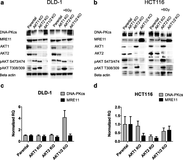
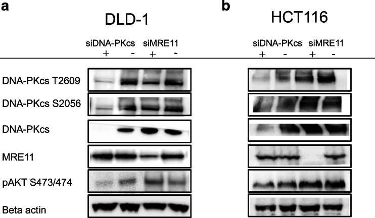
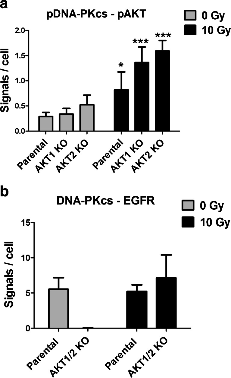
In response to ionizing radiation, several signaling cascades in the cell are activated to repair the DNA breaks, prevent apoptosis, and keep the cells proliferating. AKT is important for survival and proliferation and may also be an activating factor for DNA-PKcs and MRE11, which are essential proteins in the DNA repair process. AKT (PKB) is hyperactivated in several cancers and is associated with resistance to radiotherapy and chemotherapy. There are three AKT isoforms (AKT1, AKT2, and AKT3) with different expression patterns and functions in several cancer tumors. The role of AKT isoforms has been investigated in relation to radiation response and their effects on DNA repair proteins (DNA-PKcs and MRE11) in colon cancer cell lines. The knockout of AKT1 and/or AKT2 affected the radiation sensitivity, and a deficiency of both isoforms impaired the rejoining of radiation-induced DNA double strand breaks. Importantly, the active/phosphorylated forms of AKT and DNA-PKcs associate and exposure to ionizing radiation causes an increase in this interaction. Moreover, an increased expression of both DNA-PKcs and MRE11 was observed when AKT expression was ablated, yet only DNA-PKcs expression influenced AKT phosphorylation. Taken together, these results demonstrate a role for both AKT1 and AKT2 in radiotherapy response in colon cancer cells involving DNA repair capacity through the nonhomologous end joining pathway, thus suggesting that AKT in combination with DNA-PKcs inhibition may be used for radiotherapy sensitizing strategies in colon cancer.


Figures

Fig. 1
Fig. 1
Protein expression and mRNA levels of DNA-PKcs and MRE11 are influenced by the AKT isoforms. Western blots were performed to study the protein expression and phosphorylation of the AKT isoforms, DNA-PKcs and MRE11 in the colorectal cancer cell lines DLD-1 (a) and HCT116 (b) and their corresponding AKT isogenic knockout. Cell-lysates were made before and after irradiation (1 h post IR 6 Gy). The mRNA level of DNA-PKcs and MRE11 were analyzed with qPCR in DLD-1 cells (c) and HCT116 cells (d) and their corresponding AKT isogenic knockouts. The data is from at least two biological replicates with each sample measured in triplicates using Beta-actin as reference in the delta-delta Ct model. The error bars represent the normalized RQ min and max
Fig. 2
Fig. 2
The phosphorylation of AKT is influenced by DNA-PKcs but not MRE11. DNA-PKcs and MRE11 expression was suppressed with siRNA against either DNA-PKcs or MRE11 in DLD-1 cells (a) and HCT116 cells (b) and lysates were made 1 h after irradiation (2 Gy) 3 days after transfection. The expression of AKT and DNA-PKcs and MRE11 was studied with western blot. The experiment was repeated at least twice and a representative western blot is shown
Fig. 3
Fig. 3
DNA-PKcs is in close proximity to phospho-Ser473/474-AKT or EGFR in DLD-1 cells. DLD-1 and its corresponding AKT isotype knockout cells were exposed to radiation (1 h post IR 10 Gy) and analyzed with the proximity ligation assay (PLA) of the particular proteins. The interaction between phospho-Thr2609-DNA-PKcs with phospho-Ser473/474-AKT A) as well as between DNA-PKcs and EGFR B) were analyzed before and after irradiation (1 h, post IR10 Gy). The error bars represents the standard deviation from five measurements. The difference was analyzed with Student’s t test where *P < 0.05, **P < 0.01, and ***P < 0.001
Fig. 4
Fig. 4
Radiation survival is dependent on AKT, DNA-PKsc and serum concentration. Radiation sensitivity was analyzed with clonogenic assay (4 Gy) in DLD-1 AKT isoform knockout cell lines treated with mock (a) or siRNA against DNA-PKcs (b) in 10 % FBS and 0.5 % FBS. The cells treated with 0.5 % FBS were first seeded in flasks with 10 % FBS and incubated for 24 h to allow attachment before changing to 0.5 % FBS. The cells were incubated in 24 h in 0.5 % FBS before radiation and kept in the same media after treatment. The error bars represents the standard deviation from at least three experiments. Student’s t test evaluated if there were any significant differences in SF between the parental and the AKT KO cell lines with *P < 0.05, **P < 0.01, and ***P < 0.001
Fig. 5
Fig. 5
DNA double strand break-rejoining rate correlates with radiation sensitivity in DLD-1 AKT isoform deficient cell lines. DNA double strand break rejoining rate, evaluated with pulsed-field gel electrophoresis after irradiation (40 Gy), in the parental and AKT isoform knockout cell lines of DLD-1 (a) and HCT116 (b) in 10 % FBS. The error bars represent the standard deviation of at least two measurements
Fig. 6
Fig. 6
DNA double strand break-rejoining rate in AKT isoform deficient DLD-1 with suppressed DNA-PKcs expression. DNA double strand break rejoining rate, evaluated with pulsed field gel electrophoresis after irradiation (40 Gy), a in DLD-1 and the AKT isoform knockout cell lines at 10 % FBS and 0.5 % FBS and b mtreated with siRNA against DNA-PKcs. c Graph representing unrejoined DNA double strand breaks 4 h post irradiation in DLD-1 parental and AKT1/2 KO cell lines treated with mock or siDNAPK-cs in 10 % or 0.5 % FBS. The error bars represent the standard deviation of at least two measurements. Student’s t-test evaluated if there were any significant differences in DNA rejoining between the parental and the AKT KO cell lines with *P < 0.05, **P < 0.01, and ***P < 0.001

Similar articles

Cited by

References

    1. Glimelius B, Gronberg H, Jarhult J, Wallgren A, Cavallin-Stahl E. A systematic overview of radiation therapy effects in rectal cancer. Acta Oncol. 2003;42(5–6):476–492. doi: 10.1080/02841860310012301. - DOI - PubMed
    1. Krishnan S, Janjan N, Skibber J, Rodriguez-Bigas M, Wolff R, Das P, et al. Phase II study of capecitabine (Xeloda) and concomitant boost radiotherapy in patients with locally advanced rectal cancer. Int. J. Radiat. Oncol. Biol. Phys. 2006;66(3):762–771. doi: 10.1016/j.ijrobp.2006.05.063. - DOI - PubMed
    1. Parkin DM, Bray F, Ferlay J, Pisani P. Global cancer statistics, 2002. CA Cancer J Clin. 2005;55(2):74–108. doi: 10.3322/canjclin.55.2.74. - DOI - PubMed
    1. Vaisman A, Varchenko M, Umar A, Kunkel TA, Risinger JI, Barrett JC, et al. The role of hMLH1, hMSH3, and hMSH6 defects in cisplatin and oxaliplatin resistance: correlation with replicative bypass of platinum-DNA adducts. Cancer Res. 1998;58(16):3579–3585. - PubMed
    1. Manning BD, Cantley LC. AKT/PKB signaling: navigating downstream. Cell. 2007;129(7):1261–1274. doi: 10.1016/j.cell.2007.06.009. - DOI - PMC - PubMed

Publication types

MeSH terms

LinkOut - more resources