

An unusual presentation of subdural empyema caused by Porphyromonas gingivalis
- PMID: 24339621
- PMCID: PMC3841642
- DOI: 10.4103/0972-2327.120447
An unusual presentation of subdural empyema caused by Porphyromonas gingivalis
Abstract
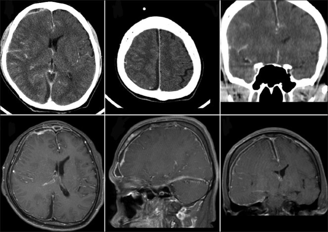
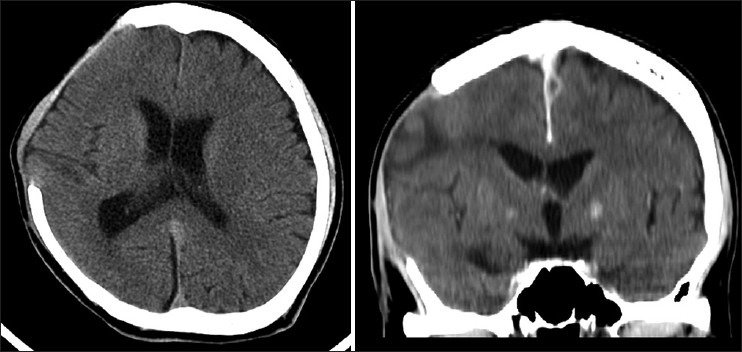
Subdural empyema is an uncommon clinical entity. The first case of Porphyromonas gingivalis subdural empyema is reported. We report a case of 34-year-old male who presented with subdural empyema and sinusitis. Through the utilization of polymerase chain reaction (PCR) tests on subdural pus, we were able to confirm the diagnosis and institute appropriate treatment. Early surgical intervention and intravenous antibiotics meant that the patient recovered fully. Infections caused by P. gingivalis should be considered in differential diagnoses of central nervous system (CNS) abscesses or subdural empyema especially in patients with precedent periodontal diseases and sinusitis.
Keywords: Porphyromonas gingivalis; periodontal disease; subdural empyema.
Conflict of interest statement
Figures
Similar articles
-
An unusual case of meningococcal meningitis complicated with subdural empyema in a 3 month old infant: a case report.Cases J. 2009 Sep 17;2:6335. doi: 10.1186/1757-1626-0002-0000006335. Cases J. 2009. PMID: 20181146 Free PMC article.
-
Subcutaneous abscess due to empyema necessitans caused by Porphyromonas gingivalis in a patient with periodontitis.IDCases. 2022 Feb 21;27:e01458. doi: 10.1016/j.idcr.2022.e01458. eCollection 2022. IDCases. 2022. PMID: 35242562 Free PMC article.
-
[Case of subdural empyema caused by Porphyromonas and Fusobacterium identified by 16S ribosomal RNA gene sequence analysis].Rinsho Shinkeigaku. 2010 May;50(5):311-4. doi: 10.5692/clinicalneurol.50.311. Rinsho Shinkeigaku. 2010. PMID: 20535979 Japanese.
-
[Subdural empyema due to gemella morbillorum as a complication of acute sinusitis].Acta Med Port. 2011 May-Jun;24(3):475-80. Epub 2011 Aug 12. Acta Med Port. 2011. PMID: 22015038 Review. Portuguese.
-
Rare Concurrent Retroclival and Pan-Spinal Subdural Empyema: Review of Literature with an Uncommon Illustrative Case.World Neurosurg. 2018 Feb;110:326-335. doi: 10.1016/j.wneu.2017.11.082. Epub 2017 Nov 23. World Neurosurg. 2018. PMID: 29174228 Review.
Cited by
-
Case of a cerebral abscess caused by Porphyromonas gingivalis in a subject with periodontitis.BMJ Case Rep. 2017 Feb 22;2017:bcr2016218845. doi: 10.1136/bcr-2016-218845. BMJ Case Rep. 2017. PMID: 28228396 Free PMC article.
-
Case report:Multiple abscesses caused by Porphyromonas gingivalis diagnosed by metagenomic next-generation sequencing.Front Med (Lausanne). 2023 Jan 26;9:1089863. doi: 10.3389/fmed.2022.1089863. eCollection 2022. Front Med (Lausanne). 2023. PMID: 36777162 Free PMC article.
References
-
- Youmans JR. Philadelphia: Saunders; 1996. Neurological surger: A Comprehensive Reference Guide to the Diagnosis and Management of Neurosurgical problems.
-
- Nathoo N, Nadvi SS, van Dellen JR, Gouws E. Intracranial subdural empyemas in the era of computed tomography: A review of 699 cases. Neurosurgery. 1999;44:529–35. - PubMed
-
- Hlavin ML, Kaminski HJ, Fenstermaker RA, White RJ. Intracranial suppuration: A modern decade of postoperative subdural empyema and epidural abscess. Neurosurgery. 1994;34:974–80. - PubMed
Publication types
LinkOut - more resources
Full Text Sources
Other Literature Sources
