. 2013 Dec 8:19:2501-7.
eCollection 2013.

Analysis of superoxide dismutase 1, dual-specificity phosphatase 1, and transforming growth factor, beta 1 genes expression in keratoconic and non-keratoconic corneas

Affiliations

Analysis of superoxide dismutase 1, dual-specificity phosphatase 1, and transforming growth factor, beta 1 genes expression in keratoconic and non-keratoconic corneas

Samira Saee-Rad et al. Mol Vis. .

Abstract

Purpose: To quantitatively assess the superoxide dismutase 1 (SOD1), transforming growth factor, beta 1 (TGF-β1), and dual-specificity phosphatase 1 (DUSP1) messenger ribonucleic acid (mRNA) expression levels as the main intracellular reactive oxygen species neutralizers, wound healing mediators, and immunomodulators (respectively) in keratoconic (KCN) and non-KCN corneas.

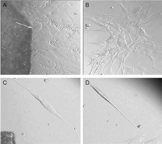
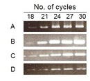
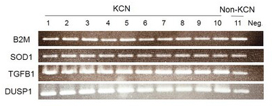
Methods: Total RNA was extracted from normal and keratoconic cultured corneal stromal fibroblasts. Semiquantitative reverse transcriptase polymerase chain reaction (RT-PCR) was used to measure the relative expression levels of mRNAs of the SOD1, TGF-β1, and DUSP1 genes.

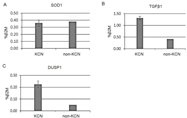
Results: The mRNA expression of TGF-β1 and DUSP1 was augmented in the KCN corneas (three- and fivefold, respectively; both p<0.05). The KCN and non-KCN samples showed no difference in comparative SOD1 mRNA levels.

Conclusions: This study demonstrated a higher level of DUSP1 and TGF-β1 expression as known molecules in the inflammatory process. These results may provide new insight into the complex molecular pathways underlying KCN for investigating other inflammatory molecules.


Figures

Figure 1
Figure 1
Primary corneal tissue culture. This figure represents primary tissue culture from sample no. 4 under light microscopy. A: The migration of cells out of the tissue started on day 7 after seeding (white arrow shows margin of tissue, 10X magnification). B: One of the cell colonies was performed after 15 days post-seeding (20X magnification). C, D: After cell culture expansion, the cultivated cells were starved for 24 h to compare the morphological difference between fibroblasts and keratocytes (C, D, respectively).
Figure 2
Figure 2
Determination of the exponential cycle of amplification. Semiquantitative reverse transcription–polymerase chain reaction (RT–PCR) mixtures were taken for five alternating cycles starting at cycles 18, 21, 24, 27, and 30 for A: beta 2 microglobulin (B2M), B: superoxide dismutase 1 (SOD1), C: dual-specificity phosphatase 1 (DUSP1), and D: transforming growth factor, beta 1 (TGF-B1). Cycle 24 was selected as an exponential cycle.
Figure 3
Figure 3
Differential messenger ribonucleic acid expression of the target genes via semiquantitative reverse transcription–polymerase chain reaction (RT-PCR; 24th cycle). This figure shows the results of the RT–PCR products in 2% agarose gel electrophoresis, stained with ethidium bromide. Lanes 1 to 10: Keratoconus (KCN) corneas; lane 11: pooled normal corneas and lane 12: negative control without RT. The fluorescent intensity of the bands was quantified with a GS-800 densitometer. The signal intensities were normalized to values obtained for beta 2 microglobulin (B2M). The messenger ribonucleic acid (mRNA) expression of transforming growth factor, beta 1 (TGF-β1), and dual-specificity phosphatase 1 (DUSP1) was increased in the KCN corneas (three- and fivefold, respectively; both p<0.05). The comparative superoxide dismutase 1 (SOD1) mRNA level showed no difference between the KCN and non-KCN samples.
Figure 4
Figure 4
The comparative mRNA expression of target genes in ten keratoconus (KCN) and pooled normal corneal tissues. This graph indicates the comparison of transcript band intensities for genes encoding superoxide dismutase 1 (SOD1), transforming growth factor, beta 1 (TGF-β1), and dual-specificity phosphatase 1 (DUSP1) between the KCN and non-KCN samples. The values were calculated relative to the beta 2 microglobulin (B2M) levels (as normalizer) and reported as mean percentages. Note that the KCN corneas showed a three- and fivefold increase in TGF-B1 and DUSP1 when compared to the pooled normal corneas. The RNA levels for SOD1 were similar for the KCN and normal corneas (p<0.05). Normal corneal samples were pooled to reduce tissue preparation errors. The error bars represent the standard deviation.

References

    1. Gillan W. The stroma and keratoconus: a review. S Afr Optom. 2007;66:87–93.
    1. Abu-Amero KK, Kalantan H, Al-Muammar AM. Analysis of the VSX1 gene in keratoconus patients from Saudi Arabia. Mol Vis. 2011;17:667–72. - PMC - PubMed
    1. Sharma R, Titiyal JS, Prakash G, Sharma N, Tandon R, Vajpayee RB. Clinical profile and risk factors for keratoplasty and development of hydrops in north Indian patients with keratoconus. Cornea. 2009;28:367–70. - PubMed
    1. Kennedy RHBW, Dyer JA. A 48-year clinical and epidemiologic study of keratoconus. Am J Ophthalmol. 1986;101:267–73. - PubMed
    1. Balasubramanian SA, Pye DC, Willcox MDP. Are proteinases the reason for keratoconus? Curr Eye Res. 2010;35:185–91. - PubMed

Publication types

MeSH terms

LinkOut - more resources