. 2013 Nov;15(11):1282-91.
doi: 10.1593/neo.131362.

Hepatitis B virus X protein inhibits tumor suppressor miR-205 through inducing hypermethylation of miR-205 promoter to enhance carcinogenesis

Affiliations

Hepatitis B virus X protein inhibits tumor suppressor miR-205 through inducing hypermethylation of miR-205 promoter to enhance carcinogenesis

Tao Zhang et al. Neoplasia. 2013 Nov.

Abstract

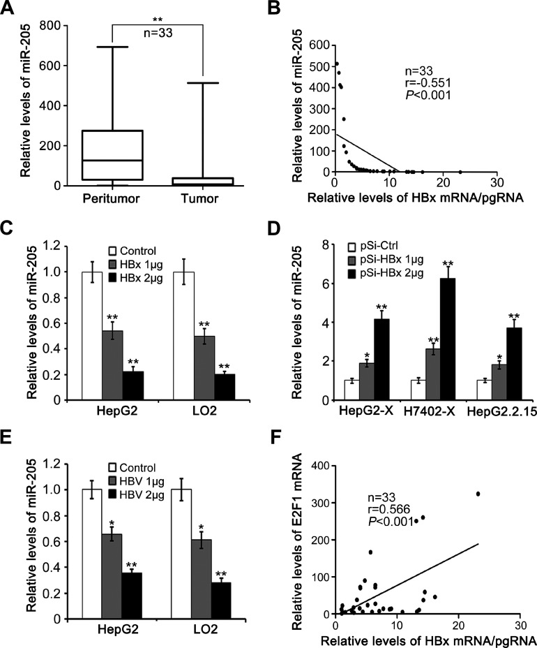
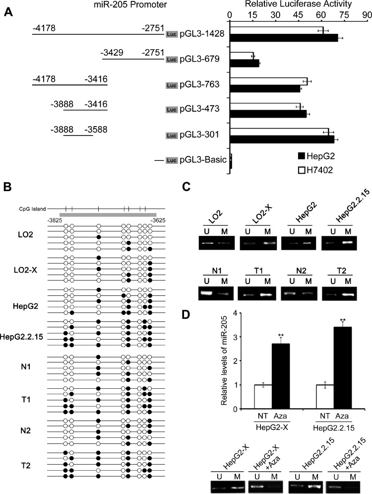
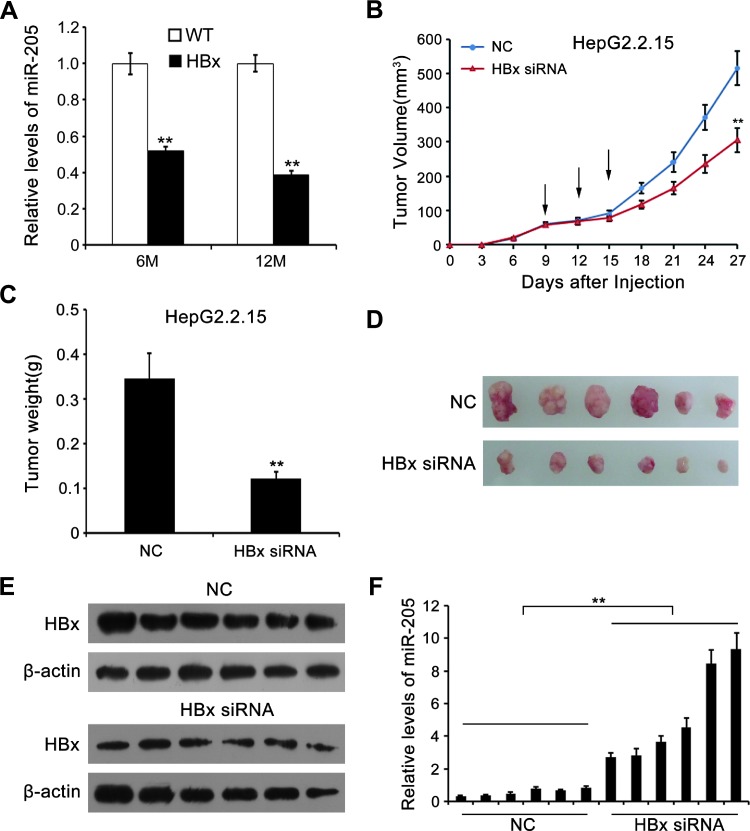
The infection of hepatitis B virus (HBV) is closely associated with the development of hepatocellular carcinoma (HCC), in which HBV X protein (HBx) plays crucial roles. MicroRNAs are involved in diverse biologic functions and in carcinogenesis by regulating gene expression. In the present study, we aim to investigate the underlying mechanism by which HBx enhances hepatocarcinogenesis. We found that miR-205 was downregulated in 33 clinical HCC tissues in comparison with adjacent noncancerous hepatic tissues. The expression levels of miR-205 were inversely correlated with those of HBx in abovementioned tissues. Then, we demonstrated that HBx was able to suppress miR-205 expression in hepatoma and liver cells. We validated that miR-205 directly targeted HBx mRNA. Ectopic expression of miR-205 downregulated HBx, whereas depletion of endogenous miR-205 upregulated HBx in hepatoma cells. Notably, our data revealed that HBx downregulated miR-205 through inducing hypermethylation of miR-205 promoter in the cells. In terms of function, the forced miR-205 expression remarkably inhibited the HBx-enhanced proliferation of hepatoma cells in vitro and in vivo, suggesting that miR-205 is a potential tumor-suppressive gene in HCC. HBx-transgenic mice showed that miR-205 was downregulated in the liver. Importantly, HBx was able to abrogate the effect of miR-205 on tumor suppression in carcinogenesis. Therefore, we conclude that HBx is able to inhibit tumor suppressor miR-205 to enhance hepatocarcinogenesis through inducing hypermethylation of miR-205 promoter during their interaction. Therapeutically, miR-205 may be useful in the treatment of HCC.


Figures

Figure 1
Figure 1
HBx is able to decrease miR-205 in hepatoma and liver cells. (A) miR-205 expression was quantified by qRT-PCR in HCC and peritumor tissues. (B) The correlation of HBx and miR-205 was determined by qRT-PCR in abovementioned HCC tissues. (C) miR-205 expression was examined by qRT-PCR in HepG2 hepatoma cells/LO2 human immortalized liver cells after transfection with pCMV-X (HBx plasmid). (D) miR-205 expression was detected by qRT-PCR after knockdown of HBx (pSi-HBx) in hepatoma cells. (E) miR-205 expression was examined by qRT-PCR in HepG2/LO2 cells after transfection with pCH-9/3091 (HBV plasmid). (F) The correlation of HBx and E2F1 was determined by qRT-PCR in clinical HCC tissues. Data are presented as means ± SD (*P < .05; **P < .01). We performed three separate experiments in triplicate.
Figure 2
Figure 2
miR-205 directly targets HBx mRNA. (A) The predicted binding sites for miR-205 in HBx mRNA by computational methods were shown. Mutations were designed in the binding sites. (B and C) The plasmid pGL3-HBx or pGL3-HBx-mut was cotransfected with synthetic miRNA mimics (miR-205) or antagomirs (anti-miR-205) into HepG2 cells. The luciferase activities of pGL3-HBx or pGL3-HBx-mut were determined at 48 hours after transfection. (D and E) The expression of HBx was examined by qRT-PCR and immunoblot analysis, respectively, after treatment with miR-205 or anti-miR-205 in the cells. Data are presented as means ± SD (*P < .05;**P < .01). We performed three separate experiments in triplicate.
Figure 3
Figure 3
HBx downregulates miR-205 through inducing DNA hypermethylation of miR-205 promoter. (A) miR-205 promoter core region was identified in hepatoma cells by luciferase reporter gene assays. (B) The methylation of miR-205 CpG sites was analyzed by bisulfitesequencing analysis in the cells, paired nontumor liver tissues (N), and HCC tissues (T). At least five independent clones were sequenced per sample. Open and filled circles represent nonmethylated and methylated CpG sites, respectively. (C) The methylation of miR-205 CpG sites was examined by MSP analysis as well. (D) miR-205 expression was measured by qRT-PCR, followed by MSP analysis in the miR-205 CpG sites in hepatoma cells treated with 5 µM Aza for 72 hours. Data are presented as means ± SD (**P < .01). We performed three separate experiments in triplicate.
Figure 4
Figure 4
miR-205 inhibits the proliferation of hepatoma cells in vitro. (A–C) MTT assay, EdU incorporation assay, and clonogenicity analysis were carried out in the cells with transfection of 100 nM miR-205, respectively. Data are presented as means ± SD (**P < .01). We performed three separate experiments in triplicate.
Figure 5
Figure 5
miR-205 inhibits the proliferation of hepatoma cells in vivo. (A) The nude mice (n = 6) were transplanted by HepG2.2.15 cells. The tumors were injected with 5 µg (100-µl total volume) of miRNA NC, miR-205, or miR-205/HBx plasmid (pCMV-X) on days 9, 12, and 15, respectively. The growth curves of the tumors in nude mice were shown. (B) The diagram showed the average weight of tumors. (C) The photos of tumors from mice transplanted with HepG2.2.15 cells were shown. (D) The expression levels of HBx were examined by Western blot analysis in the tumor tissues from mice. Data are presented as means ± SD (**P < .01). We performed three separate experiments in triplicate.
Figure 6
Figure 6
HBx promotes the growth of tumor through suppressing miR-205 in vivo. (A) miR-205 expression was examined by qRT-PCR in 6- and 12-month-old HBx-transgenic mice. (B) The nude mice (n = 6) were transplanted by HepG2.2.15 cells. The tumors were injected with siRNA control (NC) and HBx siRNA (5 µg/100 µl) on days 9, 12, and 15, respectively. The growth curves of the tumors in nude mice were shown. (C) The diagram showed the average weight of tumors. (D) The photos of tumors from mice were shown. (E and F) The expression levels of HBx and miR-205 were examined by Western blot analysis and qRT-PCR in the tumor tissues from mice, respectively. Data are presented as means ± SD (**P < .01). We performed three separate experiments in triplicate.

References

    1. Tsai WL, Chung RT. Viral hepatocarcinogenesis. Oncogene. 2010;29:2309–2324. - PMC - PubMed
    1. Wang Y, Lu Y, Toh ST, Sung WK, Tan P, Chow P, Chung AY, Jooi LL, Lee CG. Lethal-7 is down-regulated by the hepatitis B virus x protein and targets signal transducer and activator of transcription 3. J Hepatol. 2010;53:57–66. - PubMed
    1. Dandri M, Locarnini S. New insight in the pathobiology of hepatitis B virus infection. Gut. 2012;61(suppl 1):i6–i17. - PubMed
    1. El-Serag HB. Epidemiology of viral hepatitis and hepatocellular carcinoma. Gastroenterology. 2012;142:1264–1273. e1261. - PMC - PubMed
    1. Neuveut C, Wei Y, Buendia MA. Mechanisms of HBV-related hepatocarcinogenesis. J Hepatol. 2010;52:594–604. - PubMed

Publication types

MeSH terms