. 2013 Dec;11(12):e1001731.
doi: 10.1371/journal.pbio.1001731. Epub 2013 Dec 10.

Escherichia coli ribosomal protein S1 unfolds structured mRNAs onto the ribosome for active translation initiation

Affiliations

Escherichia coli ribosomal protein S1 unfolds structured mRNAs onto the ribosome for active translation initiation

Mélodie Duval et al. PLoS Biol. 2013 Dec.

Abstract

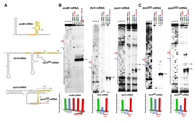
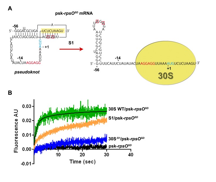
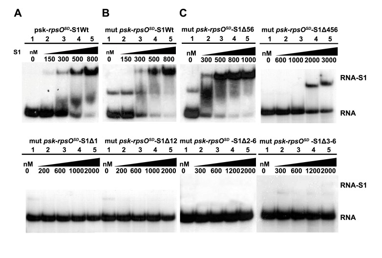
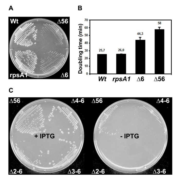
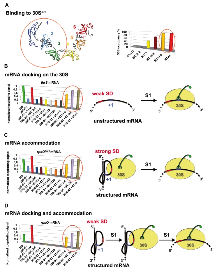
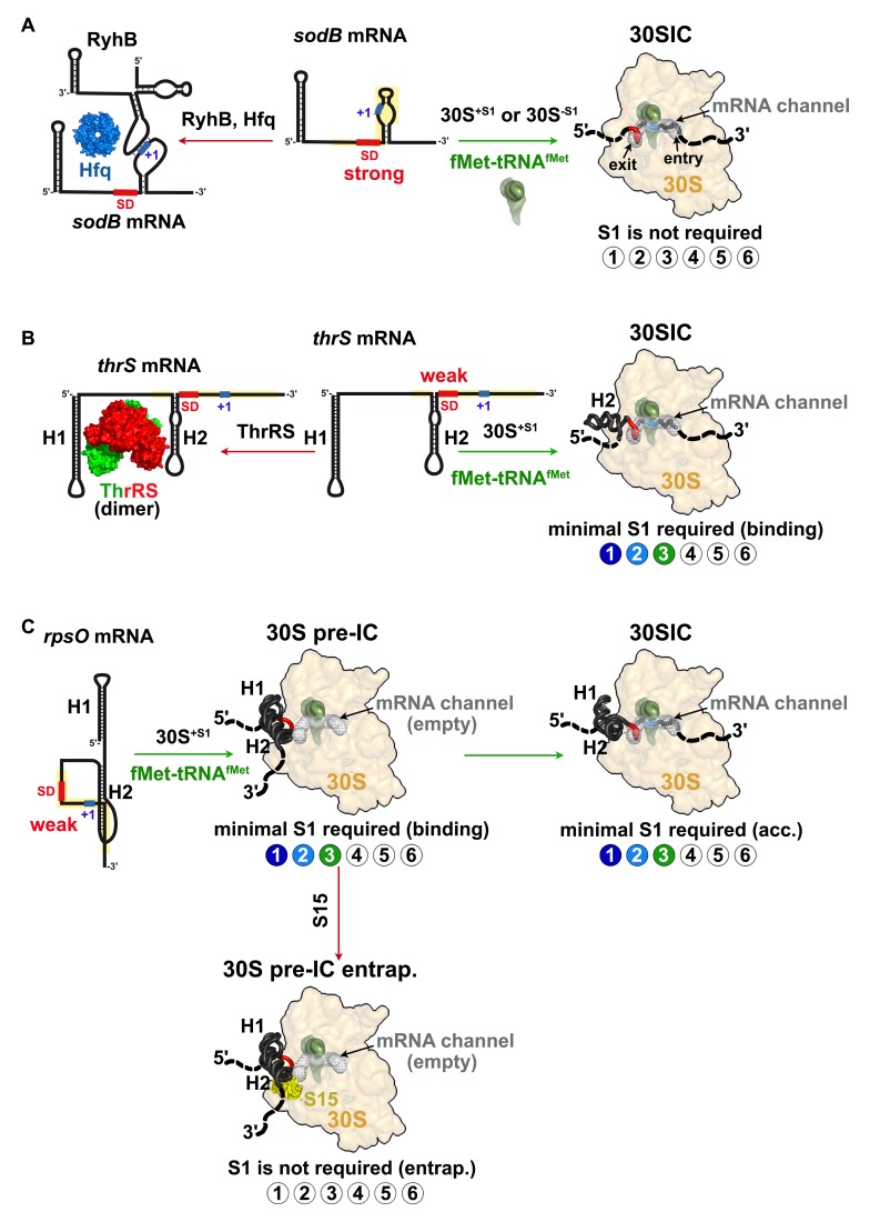
Regulation of translation initiation is well appropriate to adapt cell growth in response to stress and environmental changes. Many bacterial mRNAs adopt structures in their 5' untranslated regions that modulate the accessibility of the 30S ribosomal subunit. Structured mRNAs interact with the 30S in a two-step process where the docking of a folded mRNA precedes an accommodation step. Here, we used a combination of experimental approaches in vitro (kinetic of mRNA unfolding and binding experiments to analyze mRNA-protein or mRNA-ribosome complexes, toeprinting assays to follow the formation of ribosomal initiation complexes) and in vivo (genetic) to monitor the action of ribosomal protein S1 on the initiation of structured and regulated mRNAs. We demonstrate that r-protein S1 endows the 30S with an RNA chaperone activity that is essential for the docking and the unfolding of structured mRNAs, and for the correct positioning of the initiation codon inside the decoding channel. The first three OB-fold domains of S1 retain all its activities (mRNA and 30S binding, RNA melting activity) on the 30S subunit. S1 is not required for all mRNAs and acts differently on mRNAs according to the signals present at their 5' ends. This work shows that S1 confers to the ribosome dynamic properties to initiate translation of a large set of mRNAs with diverse structural features.


Conflict of interest statement

The authors have declared that no competing interests exist.

Figures

Figure 1
Figure 1. Formation of simplified initiation complexes involving three different mRNAs.
(A) Secondary structure models of sodB, thrS, and rpsO mRNAs including their 5′UTR and the RBSs. The secondary structure of sodB mRNA is derived from , of thrS mRNA from , and of rpsO from . The SD sequence is in red, the AUG codon in blue, and the RBS in yellow. The mutations at the SD of thrS and rpsO are specified. (B) Effect of S1 on the formation of the initiation complex (30SIC) analyzed by toeprinting assays using different mRNAs (sodB, thrS, rpsO). Lanes 1 and 2, incubation controls of the mRNA alone (lane 1) or bound to wild-type S1 (S1Wt, lane 2); lanes 3 and 4, the 30SIC was formed with the mRNA, the initiator tRNA, and either the wild-type 30S (lane 3, 30SWt), or the 30S lacking S1 (lane 4, 30S-S1); lanes 5–7, the 30SIC was formed with 30S-S1 pre-incubated with increasing concentrations of S1 (30S-S1+S1Wt): lane 5, 200 nM; lane 6, 500 nM; lane 7, 1 µM. Lanes A, C, G, U, sequencing ladders. Below the gels, the quantification of the toeprint was normalized according to the total amount of radioactivity (full-length extension and +16 product bands) using the SAFA software . The data represented the yield of mRNA bound to 30SWt (green), 30S-S1 (blue), and 30S-S1+S1 (red). (C) Effect of S1 on the 30SIC formation analyzed by toeprinting with thrSSD and rpsOSD mRNAs, which contained an enhanced SD sequence. Same legend as in panel B.
Figure 2
Figure 2. Ribosomal protein S1 induces melting of rpsO pseudoknot structure.
(A) Two 2-APs were introduced at positions A-40 and A-42 of the pseudoknot structure of rpsOSD mRNA (psk-rpsOSD) carrying an enhanced SD sequence (nucleotides squared in red). In the pseudoknot structure, the two adenines form Watson–Crick base pairs with residues of the coding sequence (in yellow). When the mRNA is placed in the 30S decoding channel, the pseudoknot is melted and these adenines become accessible. The initiation codon AUG is in blue. (B) The spectra show the 2-AP fluorescence emission of the corresponding modified adenines upon injection of wild-type 30S (30S WT/psk-rpsOSD, in green), of 30S lacking S1 (30S−S1/psk-rpsOSD, in blue), or of S1 alone (S1/psk-rpsOSD, in orange). The fitting of the experimental curves was performed with graphpad PRISM software.
Figure 3
Figure 3. Domains 1 to 3 of r-protein S1 are essential for the recognition of rpsO mRNA.
(A) Gel retardation assays were performed on complexes formed with the 5′ end-labeled rpsOSD mRNA (psk-rpsOSD) and wild-type r-protein S1 (S1Wt). Lane 1, incubation control of psk-rpsOSD alone; lanes 2–5, complex formation was performed with various concentrations of S1Wt as indicated on the top of the autoradiography. The addition of S1 causes fuzzy bands due to the dissociation of the complex during the migration. (B) Gel retardation assays were done on complexes formed with the 5′ end-labeled mutant C-14G psk-rpsOSD (mut psk-rpsOSD) and S1Wt. Lane 1, incubation control of mut psk-rpsOSD alone; lanes 2–5, complex formation was performed with various concentrations of S1Wt as indicated. (C) Gel retardation assays were done on complexes formed with mut psk-rpsOSD and various truncated forms of r-protein S1. The protein was deleted of either domains 5 and 6 (Δ56), domains 4 to 6 (Δ4–6), domain 1 (Δ1), domains 1 and 2 (Δ12), domains 2 to 6 (Δ2–6), or domains 3 to 6 (Δ3–6). The positions of the complex (RNA-S1) and of the free RNA (RNA) are given. Same legend as in panel B.
Figure 4
Figure 4. Effect of successive deletion in rpsA performed at the original rpsA locus on cell growth.
(A) Growth was compared between WT strains (Wt, rpsA1) and strains carrying deletions of domains 6 (Δ6) and of domains 5 and 6 (Δ56) in rpsA on LB plates at 37°C. The E. coli strains are AnK02 (WT), MS77 (rpsA1), MS78 (Δ6), and MS79 (Δ56). (B) Measurements of the doubling times of various strains. The growth was done in LB medium at 37°C. The strains were identical to those of the panel A. (C) The growth was compared in strains carrying deletions of domains 5 and 6 (Δ56), 4 to 6 (Δ4–6), 3 to 6 (Δ3–6), 2 to 6 (Δ2–6) in rpsA. They were complemented with the plasmid pNK34, which carries WT rpsA under the control of the hybrid trc promoter with the lac operator. The experiments were done in the presence of IPTG (+IPTG) or in the absence of IPTG (−IPTG). Strains are MS79pNK34 (Δ56), MS84pNK34 (Δ4–6), MS83pNK34 (Δ3–6), and MS82pNK34 (Δ2–6).
Figure 5
Figure 5. Effect of S1 variants on 30S binding and formation of the simplified initiation complex.
(A) Domains 1 to 3 of r-protein S1 are required for efficient 30S−S1 binding. (Left panel) A model of r-protein S1 was built based on the structure of domains 4 to 6 analyzed by NMR and SAXS experiments ,. Each OB-fold domains are represented in different colors. (Right panel) Direct binding of r-protein S1 variants to 30S was visualized by Western blot analysis and quantified (see Text S1). Wild-type S1 (S1wt); S1 was deleted of domains 1 and 2 (S1Δ12), of domains 2 to 6 (S1Δ2–6), of domain 1 (S1Δ1), of domains 3 to 6 (S1Δ3–6), and of domains 4 to 6 (S1Δ4–6). (B) Domains 1 to 3 of S1 are essential for thrS mRNA docking on the 30S. Formation of the 30S initiation complex (30SIC) with thrS mRNA was probed by toeprinting. The toeprint at position +16 was quantified and normalized to the full-length RNA. The 30SIC was done with the 30S, with the 30S-S1 lacking S1, and with the 30S-S1 complemented with either wild-type S1 (S1Wt) or with the truncated forms of S1: deletion of domain 1 (S1Δ1), domains 1 and 2 (S1Δ12), domains 1, 2, and 6 (S1Δ126), domains 1 and 6 (S1Δ16), domains 5 and 6 (S1Δ56), domain 6 (S1Δ6), domains 4 to 6 (S1Δ4–6), domains 3 to 6 (S1Δ3–6), and domains 2 to 6 (S1Δ2–6). (C) Toeprinting assays performed with rpsOSD mRNA show that domains 1 to 3 of S1 are required to accommodate mRNA into the decoding channel. (D) Toeprinting assays performed with rpsO mRNA demonstrate that domains 1 to 3 of S1 are essentiel for the docking and the accommodation steps. (C and D) The legends are as in panel B. (B–D) A schematic drawing illustrates the key roles of S1 in the different steps of the formation of the 30SIC involving thrS, rpsOSD, and rpsO mRNAs. The 30S is colored in yellow, and the initiator tRNA is in green. The SD sequence and the AUG codon are colored in red and blue, respectively.
Figure 6
Figure 6. Different mechanisms of action of S1 r-protein.
(A) Formation of the 30S initiation complex (30SIC) with E. coli sodB mRNA is S1-independent. The mRNA contains a weakly structured RBS (in yellow) and a strong SD sequence (in red) . Under iron depletion, binding of RyhB and Hfq to sodB mRNA occludes the 30S binding and causes rapid degradation of the mRNA ,. (B) A single step pathway to form the initiation complex with thrS mRNA. The bipartite RBS comprised an unstructured SD sequence and a U-rich sequence upstream of the H2 domain. The three first domains of S1 are essential and sufficient for the docking of thrS mRNA to the 30S. The free ThrRS binds to the H1 and H2 hairpin motifs to prevent the 30S binding (adapted from [34]). (C) A two-step pathway to form the 30SIC involving E. coli rpsO mRNA. The pseudoknot structure is recognized by the 30S and by r-protein S15 (in yellow). Domains 1 to 3 of S1 are required both for the docking and the accommodation of rpsO mRNA on the 30S to relocate the initiation codon into the P-site. The free S15 binds and stabilizes the pseudoknot on the 30S and prevents the initiation codon to reach the decoding center. The six OB-fold domains of S1 are schematized by circles: colored circles are the active domains of S1, and white circles represented the domains that are not required for the translation of a given mRNA. The color code is as found in Figure 5A.

Similar articles

Cited by

References

    1. Milón P, Maracci C, Filonava L, Gualerzi CO, Rodnina MV (2012) Real-time assembly landscape of bacterial 30S translation initiation complex. Nat Struct Mol Biol 19: 609–615. - PubMed
    1. Studer SM, Joseph S (2006) Unfolding of mRNA secondary structure by the bacterial translation initiation complex. Mol Cell 22: 105–115. - PubMed
    1. Yusupova G, Yusupov MM, Cate JH, Noller HF (2001) The path of messenger RNA through the ribosome. Cell 106: 233–241. - PubMed
    1. Jenner L, Romby P, Rees B, Schulze-Briese C, Springer M, et al. (2005) Translational operator of mRNA on the ribosome: how repressor proteins exclude ribosome binding. Science 308: 120–123. - PubMed
    1. Yusupova G, Jenner L, Rees B, Moras D, Yusupov M (2006) Structural basis for messenger RNA movement on the ribosome. Nature 444: 391–394. - PubMed

Publication types

MeSH terms