. 2013;9(12):e1003803.
doi: 10.1371/journal.ppat.1003803. Epub 2013 Dec 5.

The TFPI-2 derived peptide EDC34 improves outcome of gram-negative sepsis

Affiliations

The TFPI-2 derived peptide EDC34 improves outcome of gram-negative sepsis

Praveen Papareddy et al. PLoS Pathog. 2013.

Abstract

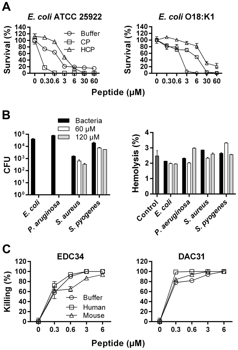
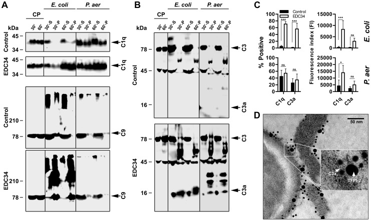
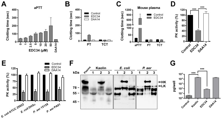
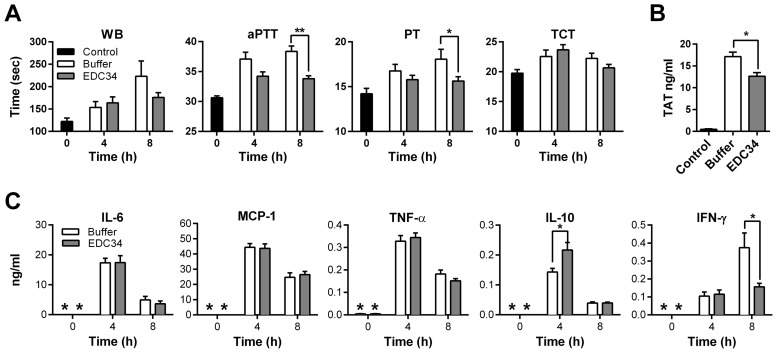
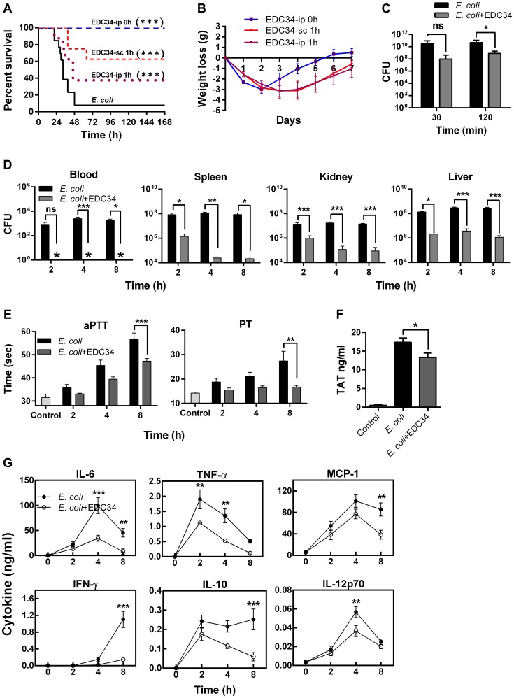
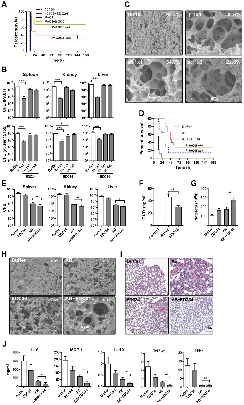
Sepsis is characterized by a dysregulated host-pathogen response, leading to high cytokine levels, excessive coagulation and failure to eradicate invasive bacteria. Novel therapeutic strategies that address crucial pathogenetic steps during infection are urgently needed. Here, we describe novel bioactive roles and therapeutic anti-infective potential of the peptide EDC34, derived from the C-terminus of tissue factor pathway inhibitor-2 (TFPI-2). This peptide exerted direct bactericidal effects and boosted activation of the classical complement pathway including formation of antimicrobial C3a, but inhibited bacteria-induced activation of the contact system. Correspondingly, in mouse models of severe Escherichia coli and Pseudomonas aeruginosa infection, treatment with EDC34 reduced bacterial levels and lung damage. In combination with the antibiotic ceftazidime, the peptide significantly prolonged survival and reduced mortality in mice. The peptide's boosting effect on bacterial clearance paired with its inhibiting effect on excessive coagulation makes it a promising therapeutic candidate for invasive Gram-negative infections.


Conflict of interest statement

I have read the journal's policy and have the following conflicts. The authors M. Malmsten and A. Schmidtchen are founders and own shares in XImmune AB, a company developing anti-inflammatory peptides for human therapy. This does not alter our adherence to all PLoS Pathogens policies on sharing data and materials.

Figures

Figure 1
Figure 1. Antimicrobial activities of EDC34.
(A) Antibacterial effects of EDC34 at the indicated concentrations were studied in viable count assays, using the bacteria E. coli ATCC 25922 (complement sensitive) and E. coli O18:K1 (complement-resistant). Analyses were performed in 10 mM Tris, 0.15 M NaCl, pH 7.4 (Buffer) or buffer containing 20% citrate plasma (CP) or heat-inactivated citrate plasma (HCP). Mean with SD is shown (n = 3). (B) Antimicrobial and hemolytic effects of EDC34 against the indicated bacteria in whole blood (n = 3, mean±SEM is presented). (C) Antimicrobial effects of EDC34 and DAC31, a mouse derived TFPI-2 C-terminal peptide against E. coli in human or mouse citrate plasma (n = 3, mean±SEM is presented).
Figure 2
Figure 2. EDC34 enhances the binding of complement proteins to bacteria.
(A) E. coli ATCC 25922 and P. aeruginosa PA01 bacteria were incubated 30–60 min with citrate plasma alone (Control) or supplemented with EDC34 (at 3 µM) at 37°C. The bacterial cells and supernatants were collected and proteins were detected by immunoblotting with antibodies recognizing C1q or C5b-9. CP, citrate plasma; S, supernatant or unbound bacteria; P, pellet or material bound to bacterial cells. (B), as in figure 2A, but antibodies against C3a were used. (C) Quantification of deposition of complement components on bacteria by flow cytometry, left panel, comparison of the mean proportion of bacteria positive for C1q/C3a binding in citrate plasma and in plasma supplemented with EDC34. Right panel, comparative degree of C1q and C3a binding to E. coli and P. aeruginosa strains, expressed as means of the fluorescence index (FI; proportion of bacteria positive for C1q/C3a multiplied by the mean intensity of C1q/C3a binding). (n = 4, mean±SEM is presented; Two-Way ANOVA Bonferroni's Multiple Comparison Test). (D) TFPI-2 and C3a binding to bacteria in fibrin slough collected from an infected chronic wound were visualized by using gold-labeled antibodies of different sizes, specific for the C-terminus of TFPI-2 (20 nm) and C3a (10 nm), respectively. Insert shows a higher magnification.
Figure 3
Figure 3. EDC34 inhibits contact activation at negatively charged surfaces.
(A) Effect of EDC34 on the contact system was assessed by measuring the aPTT in human citrate plasma. The control peptide DAA14 was used at 50 µM. (B) Measurement of effects of EDC34 (50 µM) and DAA14 (50 µM) on the extrinsic (PT) and common pathway (TCT) of coagulation. (C) Measurement of aPTT, PT and TCT clotting times of mouse citrate plasma in absence (Control) or presence of 50 µM EDC34. (D) Plasma kallikrein activity induced by negatively charged kaolin in human citrate plasma in absence (Control) or presence of EDC34 (50 µM) or DAA14 (50 µM), was determined by a chromogenic substrate assay. Data are presented as mean±SEM relative to control without peptides (n = 4; One-Way ANOVA Bonferroni's Multiple Comparison Test). (E) Plasma kallikrein activity in human citrate plasma induced by the indicated bacteria in absence (Control) or presence of the peptides EDC34 (50 µM) or DAA14 (50 µM) was measured by a chromogenic substrate assay. Data are presented as mean±SEM relative to control without peptides (n = 5). (F) Western blot analysis of HK and HK degradation products: 1, No peptide; 2, EDC34; 3, DAA14. (G) Bradykinin release in human citrate plasma incubated with kaolin in absence (Control) or presence of EDC34 (50 µM) or DAA14 (50 µM) (n = 3, mean±SEM is presented; One-Way ANOVA Bonferroni's Multiple Comparison Test).
Figure 4
Figure 4. EDC34 is anti-coagulative, but not anti-inflammatory during LPS-shock.
C57BL/6 mice were injected i. p. with 5 mg/kg E. coli LPS O111:B4 and treated after a period of 30 min with buffer (Buffer) or EDC34 (0.5 mg, i. p.). Mice were sacrificed 4 and 8 h after LPS injection and (A) clotting times of whole blood (WB) or in citrate plasma (aPTT, PT, TCT), (n = 9/group, mean±SEM is presented; Two-Way ANOVA Bonferroni's Multiple Comparison Test), (B) Thrombin-antithrombin complexes (TAT) (n = 8/group, mean±SEM is presented; One-Way ANOVA Bonferroni's Multiple Comparison Test) and (C) cytokines were determined (in C, * indicates below detection level) (Control; n = 8, Buffer and EDC34; n = 9/group, mean±SEM is presented; Two-Way ANOVA Bonferroni's Multiple Comparison Test).
Figure 5
Figure 5. EDC34 is antimicrobial and anti-coagulative in an E. coli infection model in vivo.
BALB/c mice were injected (i. p.) with E. coli DH5-α (2×10⁸ cfu) and treated with EDC34 (0.5 mg) as indicated in A and B. (A) Survival is presented (E. coli infection; n = 13, treatment with EDC34 i.p., or sc; n = 8, Log-rank Mantel-Cox test) and (B) weight was determined daily. As only one animal survived in the untreated group (after day 2), the weight curve for untreated mice is not shown (EDC34-ip. 0 h vs. EDC34-sc. 1 h (***); EDC34-ip. 0 h vs. EDC34-ip. 1 h (*); and EDC34-sc. 1 h vs. EDC34-ip. 1 h (ns), mean±SEM is presented; Two-Way ANOVA Bonferroni's Multiple Comparison Test). (C–G) BALB/c mice were injected (i. p.) with E. coli DH5-α (1.5×10⁸ cfu) and immediately treated with EDC34 (0.5 mg, i. p.). The following parameters were determined: (C) Cfu in peritoneal wash fluid (n = 15/group, mean±SEM is presented; Two-Way ANOVA Bonferroni's Multiple Comparison Test), (D) cfu in blood and indicated organs (n = 9/group, mean±SEM is presented; Two-Way ANOVA Bonferroni's Multiple Comparison Test), (E) aPTT and PT in citrate plasma (n = 9/group, mean±SEM is presented; Two-Way ANOVA Bonferroni's Multiple Comparison Test), (F) TAT complexes (8 h post-infection, n = 8/group, mean±SEM is presented; One-Way ANOVA Bonferroni's Multiple Comparison Test), and (G) cytokines (Control, 0 h; n = 7, E. coli and EDC34 treatment; n = 9/group, mean±SEM is presented; Two-Way ANOVA Bonferroni's Multiple Comparison Test).
Figure 6
Figure 6. Effects of EDC34 in a P. aeruginosa infection model.
(A) C57BL/6 mice were injected (i. p.) with P. aeruginosa PAO1 or 15159 followed by i. p. injection of EDC34 (0.5 mg) or buffer only (n = 9/group; log-rank Mantel-Cox test). (B) C57BL/6 mice were injected i. p. with P. aeruginosa PA01 or 15159 bacteria and treated immediately after inoculation with 0.5 mg of EDC34 injected i. p. or s. c. at 1 h (1×1) or 1 and 7 h (1×2) after bacterial infection. (For P. aeruginosa PA01 infection model, control; n = 18, i. p. 1×1; n = 9, s. c. 1×1; n = 13, s. c. 1×2; n = 8; One-Way ANOVA Bonferroni's Multiple Comparison Test) (For P. aeruginosa 15159 infection model n = 8/group, mean±SEM is presented). Cfu were determined 12 h post-infection. (C) Animal lungs of P. aeruginosa PA01 infected animals (see B for details) were analyzed by scanning electron microscopy 12 h post-infection (n = 3, and a representative lung section is shown). Percentage of fibrin deposition is indicated. (D) C57BL/6 mice were injected i. p. with P. aeruginosa 15159, followed by s. c. treatment after 1.5 h with buffer (Buffer), ceftazidime (AB) (300 mg/kg) or EDC34 (0.5 mg) or a combination of ceftazidime (300 mg/kg) and EDC34 (0.5 mg). The injections were repeated 4.5 h after bacterial injection. Survival was determined for 7 days (n = 10/group, P = 0.0002; log-rank Mantel-Cox test). (E–J) C57BL/6 mice were injected i. p. with P. aeruginosa 15159, followed by s. c. treatment after 1.5 h with ceftazidime (AB) (300 mg/kg) or EDC34 (0.5 mg) or a combination of ceftazidime (300 mg/kg) and EDC34 (0.5 mg). The injections were repeated 4.5 h post-infection. (E) Bacterial cfu were determined 10 h post-infection (Buffer; n = 10, ceftazidime (AB); n = 13, combination (AB+EDC34); n = 12). (F) Thrombin-antithrombin complexes in plasma were analyzed from a separate in vivo experiment. Mean±SEM is presented and compared by Mann-Whitney U-test (Control; n = 3, Buffer; n = 22, EDC34; n = 18). (G) Platelets were analyzed in blood from animals of the above experiment. (H) From the above experiment, lungs of the animals were collected 10 h post-infection and analyzed by SEM (n = 3, and a representative lung section is shown). Percentage of fibrin deposition is indicated. (I) A representative lung section, stained with hematoxylin and eosin, taken 10 h post-infection is shown. For scoring and statistics of a larger set of samples, see Supplementary Fig. S6. (J) Cytokines were analyzed in blood from the above experiment.

Similar articles

Cited by

References

    1. Riedemann NC, Guo RF, Ward PA (2003) The enigma of sepsis. J Clin Invest 112: 460–467. - PMC - PubMed
    1. de Jong HK, van der Poll T, Wiersinga WJ (2010) The systemic pro-inflammatory response in sepsis. J Innate Immun 2: 422–430. - PubMed
    1. Esmon CT, Xu J, Lupu F (2011) Innate immunity and coagulation. J Thromb Haemost 9 Suppl 1: 182–188. - PMC - PubMed
    1. Stearns-Kurosawa DJ, Osuchowski MF, Valentine C, Kurosawa S, Remick DG (2011) The pathogenesis of sepsis. Annu Rev Pathol 6: 19–48. - PMC - PubMed
    1. Marti-Carvajal AJ, Sola I, Lathyris D, Cardona AF (2012) Human recombinant activated protein C for severe sepsis. Cochrane Database Syst Rev 3: CD004388. - PubMed

Publication types

MeSH terms