Review
. 2013 Dec 10:7:233.
doi: 10.3389/fnins.2013.00233.

The role of the striatum in social behavior

Affiliations
Review

The role of the striatum in social behavior

Raymundo Báez-Mendoza et al. Front Neurosci. .

Abstract

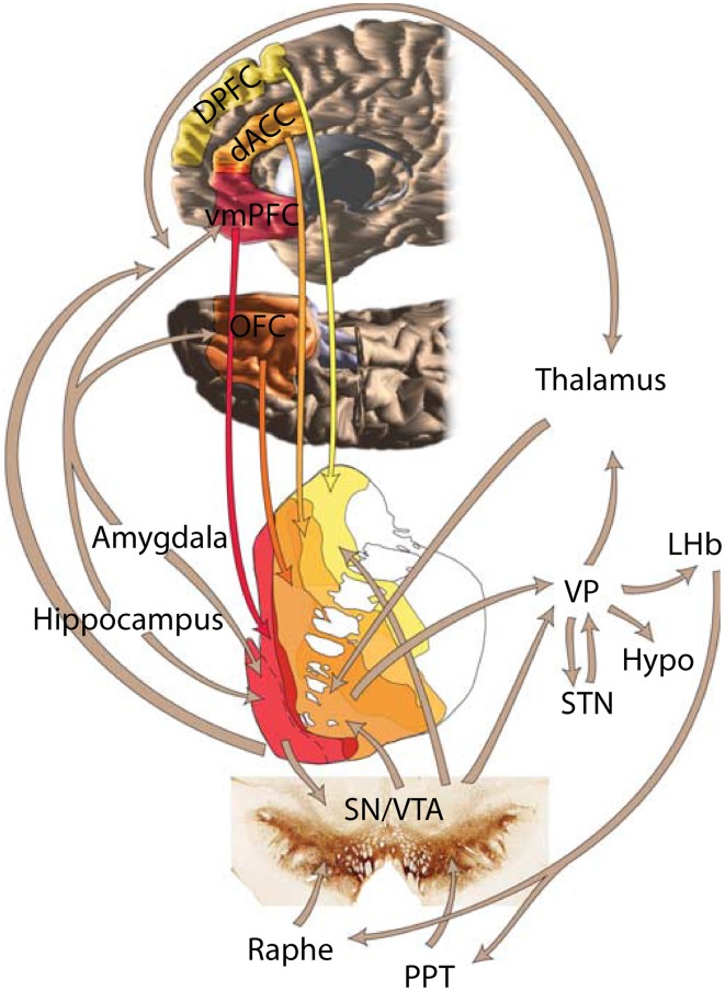
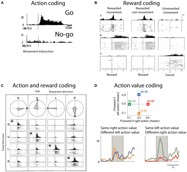
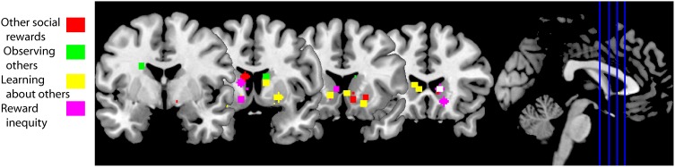
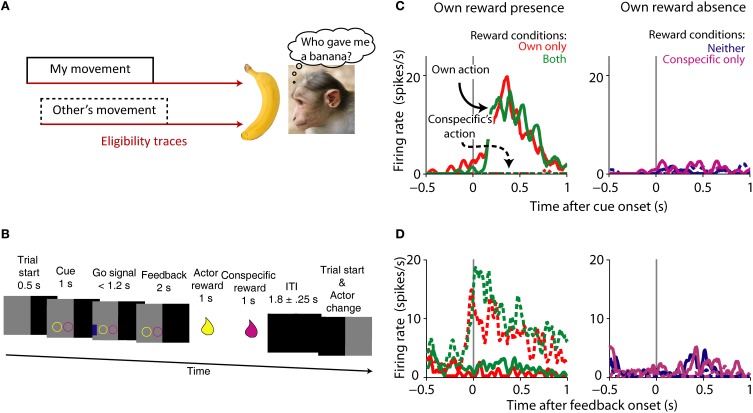
Where and how does the brain code reward during social behavior? Almost all elements of the brain's reward circuit are modulated during social behavior. The striatum in particular is activated by rewards in social situations. However, its role in social behavior is still poorly understood. Here, we attempt to review its participation in social behaviors of different species ranging from voles to humans. Human fMRI experiments show that the striatum is reliably active in relation to others' rewards, to reward inequity and also while learning about social agents. Social contact and rearing conditions have long-lasting effects on behavior, striatal anatomy and physiology in rodents and primates. The striatum also plays a critical role in pair-bond formation and maintenance in monogamous voles. We review recent findings from single neuron recordings showing that the striatum contains cells that link own reward to self or others' actions. These signals might be used to solve the agency-credit assignment problem: the question of whose action was responsible for the reward. Activity in the striatum has been hypothesized to integrate actions with rewards. The picture that emerges from this review is that the striatum is a general-purpose subcortical region capable of integrating social information into coding of social action and reward.

Keywords: agency; human; macaque; rat; social interactions; social neurophysiology; value; vole.


Figures

Figure 1
Figure 1
Depiction of the brain's reward circuit highlighting the role of the striatum and its anatomical connections. Abbreviations: dACC, dorsal anterior cingulate cortex; DPFC, dorsal prefrontal cortex; vmPFC, ventromedial prefrontal cortex; VP, ventral pallidum; LHb, lateral habenula; Hypo, hypothalamus; STN, subthalamic nucleus; SN, substantia nigra; VTA, ventral tegmental area; PPT, pedunculopontine tegmentum. Based on Haber and Knutson (2010), reproduced with permission.
Figure 2
Figure 2
Action and reward coding by striatal neurons. (A) Example striatal neuron active before movement (go) and silent before no-movement (no-go). Based on Schultz and Romo (1988), reproduced with permission. (B) Example striatal neurons coding reward. First row depicts a neuron with phasic active after juice reward delivery independent of the action to obtain reward. Second row depicts a neuron with tonic activity after juice reward delivery. Third row shows a neuron with tonic activity after no reward is delivered. Based on Hollerman et al. (1998), reproduced with permission. (C) Example caudate neuron coding the conjunction of action and reward. This neuron is active during the presentation of a cue indicating the saccade necessary to complete the trial if the trial will be rewarded (rewarded direction is highlighted by a bulls eye). R, right; U, up; L, left; D, down. Polar plots show the average response for each cue and direction. Based on Kawagoe et al. (1998), reproduced with permission. (D) (Top) Depiction of the probability of larger rewards associated with left or right actions on each condition block. Colored numbers refer to the probability associated with left-right actions. (Bottom) Example striatal neuron coding right action value. Based on Samejima et al. (2005), reproduced with permission.
Figure 3
Figure 3
fMRI studies of social behaviors in which the striatum is active. Peak activation coordinates in the striatum of the fMRI studies cited in this review color-coded for each section as illustrated in the legend. Studies using a region of interest analysis strategy were not included in this image. These striatal responses are compatible with a general activation in response to social behaviors, including social rewards. A functional subdivisions according to types of social rewards need to await further experiments. Studies aggregated in “Other social rewards”: (Rilling et al., ; Moll et al., ; Izuma et al., ; Mobbs et al., ; Acevedo et al., ; Fareri et al., ; Korn et al., 2012). Studies clustered in “Observing others”: (Bartels and Zeki, , ; Aron, ; Acevedo et al., 2012). Studies in “Learning about others”: (Delgado et al., ; King-Casas et al., ; Baumgartner et al., ; Burke et al., ; Phan et al., ; Xiang et al., ; Fouragnan et al., 2013). Studies in “Reward inequity”: (Moll et al., ; Fliessbach et al., ; Hsu et al., ; Tricomi et al., 2010).
Figure 4
Figure 4
Agency credit assignment cartoon and striatal neurons coding social action and own reward. (A) Once the monkey receives a banana it needs to know which action produced reward to assign credit. The action can be its own (solid lines) or someone else's (dashed lines). Many actions take place before reward is delivered, therefore looking at a memory of each action or eligibility trace (brown arrows) can solve the agency credit assignment problem. (B) Task sequence for the actor: shape of conditioned cue predicted absence or presence of reward for each animal. Appearance of a subsequent blue go signal was followed by key release, stimulus touch and reward for actor, and later for conspecific. After the ITI the monkeys switched roles as actor and passive. (C) Single striatal neuron coding own action and own reward. Note the higher neuronal activity during own action and own reward compared to own reward absence and conspecific's actions. (D) Single striatal neuron coding social action and own reward. This neuron is active during conspecific's actions that will result in own reward, a complement to the neuron shown in (A). Monkey picture by smerikal (Flickr), reproduced with permission. Panels (B–D) based on Báez-Mendoza et al. (2013), reproduced with permission.

Similar articles

Cited by

References

    1. Acevedo B. P., Aron A., Fisher H. E., Brown L. L. (2012). Neural correlates of long-term intense romantic love. Soc. Cogn. Affect. Neurosci. 7, 145–159 10.1093/scan/nsq092 - DOI - PMC - PubMed
    1. Ambrose L. M., Unterwald E. M., Van Bockstaele E. J. (2004). Ultrastructural evidence for co-localization of dopamine D2 and μ-opioid receptors in the rat dorsolateral striatum. Anat. Rec. A Discov. Mol. Cell. Evol. Biol. 279A, 583–591 10.1002/ar.a.20054 - DOI - PubMed
    1. Anderson J. R. (1998). Social stimuli and social rewards in primate learning and cognition. Behav. Process. 42, 159–175 10.1016/S0376-6357(97)00074-0 - DOI - PubMed
    1. Apicella P., Ljungberg T., Scarnati E., Schultz W. (1991a). Responses to reward in monkey dorsal and ventral striatum. Exp. Brain Res. 85, 491–500 10.1007/BF00231732 - DOI - PubMed
    1. Apicella P., Scarnati E., Schultz W. (1991b). Tonically discharging neurons of monkey striatum respond to preparatory and rewarding stimuli. Exp. Brain Res. 84, 672–675 10.1007/BF00230981 - DOI - PubMed

LinkOut - more resources