Review
. 2013 Dec 10:4:437.
doi: 10.3389/fimmu.2013.00437.

Granuloma formation in pulmonary sarcoidosis

Affiliations
Review

Granuloma formation in pulmonary sarcoidosis

Caroline E Broos et al. Front Immunol. .

Abstract

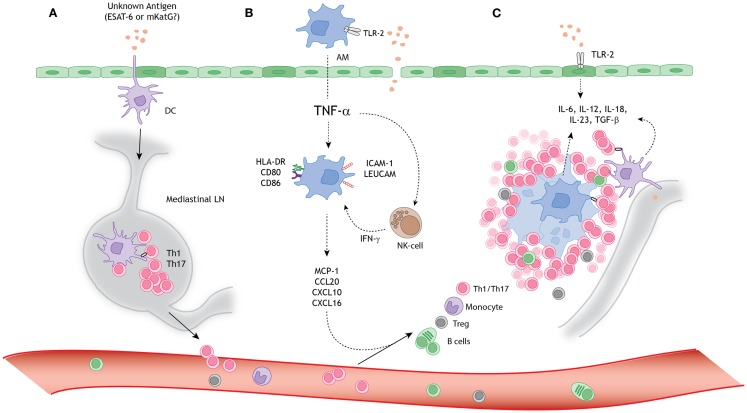
Sarcoidosis is a granulomatous disorder of unknown cause, affecting multiple organs, but mainly the lungs. The exact order of immunological events remains obscure. Reviewing current literature, combined with careful clinical observations, we propose a model for granuloma formation in pulmonary sarcoidosis. A tight collaboration between macrophages, dendritic cells, and lymphocyte subsets, initiates the first steps toward granuloma formation, orchestrated by cytokines and chemokines. In a substantial part of pulmonary sarcoidosis patients, granuloma formation becomes an on-going process, leading to debilitating disease, and sometimes death. The immunological response, determining granuloma sustainment is not well understood. An impaired immunosuppressive function of regulatory T cells has been suggested to contribute to the exaggerated response. Interestingly, therapeutical agents commonly used in sarcoidosis, such as glucocorticosteroids and anti-TNF agents, interfere with granuloma integrity and restore the immune homeostasis in autoimmune disorders. Increasing insight into their mechanisms of action may contribute to the search for new therapeutical targets in pulmonary sarcoidosis.

Keywords: T helper 1 cells; T helper 17 cells; dendritic cells; formation; granuloma; integrity; pulmonary sarcoidosis; regulatory T cells.


Figures

Figure 1
Figure 1
A schematic model for granuloma formation in pulmonary sarcoidosis. An unknown airborne-antigen activates (A) interstitial dendritic cells (DCs), (B) alveolar macrophages (AMs), and (C) alveolar epithelial cells type II (AEC-II) (dark green), simultaneously. This process is initiated by toll-like receptor-2 (TLR-2) ligands, possibly Mycobacterium tuberculosis-derived ESAT-6 or mKatG. (A) The interstitial DCs pick up the putative antigen and migrate toward the mediastinal lymph nodes (LNs), where they initiate differentiation and clonal expansion of T helper (Th)1 and 17 cells. (B) Simultaneously, AMs produce tumor necrosis factor-α (TNF-α), which initiates upregulation of activation (HLA-DR and CD80/86) and adhesion (ICAM-1 and LeuCAM) molecules. Macrophages produce chemokine ligands (MCP-1, CCL20, CXCL10, and CXCL16) under stimulation of both TNF-α and natural-killer (NK) cell-derived interferon-γ (INF-γ), thereby attracting Th1/17 cells, monocytes, regulatory T cells (Tregs), and B cells. (C) The lung environment is characterized by the presence of Th1 and Th17 favoring cytokines, such as IL-6, IL-12, IL-18, IL-23, and TGF-β, produced by macrophages, perilymphatic DCs, and AEC-II. Persistent stimulation, mediated by antigen presenting cells (APCs), leads to continuous cellular recruitment to the site of inflammation, which leads to granuloma formation. Tregs infiltrating the granuloma fail to diminish the exaggerated immune response, thereby contributing to granuloma persistence and integrity.

Similar articles

Cited by

References

    1. Hunninghake GW, Costabel U, Ando M, Baughman R, Cordier JF, du Bois R, et al. ATS/ERS/WASOG statement on sarcoidosis. American thoracic society/European respiratory society/World Association of sarcoidosis and other granulomatous disorders. Sarcoidosis Vasc Diffuse Lung Dis (1999) 16(2):149. - PubMed
    1. Guirado E, Schlesinger LS. Modeling the Mycobacterium tuberculosis granuloma-the critical battlefield in host immunity and disease. Front Immunol (2013) 4:98.10.3389/fimmu.2013.00098 - DOI - PMC - PubMed
    1. Moore JWJ, Moyo D, Beattie L, Andrews PS, Timmis J, Kaye PM. Functional complexity of the Leishmania granuloma and the potential of in silico modeling. Front Immunol (2013) 4:98.10.3389/fimmu.2013.00035 - DOI - PMC - PubMed
    1. Newman LS, Rose CS, Bresnitz EA, Rossman MD, Barnard J, Frederick M, et al. A case control etiologic study of sarcoidosis: environmental and occupational risk factors. Am J Respir Crit Care Med (2004) 170(12):1324–3010.1164/rccm.200402-249OC - DOI - PubMed
    1. Eishi Y, Suga M, Ishige I, Kobayashi D, Yamada T, Takemura T, et al. Quantitative analysis of mycobacterial and propionibacterial DNA in lymph nodes of Japanese and European patients with sarcoidosis. J Clin Microbiol (2002) 40(1):198–20410.1128/JCM.40.1.198-204.2002 - DOI - PMC - PubMed