

doi: 10.1063/1.4771544.
eCollection 2012.
Dielectrophoretic properties of engineered protein patterned colloidal particles
Affiliations
- PMID: 24339848
- PMCID: PMC3555509
- DOI: 10.1063/1.4771544
Item in Clipboard
Dielectrophoretic properties of engineered protein patterned colloidal particles
Biomicrofluidics.
.
Abstract
This work determines the dielectrophoretic response of surface modified polystyrene and silica colloidal particles by experimentally measuring their Clausius-Mossotti factors. Commercial charged particles, fabricated ones coated with fibronectin, and Janus particles that have been grafted with fibronectin on one side only were investigated. We show that the dielectrophoretic response of such particles can be controlled by the modification of the chemistry or the anisotropy of their surface. Moreover, by modelling the polarizabilities of those particles, the dielectric parameters of the particles and the grafted layer of protein can be measured.
Figures
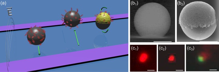
Schematic representation, SEM, and fluorescence images of the investigated particles. (a) Dielectrophoretic responses of isotropic and anisotropic particles coated with fibronectin. (b) SEM images of plain PS (b1) and Janus particles (100 nm Au/1 μm PS) (b2). (c) Fluorescence images of 1 μm PS particle coated with fibronectin (adsorption (c1) or covalent coupling (c2)) and JP ((c3) green: fluorescence stained PS particle side, red: fibronectin on Au side). The difference is size between adsorbed (c1) and covalent coupling (c2), however, the same size, may be the difference in the scattered fluorescence intensity of those particles. The present work shows a much higher surface concentration of fluorescent fibrinogen in the adsorption coupling than the one in the covalent coupling. The scale bar indicates 1 μm.
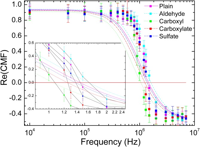
for 400 nm PS functionalized particles: Aldehyde, carboxyl, carboxylate, and sulfate. Experimental values are represented by dots for all tested frequencies whereas fitted functions are represented in dashed lines.
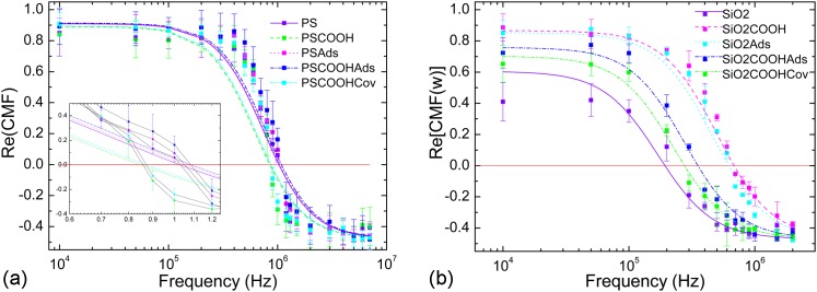
experimentally determined for (a) PS and (b) functionalized particles. The symbol Ads stands for adsorption coupling and Cov for covalent coupling method. Experimental values are represented by dots for all tested frequencies whereas fitted functions are represented in dashed lines.
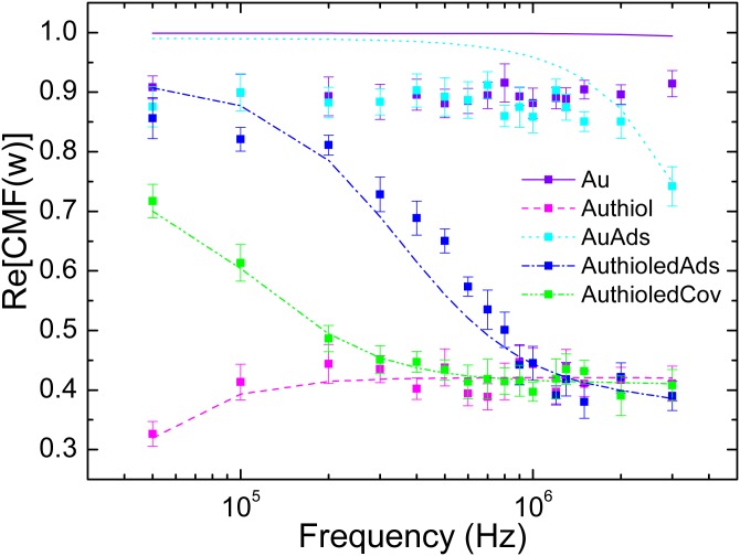
experimentally determined for functionalized Au particles. Experimental values are represented by dots for all tested frequencies whereas fitted functions are represented in dashed lines. Particles were first coated with thiols and then with proteins according to adsorption and covalent coupling protocols.
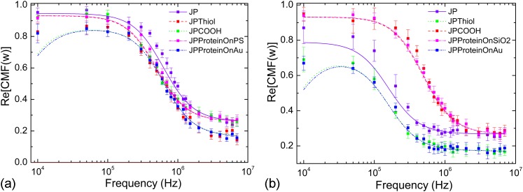
experimentally determined for (a) PS and (b) JP particles. The symbols JP, JP-Thiol, and JPCOOH stand, respectively, for plain JPs (PS or with 100 nm Ti/Au), JPs whom Au side has been alkylated, and JPs that present carboxylate COOH function on their PS or side. The symbols JPProteinOn stand for JPs that have been selectively covalently coupled with fibronectin on the specified side. Experimental values are represented by dots for all tested frequencies whereas fitted functions are represented in dashed lines.
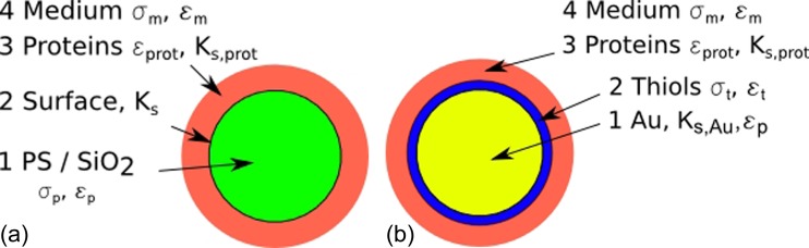
Core-shell model for (a): particles functionalized with proteins and (b): Au particles functionalized with thiols and proteins.
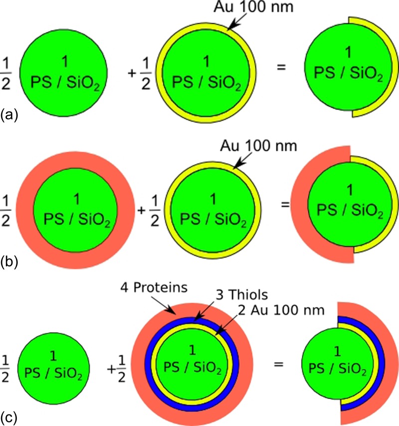
Models and expressions used to compute the CMF of Janus particles, (a) plain Janus particles, (b) Janus particles with proteins grafted on their dielectric side (PS or ), and (c) Janus particles with proteins on their Au side.
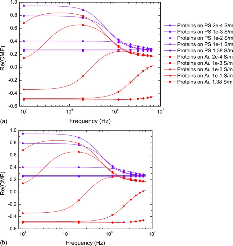
Real part of the Clausius-Mossotti factor of several JPs suspended in increasing media conductivities for (a): 1 μm PS/100 nm Au JPs selectively functionalized with fibronectin and (b):1 μm Au particles selectively functionalized with thiols and proteins. An unusual behaviour is predicted in high conductivity medium where JPs could present dual p-DEP and n-DEP motion.
Similar articles
-
Measurement of the real part of the Clausius-Mossotti factor of dielectrophoresis for Brownian particles.Electrophoresis. 2020 Jan;41(1-2):137-147. doi: 10.1002/elps.201900345. Epub 2019 Nov 11. Electrophoresis. 2020. PMID: 31661554
-
Dielectrophoretic Manipulation of Janus Particle in Conductive Media for Biomedical Applications.Biotechnol J. 2020 Dec;15(12):e2000343. doi: 10.1002/biot.202000343. Epub 2020 Nov 1. Biotechnol J. 2020. PMID: 33067912
-
Polarization behavior of polystyrene particles under direct current and low-frequency (<1 kHz) electric fields in dielectrophoretic systems.Electrophoresis. 2016 Feb;37(4):635-44. doi: 10.1002/elps.201500338. Epub 2015 Dec 15. Electrophoresis. 2016. PMID: 26531799
-
Synthesis, Transformation, and Utilization of Monodispersed Colloidal Spheres.Acc Chem Res. 2019 Dec 17;52(12):3475-3487. doi: 10.1021/acs.accounts.9b00490. Epub 2019 Dec 3. Acc Chem Res. 2019. PMID: 31793763 Free PMC article. Review.
-
Janus colloidal particles: preparation, properties, and biomedical applications.ACS Appl Mater Interfaces. 2013 Mar;5(6):1857-69. doi: 10.1021/am302528g. Epub 2013 Mar 7. ACS Appl Mater Interfaces. 2013. PMID: 23394306 Review.
Cited by
-
High-throughput particle manipulation by hydrodynamic, electrokinetic, and dielectrophoretic effects in an integrated microfluidic chip.Biomicrofluidics. 2013 Mar 20;7(2):24106. doi: 10.1063/1.4795856. eCollection 2013. Biomicrofluidics. 2013. PMID: 24404011 Free PMC article.
-
Dielectrophoresis: Developments and applications from 2010 to 2020.Electrophoresis. 2021 Mar;42(5):539-564. doi: 10.1002/elps.202000156. Epub 2020 Dec 28. Electrophoresis. 2021. PMID: 33191521 Free PMC article. Review.
-
Comprehensive analysis of human cells motion under an irrotational AC electric field in an electro-microfluidic chip.PLoS One. 2014 Apr 15;9(4):e95231. doi: 10.1371/journal.pone.0095231. eCollection 2014. PLoS One. 2014. PMID: 24736275 Free PMC article.
-
Measurement of the Imaginary Part of the Clausius-Mossotti Factor of Particle/Cell via Dual Frequency Electrorotation.Micromachines (Basel). 2020 Mar 22;11(3):329. doi: 10.3390/mi11030329. Micromachines (Basel). 2020. PMID: 32235798 Free PMC article.
-
Study on non-bioparticles and Staphylococcus aureus by dielectrophoresis.RSC Adv. 2020 Jan 15;10(5):2598-2614. doi: 10.1039/c9ra05886a. eCollection 2020 Jan 14. RSC Adv. 2020. PMID: 35496126 Free PMC article.
References
LinkOut - more resources
Full Text Sources