. 2013 Dec 5;8(12):e80944.
doi: 10.1371/journal.pone.0080944. eCollection 2013.

Evaluation of the toxic potential of graphene copper nanocomposite (GCNC) in the third instar larvae of transgenic Drosophila melanogaster (hsp70-lacZ)Bg(9.)

Affiliations

Evaluation of the toxic potential of graphene copper nanocomposite (GCNC) in the third instar larvae of transgenic Drosophila melanogaster (hsp70-lacZ)Bg(9.)

Yasir Hasan Siddique et al. PLoS One. .

Erratum in

  • PLoS One. 2014;9(3):e93127

Abstract

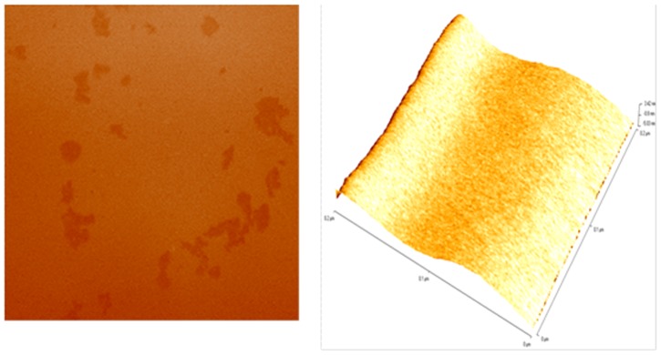
Graphene, a two-dimensional carbon sheet with single-atom thickness, have attracted the scientific world for its potential applications in various field including the biomedical areas. In the present study the graphene copper nanocomposite (GCNC) was synthesized, characterized and evaluated for its toxic potential on third instar larvae of transgenic Drosophila melanogaster (hsp70-lacZ)Bg(9) . The synthesized GCNC was analyzed by X-ray diffraction (XRD), scanning/transmission electron microscopy (SEM/TEM), atomic force microscopy (AFM), and fourier transform infrared spectroscopy (FTIR). The GCNC in 0.1% DMSO was sonicated for 10 min and the final concentration of 0.033, 0.099, 0.199 and 3.996 µg/µl of diet were established. The third instar larvae were allowed to feed on it separately for 24 and 48 hrs. The hsp70 expression was measured by O-nitrophenyl-β-D-galactopyranoside assay, tissue damage by trypan blue exclusion test and β-galactosidase activity was monitored by in situ histochemical β-galactosidase staining. Oxidative stress was monitored by performing lipid peroxidation assay and total protein estimation. Ethidium bromide/acridine orange staining was performed on midgut cells for apoptotic index and the comet assay was performed for the DNA damage. The results of the present study showed that the exposure of 0.199 and 3.996 µg/µl of GCNC were toxic for 24 hr of exposure and for 48 hr of exposure: 0.099, 0.199 and 3.996 µg/µl of GCNC was toxic. The dose of 0.033 µg/µl of GCNC showed no toxic effects on its exposure to the third instar larvae for 24 hr as well as 48 hrs. This dose can be considered as No Observed Adverse Effect Level (NOAEL).


Conflict of interest statement

Competing Interests: The authors have declared that no competing interests exist.

Figures

Figure 1
Figure 1. UV-Vis spectra for GO (a) and graphene-copper nanocomposite (b).
Figure 2
Figure 2
Figure 2a. XRD pattern of GCNC. Figure 2b. Scanning electron micrograph of GCNC. Figure 2c. Energy disperse spectrum of GCNC. Figure 2d. Transmission electron micrograph of the GCNC.
Figure 3
Figure 3. Atomic micrograph of the GCNC.
Figure 4
Figure 4. FT-IR spectrum of GCNC.
Figure 5
Figure 5. β-galactosidase activity measured in transgenic Drosophila melanogaster(hsp70-lacZ)Bg9 third instar larvae exposed to different doses of Graphene copper nano composite (GCNC) for 24 and 48 hrs.
*significant at p<0.05 with respect to untreated [GCNC = Graphene copper nano composite; GONP = Graphene oxide nano particle; CONP = Cuprous oxide nano particle; NC = Negative control; DMSO = Dimethyl sulphoxide; OD = Optical Density; SE = Standard error].
Figure 6
Figure 6. blue staining in the tissues of third instar larvae of transgenic Drosophila melanogaster (hsp70-lacZ)Bg9 for untreated (a) and the larvae exposed to different doses of graphene copper nanocomposite (GCNC) for 48 hr of duration [0.099 µg/µl (b) 0.199 µg/µl (c) 3.996 µg/µl (d)].
[BG- Brain ganglia, SG- Salivary gland, PV- Proventriculus, FG- Foregut, MG-Midgut, HG- Hindgut, MT- Malpighian tubule, GC- Gastric caeca].
Figure 7
Figure 7. β-galactosidase staining pattern in the tissues of third instar larvae of transgenic Drosophila melanogaster (hsp70-lacZ)Bg9 for untreated (a) and the larvae exposed to different doses of graphene copper nanocomposite (GCNC) for 48 hr of duration [0.099 µg/µl (b) 0.199 µg/µl (c) 3.996 µg/µl(d)].
[BG- Brain ganglia, SG- Salivary gland, PV- Proventriculus, FG- Foregut, MG-Midgut, HG- Hindgut, MT- Malpighian tubule, GC- Gastric caeca].
Figure 8
Figure 8. Lipid peroxidation in the third instar larvae of transgenic Drosophila melanogaster (hsp70-lacZ)Bg9 exposed to different doses of Graphene copper nano composite (GCNC) for 24 and 48 hrs.
*significant at p<0.05 with respect to untreated[GCNC = Graphene copper nano composite; GONP = Graphene oxide nano particle; CONP = Cuprous oxide nano particle; NC = Negative control; DMSO = Dimethyl sulphoxide; OD = Optical Density; SE = Standard error].
Figure 9
Figure 9. Protein content in the third instar larvae of transgenic Drosophila melanogaster(hsp70-lacZ)Bg9 exposed to different doses of Graphene copper nano composite (GCNC) for 24 and 48 hrs.
*significant at p<0.05 with respect to untreated [GCNC = Graphene copper nano composite; GONP = Graphene oxide nano particle; CONP = Cuprous oxide nano particle; NC = Negative control; DMSO = Dimethyl sulphoxide; SE = Standard error].
Figure 10
Figure 10. Drosophila melanogaster(hsp70-lacZ)Bg9 mid gut cells (a) Normal cell; (b) Apoptotic cell and (c) Comet assay performed in gut cell exposed to 3.996 µg/µl of GCNC for 48 hrs of duration.
Figure 11
Figure 11. Apoptotic index measure in the midgut cells of the third instar larvae of transgenic Drosophila melanogaster(hsp70-lacZ)Bg9 exposed to different doses of graphene copper nano composite (GCNC) for 24 and 48 hrs.
*significant at p<0.05 with respect to untreated [GCNC = Graphene copper nano composite; GONP = Graphene oxide nano particle; CONP = Cuprous oxide nano particle; NC = Negative control; DMSO = Dimethyl sulphoxide; SE = Standard error].
Figure 12
Figure 12. Comet assay performed on the midgut cells of the third instar larvae of transgenic Drosophila melanogaster(hsp70-lacZ)Bg9 exposed to different doses of Graphene copper nano composite (GCNC) for 24 and 48 hrs.
*significant at p<0.05 with respect to untreated [GCNC = Graphene copper nano composite; GONP = Graphene oxide nano particle; CONP = Cuprous oxide nano particle; NC = Negative control; DMSO = Dimethyl sulphoxide; SE = Standard error].

References

    1. Chang Y, Yang ST, Liu JH, Dong E, Wang Y, et al. (2011) Invitro toxicity evaluation of graphene oxide on A549 cells. Toxicology Letters 200: 201–210. - PubMed
    1. Pumera M (2009) Electrochemistry of graphene: new horizons for sensing and energy storage. The Chemical Record 9: 211–23. - PubMed
    1. Willemse CM, Tlhomelang K, Jahed N, Baker PG, Iwuoha EI (2011) Metallo-graphene nanocomposite electrocatalytic platform for the determination of toxic metal ions. Sensors 11: 3970–3987. - PMC - PubMed
    1. Aillon KL, Xie YM, El-Gendy N, Berkland CJ, Forrest ML (2009) Effects of nanomaterial physicochemical properties on in vivo toxicity. Advanced Drug Delivery Reviews 61: 457–466. - PMC - PubMed
    1. Cao A, Liu Z, Chu S, Wu M, Ye Z, et al. (2010) A Facile One-step Method to Produce Graphene–CdS Quantum Dot Nanocomposites as Promising Optoelectronic Materials. Advanced Materials 22: 103–106. - PubMed

LinkOut - more resources