

Learning reflectance confocal microscopy of melanocytic skin lesions through histopathologic transversal sections
- PMID: 24339910
- PMCID: PMC3855214
- DOI: 10.1371/journal.pone.0081205
Learning reflectance confocal microscopy of melanocytic skin lesions through histopathologic transversal sections
Abstract
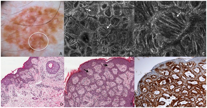
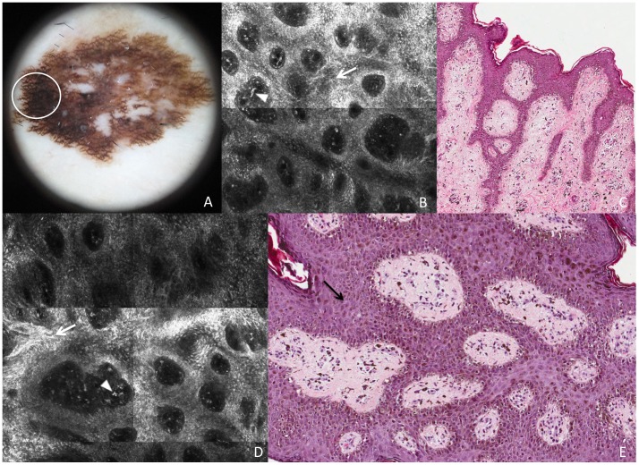
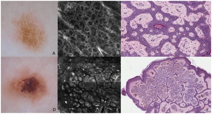
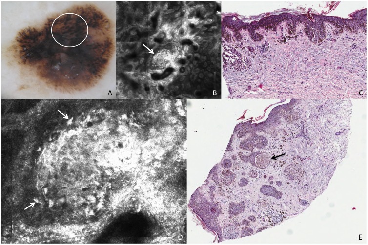
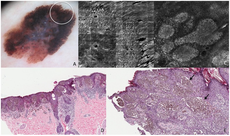
Histopathologic interpretation of dermoscopic and reflectance confocal microscopy (RCM) features of cutaneous melanoma was timidly carried out using perpendicular histologic sections, which does not mimic the same plane of the image achieved at both techniques (horizontal plane). The aim of this study was to describe the transverse histologic sections research technique and correlate main dermoscopic features characteristic of cutaneous melanoma (atypical network, irregular globules and pseudopods) with RCM and histopathology in perpendicular and transverse sections in order to offer a more precise interpretation of in vivo detectable features. Four melanomas and 2 nevi with different dermoscopic clues have been studied. Lesion areas that showed characteristic dermoscopic features were imaged by dermoscopy and confocal microscopy and directly correlated with histopathology in perpendicular and transverse sections. We presented the possibility to perform transverse sections as a new approach to understand RCM features. Atypical network showed different aspects in the 2 melanomas: in one case it was characterized by pleomorphic malignant melanocytes with tendency to form aggregates, whereas in the other elongated dendritic cells crowded around dermal papillae, some of them forming bridges that resembled the mitochondrial aspect at confocal and histopathology transversal sections. Pigment globules in melanomas and nevi differed for the presence of large atypical cells in the former, and pseudopods showed up as elongated nests protruded toward the periphery of the lesion. Transverse histologic research sections have a consistent dermoscopic and confocal correlate, and it may represent an help in confocal feature interpretation and an advance in improving melanoma diagnosis and knowledge of the biology of melanocytic lesions.
Conflict of interest statement
Figures
Similar articles
-
Role of In Vivo Reflectance Confocal Microscopy in the Analysis of Melanocytic Lesions.Acta Dermatovenerol Croat. 2018 Apr;26(1):64-67. Acta Dermatovenerol Croat. 2018. PMID: 29782304 Review.
-
In vivo confocal microscopic and histopathologic correlations of dermoscopic features in 202 melanocytic lesions.Arch Dermatol. 2008 Dec;144(12):1597-608. doi: 10.1001/archderm.144.12.1597. Arch Dermatol. 2008. PMID: 19075142
-
A comparative dermoscopic and reflectance confocal microscopy study of naevi and melanoma with negative pigment network.J Eur Acad Dermatol Venereol. 2019 Dec;33(12):2273-2282. doi: 10.1111/jdv.15784. Epub 2019 Aug 19. J Eur Acad Dermatol Venereol. 2019. PMID: 31283045
-
Correlation of dermoscopic structures of melanocytic lesions to reflectance confocal microscopy.Arch Dermatol. 2007 Feb;143(2):176-85. doi: 10.1001/archderm.143.2.176. Arch Dermatol. 2007. PMID: 17309998 Clinical Trial.
-
Differentiating between early melanomas and melanocytic nevi: A state-of-the-art review.Pathol Res Pract. 2023 Sep;249:154734. doi: 10.1016/j.prp.2023.154734. Epub 2023 Aug 4. Pathol Res Pract. 2023. PMID: 37573619 Review.
Cited by
-
Case Report: melanoma and melanocytic nevus differentiation with reflectance confocal microscopy.F1000Res. 2015 Jul 15;4:257. doi: 10.12688/f1000research.6793.1. eCollection 2015. F1000Res. 2015. PMID: 26236471 Free PMC article.
-
Reflectance confocal endomicroscope with optical axial scanning for in vivo imaging of the oral mucosa.Biomed Opt Express. 2014 Oct 1;5(11):3781-91. doi: 10.1364/BOE.5.003781. eCollection 2014 Nov 1. Biomed Opt Express. 2014. PMID: 25426310 Free PMC article.
-
An intuitive explanation of dermoscopic structures by digitally reconstructed pathological horizontal top-down view images.Sci Rep. 2019 Dec 27;9(1):19875. doi: 10.1038/s41598-019-56522-8. Sci Rep. 2019. PMID: 31882764 Free PMC article.
-
Diagnosis and Management of Lentigo Maligna: Clinical Presentation and Comprehensive Review.J Skin Cancer. 2021 Jul 24;2021:7178305. doi: 10.1155/2021/7178305. eCollection 2021. J Skin Cancer. 2021. PMID: 34350036 Free PMC article. Review.
-
Optical imaging guided- 'precision' biopsy of skin tumors: a novel approach for targeted sampling and histopathologic correlation.Arch Dermatol Res. 2021 Sep;313(7):517-529. doi: 10.1007/s00403-020-02126-6. Epub 2020 Aug 25. Arch Dermatol Res. 2021. PMID: 32844312 Free PMC article.
References
-
- Argenziano G, Soyer HP, Chimenti S, Talamini R, Corona R, et al. (2003) Dermoscopy of pigmented skin lesions: results of a consensus meeting via the Internet. Journal of the American Academy of Dermatology 48: 679–693. - PubMed
-
- Pehamberger H, Steiner A, Wolff K (1987) In vivo epiluminescence microscopy of pigmented skin lesions. I. Pattern analysis of pigmented skin lesions. Journal of the American Academy of Dermatology 17: 571–583. - PubMed
-
- Guitera P, Menzies SW, Longo C, Cesinaro AM, Scolyer RA, et al. (2012) In vivo confocal microscopy for diagnosis of melanoma and basal cell carcinoma using a two-step method: analysis of 710 consecutive clinically equivocal cases. The Journal of investigative dermatology 132: 2386–2394. - PubMed
-
- Longo C, Zalaudek I, Argenziano G, Pellacani G (2012) New directions in dermatopathology: in vivo confocal microscopy in clinical practice. Dermatologic clinics 30: 799–814. - PubMed
-
- Pellacani G, Longo C, Malvehy J, Puig S, Carrera C, et al. (2008) In vivo confocal microscopic and histopathologic correlations of dermoscopic features in 202 melanocytic lesions. Archives of dermatology 144: 1597–1608. - PubMed
Publication types
MeSH terms
LinkOut - more resources
Full Text Sources
Other Literature Sources
Medical
