

CT angiography of the head-and-neck vessels acquired with low tube voltage, low iodine, and iterative image reconstruction: clinical evaluation of radiation dose and image quality
- PMID: 24339936
- PMCID: PMC3855260
- DOI: 10.1371/journal.pone.0081486
CT angiography of the head-and-neck vessels acquired with low tube voltage, low iodine, and iterative image reconstruction: clinical evaluation of radiation dose and image quality
Abstract
Objectives: We aimed to assess the effectiveness and feasibility of head-and-neck Computed Tomography Angiography (CTA) with low tube voltage and low concentration contrast media combined with iterative reconstruction algorithm.
Methods: 92 patients were randomly divided into group A and B: patients in group A received a conventional scan with 120 kVp and contrast media of 320 mgI/ml. Patients in group B, 80 kVp and contrast media of 270 mgI/ml were used along with iterative reconstruction algorithm techniques. Image quality, radiation dose and the effectively consumed iodine amount between two groups were analyzed and compared.
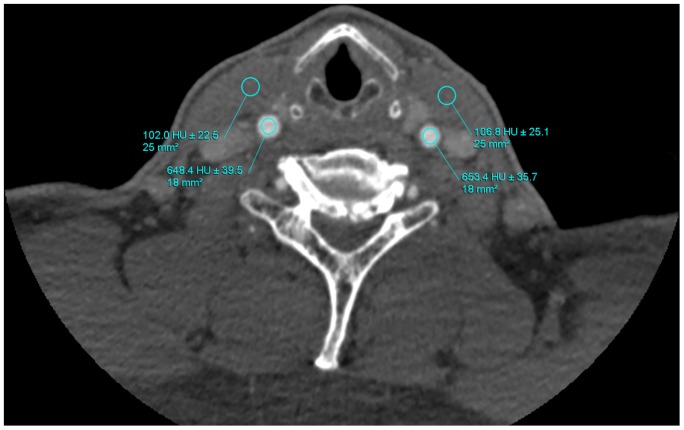
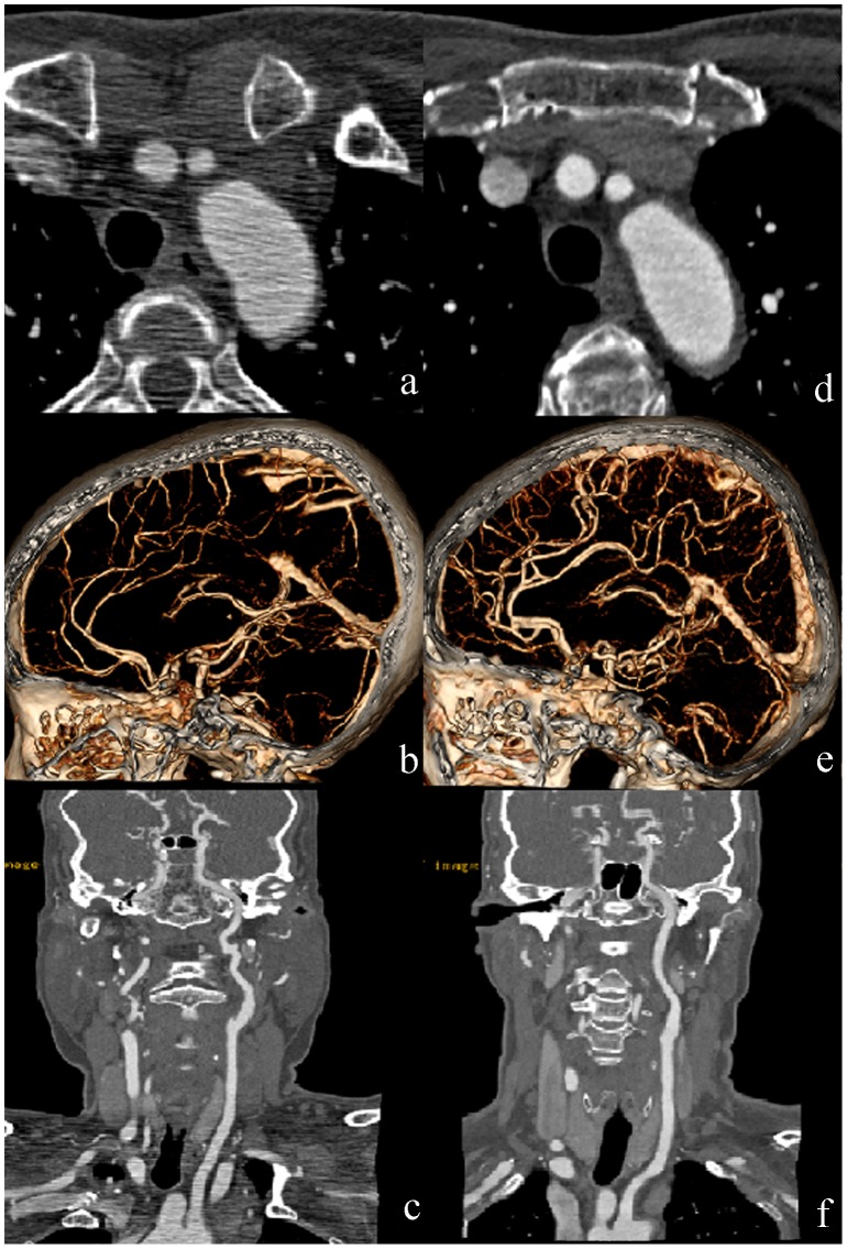
Results: Image quality of CTA of head-and-neck vessels obtained from patients in group B was significantly improved quantitatively and qualitatively. In addition, CT attenuation values in group B were also significantly higher than that in group A (p<0.001). Furthermore, compared with the protocol whereby 120 kVp and 320 mgI/dl were administrated, the mean radiation dose and consumed iodine amount in protocol B were also reduced by 50% and 15.6%, respectively (p<0.001).
Conclusions: With the help of iterative reconstruction algorithm techniques, the head-and-neck CTA with diagnostic quality can be adequately acquired with low tube voltage and low concentration contrast media. This method could be potentially extended to include any part of the body to reduce the risks related to ionizing radiation.
Conflict of interest statement
Figures
References
-
- Brenner DJ, Hall EJ (2007) Computed tomography–an increasing source of radiation exposure. N Engl J Med 357: 2277–2284. - PubMed
MeSH terms
Substances
LinkOut - more resources
Full Text Sources
Other Literature Sources
