

Circulating endothelial cells in patients with venous thromboembolism and myeloproliferative neoplasms
- PMID: 24339944
- PMCID: PMC3855326
- DOI: 10.1371/journal.pone.0081574
Circulating endothelial cells in patients with venous thromboembolism and myeloproliferative neoplasms
Abstract
Background: Circulating endothelial cells (CEC) may be a biomarker of vascular injury and pro-thrombotic tendency, while circulating endothelial progenitor cells (CEP) may be an indicator for angiogenesis and vascular remodelling. However, there is not a universally accepted standardized protocol to identify and quantify these cells and its clinical relevancy remains to be established.
Objectives: To quantify CEC and CEP in patients with venous thromboembolism (VTE) and with myeloproliferative neoplasms (MPN), to characterize the CEC for the expression of activation (CD54, CD62E) and procoagulant (CD142) markers and to investigate whether they correlate with other clinical and laboratory data.
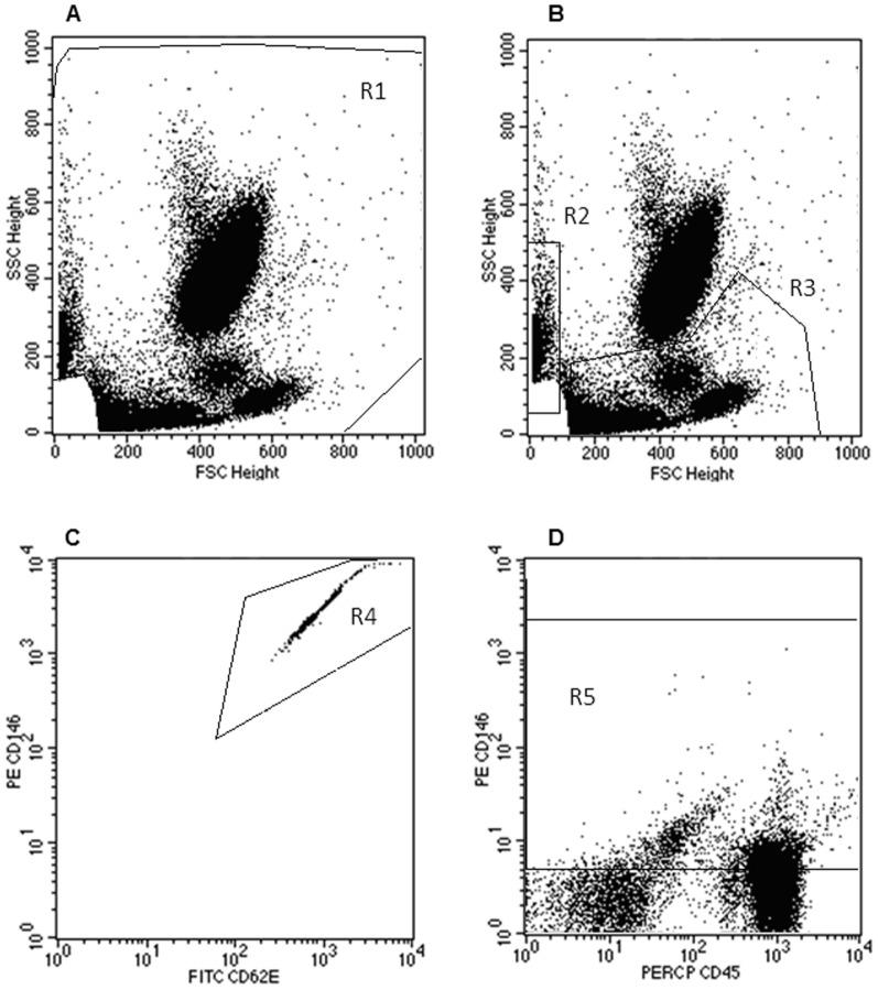
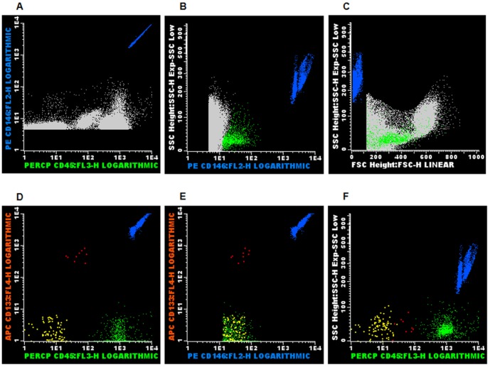
Patients and methods: Sixteen patients with VTE, 17 patients with MPN and 20 healthy individuals were studied. The CEC and CEP were quantified and characterized in the blood using flow cytometry, and the demographic, clinical and laboratory data were obtained from hospital records.
Results: We found the CEC counts were higher in both patient groups as compared to controls, whereas increased numbers of CEP were found only in patients with MPN. In addition, all disease groups had higher numbers of CD62E+ CEC as compared to controls, whereas only patients with VTE had increased numbers of CD142+ and CD54+ CEC. Moreover, the numbers of total and CD62+ CEC correlated positively with the white blood cells (WBC) counts in both groups of patients, while the numbers of CEP correlated positively with the WBC counts only in patients with MPN. In addition, in patients with VTE a positive correlation was found between the numbers of CD54+ CEC and the antithrombin levels, as well as between the CD142+ CEC counts and the number of thrombotic events.
Conclusions: Our study suggests that CEC counts may reveal endothelial injury in patients with VTE and MPN and that CEC may express different activation-related phenotypes depending on the disease status.
Conflict of interest statement
Figures
Similar articles
-
Soluble endothelial cell molecules and circulating endothelial cells in patients with venous thromboembolism.Blood Coagul Fibrinolysis. 2017 Dec;28(8):589-595. doi: 10.1097/MBC.0000000000000650. Blood Coagul Fibrinolysis. 2017. PMID: 28661913
-
Circulating endothelial cells in essential thrombocythemia and polycythemia vera: correlation with JAK2-V617F mutational status, angiogenic factors and coagulation activation markers.Int J Hematol. 2010 Jun;91(5):792-8. doi: 10.1007/s12185-010-0596-7. Epub 2010 May 15. Int J Hematol. 2010. PMID: 20473593
-
Risk factors for vascular complications and treatment patterns at diagnosis of 2389 PV and ET patients: Real-world data from the Swedish MPN Registry.Eur J Haematol. 2017 Jun;98(6):577-583. doi: 10.1111/ejh.12873. Epub 2017 Apr 3. Eur J Haematol. 2017. PMID: 28251679
-
The paradox of platelet activation and impaired function: platelet-von Willebrand factor interactions, and the etiology of thrombotic and hemorrhagic manifestations in essential thrombocythemia and polycythemia vera.Semin Thromb Hemost. 2006 Sep;32(6):589-604. doi: 10.1055/s-2006-949664. Semin Thromb Hemost. 2006. PMID: 16977569 Review.
-
Pathogenesis of thrombosis in essential thrombocythemia and polycythemia vera: the role of neutrophils.Semin Hematol. 2005 Oct;42(4):239-47. doi: 10.1053/j.seminhematol.2005.05.023. Semin Hematol. 2005. PMID: 16210037 Review.
Cited by
-
Thrombosis in Philadelphia negative classical myeloproliferative neoplasms: a narrative review on epidemiology, risk assessment, and pathophysiologic mechanisms.J Thromb Thrombolysis. 2018 May;45(4):516-528. doi: 10.1007/s11239-018-1623-4. J Thromb Thrombolysis. 2018. PMID: 29404876 Review.
-
Comparative Mutational Profiling of Hematopoietic Progenitor Cells and Circulating Endothelial Cells (CECs) in Patients with Primary Myelofibrosis.Cells. 2021 Oct 15;10(10):2764. doi: 10.3390/cells10102764. Cells. 2021. PMID: 34685741 Free PMC article.
-
Circulating Endothelial Cell Levels Correlate with Treatment Outcomes of Splanchnic Vein Thrombosis in Patients with Chronic Myeloproliferative Neoplasms.J Pers Med. 2022 Feb 27;12(3):364. doi: 10.3390/jpm12030364. J Pers Med. 2022. PMID: 35330364 Free PMC article.
-
Liquid Biopsy and Potential Liquid Biopsy-Based Biomarkers in Philadelphia-Negative Classical Myeloproliferative Neoplasms: A Systematic Review.Life (Basel). 2021 Jul 10;11(7):677. doi: 10.3390/life11070677. Life (Basel). 2021. PMID: 34357048 Free PMC article. Review.
-
Expression of CD markers in JAK2V617F positive myeloproliferative neoplasms: Prognostic significance.Oncol Rev. 2018 Oct 2;12(2):373. doi: 10.4081/oncol.2018.373. eCollection 2018 Jul 4. Oncol Rev. 2018. PMID: 30405895 Free PMC article.
References
-
- Wu KK, Thiagarajan P (1996) Role of endothelium in thrombosis and hemostasis. Annu Rev Med 47: 315–331. - PubMed
-
- Aird WC (2007) Phenotypic heterogeneity of the endothelium: II. Representative vascular beds. Circ Res 100: 174–190. - PubMed
-
- Cines DB, Pollak ES, Buck CA, Loscalzo J, Zimmerman GA, et al. (1998) Endothelial cells in physiology and in the pathophysiology of vascular disorders. Blood 91: 3527–3561. - PubMed
-
- Bevilacqua MP (1993) Endothelial-leukocyte adhesion molecules. Annual review of immunology 11: 767–804. - PubMed
Publication types
MeSH terms
Substances
LinkOut - more resources
Full Text Sources
Other Literature Sources
